
Abstract
This study estimates the influence of five Al2O3 nanopowders on the growth of Scenedesmus quadricauda depending on the physical and chemical properties of the alumina nanoparticles and on the concentration of the nanopowder. The agglomeration and crystallisation levels of the alumina nanopowders as well as the zeta potential, the conductivity and the pH of nanopowders-containing growth media are considered. The strongest algal growth inhibiting effect was accompanied by the highest degree of agglomeration and the highest crystallisation level as well as the smallest specific surface area.
Keywords
Introduction
The extremely wide range of applications of nanomaterials, including in cosmetics (TiO2, ZnO), polishing materials (Al2O3), catalysis (rare earth metal oxides), fuel cells (CeO2), electronics (SnO2), medicine (e.g. implants), production of conducting pastes, and paints,1 results in rising levels of their production and – in consequence – their emission to the environment. The lack of knowledge regarding the fate and the reactivity of various types of nanoparticles in natural systems and living organisms makes it difficult to estimate the impact of nanomaterials on the environment.2
There is a large amount of research data evidencing the influence of nanoparticles on living cells. It has been found that titanium dioxide acts as an antibacterial agent showing photocatalytic behaviour. MgO and Mg nanoparticles have been found to be active against both Gram-positive and Gram-negative bacteria and bacterial spores. Silver compounds have been seen to be effective antimicrobials in the case of wastewater coliforms as well as Staphylococcus aureus, Klebsiella pneumoniae, and Pseudomonas aruginosa.3 Antibacterial and antifungal activity of silver nanoparticles was observed by Jastrzębska et al.4 Luminescent silica particles at concentrations of over 0·1 mg L−1 show a toxic effect on cell DNA.5 Interactions of ZnO, TiO2 and CuO nanoparticles with the microalga Pseudokirchneriella subcapitata result in the prolongation of the lag phase of algal growth and the formation of characteristic aggregates entrapping algal cells in the presence of nano-TiO2.6 EC50/72 h of ZnO nanopowder was 0·04 mg L−1. A large amount of literature data evidences the toxicity of nanoparticles to animal cells.7 Some toxicity tests for the determination of nanoparticle toxicity, using the alga Pseudokirchinella subcapitata, the crustacean Daphnia magna or Thamnocephalus platyurus, the Protozoa Tetrahymena termophila, and the bacteria Vibrio fischeri, have been carried out.8 The influence of metal oxide nanoparticles on marine phytoplankton – Thalassiosira pseudonana, Skeletonema marinoi, Dunaliella tertiolecta and Isochrysis galbana was also observed.9
Various mechanisms of interactions between nanoparticles and living cells have been studied. It has been demonstrated that nanoparticles may provoke alterations of membranes, cell structures and protective mechanisms. Nanoparticles are able to cross into animal cell membranes and become internalised. Inside the cell, nanoparticles may be bound to various cell structures (endoplasmic reticulum, Golgi system) and interfere with the metabolic processes of the cell, possibly by forming reactive oxygen species.10 Protista use endocytosis and phagocytosis processes. In unicellular protozoans, some nanotubes have been taken up and localised in mitochondria.7 Some data indicate that inorganic oxidic nanoparticles are able to interact with cells of plants and green algae. Probable mechanisms for plant root adsorption and incorporation into the cell wall or cell membrane have been proposed, but no mechanism of internalisation via ion transporting systems has been postulated because of particle sizes and particles not fitting the binding sites.7 Some chemical changes have been observed in the membranes of nanoparticle exposed bacterial cells, with regard to changes in membrane fatty acids. Pseudomonas putida cells exposed to fullerene nanoparticles show a higher level of cyclopropane fatty acids compared with unexposed cells. In Bacillus subtilis, an increase in iso- and anteiso-branched fatty acid as well as monounsaturated fatty acid content has been observed.2
As a consequence of the current practice of discharging nanoparticle wastes, a lot of them are introduced into wastewater streams or transported to wastewater treatment systems, which results in the contamination of aquatic environments.11 The estimation of the nanomaterial toxicity to algae is of significant importance due to the fact that algae form the basis of aquatic food chains, as a result of which nanoparticles can impact on entire aquatic ecosystems.
It has been demonstrated that the toxicity of nanoparticles depends not only on their concentration in the environment but also on their properties and the ambient solution conditions.12
The aim of this research work was to estimate the influence of prepared Al2O3 nanopowders, differing in physicochemical properties, on the growth of the common European green alga Scenedesmus quadricauda.
Experimental procedure
The aluminium oxide nanopowders used in this work, marked A–D, were prepared from alumina precursors obtained by exposure of triethylaluminium hexane solution to oxygen and moisture from air.13 Obtained in this manner, the nanoalumina precursors were then calcined at 700°C giving Al2O3 nanopowders. Changing the time of triethylaluminium exposure to oxygen and moisture, alumina nanopowders of various morphologies were obtained. Nanopowder E, with larger grains, was a commercial one.
A scanning electron microscope (SEM), Zeiss LEO 1530, was used to study the structure of the nanopowders. They were characterised qualitatively and quantitatively. Prior to the examinations, the powders were evaporated with a thin carbon film.
The qualitative description of the powders was prepared using the MicroMeter computer program. The individual parameters were measured on cross-sections of several single particles and then averaged to obtain their mean values E(x).14, 15
The phase composition of the powders was analysed in a PHILIPS 1830 diffractometer with a copper lamp (Cu Kα = 1·54056 Å), operated with stepwise adjustment of the 2θ angle.
The specific surface area was measured by the BET method using a Quadrasorb device (Quantachrome).
The zeta potential of the powders was determined using a Zetasizer 3000 instrument (Malvern).
A Mettler Toledo MPC 227 meter was used to measure conductivity and pH.
S. quadricauda cultures were grown at room temperature, under continuous artificial lighting using the Jankowski L5 growth medium (KNO3 – 0·1 g L−1; Ca(NO3)2 – 0·1 g L−1; KH2PO4 – 0·04 g L−1; MgSO4.7H2O – 0·03 g L−1; citric acid – 0·003 g L−1; ferric citrate – 0·003 g L−1; distilled water – 1000 mL) supplied with 1 mL L−1 of a microelements solution (H3BO3 – 2·86 g L−1; MnCl2.7H2O – 1·81 g L−1; ZnSO4.7H2O – 0·222 g L−1; MoO3 – 0·176 g L−1; NH4VO3 – 0·23 g L−1; CuSO4.7H2O – 0·01 g L−1; Co(NO3)2.H2O – 0·02 g L−1).
Nanopowders were added to each 200 mL culture, in amounts of 500 and 1000 mg L−1. Two parallel series of algal cultures were grown for each concentration of the nanopowders; the results are presented as average values for both experimental series. The atomic absorption spectroscopy procedure was used to determine the solubility of Al2O3 in growth medium after 7 days storage of the nanopowder–medium mixtures at room temperature.
The number of algal cells was determined by direct counting in a Sedgwick-Rafter chamber, using a light microscope, at the beginning of the experiment and after 7 and 21 days. Cultures grown in the presence of nanopowders were compared with control samples without any nanopowder added. The statistical analysis, including standard deviation and variation coefficient was accomplished.
Results
Stereometric examinations of the nanopowders showed that they had nanometric grain sizes and formed agglomerates. The average grain size in the commercial powder E was estimated to be 600 nm with grain sizes ranging from 100 nm to 3 μm. The grain size of the prepared nanopowders A–D ranged from 10 to 100 nm, the average size not exceeding 45 nm. For nanopowder A, characterised by the greatest average grain size (37 nm), grain sizes ranged from 20 to 50 nm. Nanopowder B had the smallest average grain size (27 nm), with grain sizes ranging from 10 to 50 nm. For nanopowders C and D, the average grain size was found to be 32 and 35 nm respectively. Morphology and grain size distribution of powders A–D are shown in Figs. 1 and 2 respectively.
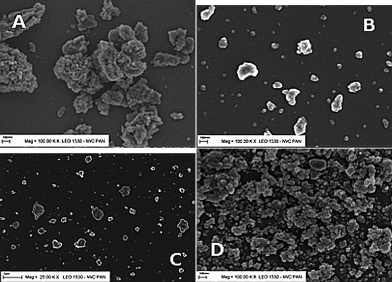
Morphology of alumina nanopowders A, B, C and D
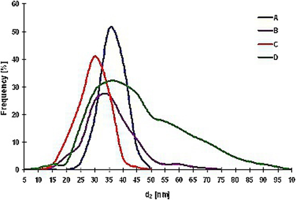
Grain size distribution of nanopowders A–D
Differences were observed in the degree of agglomeration of the prepared nanopowders. Powder A was characterised by the highest degree of agglomeration, with agglomerates of ∼1 μm. The average size of the agglomerates in powder C was found to be ∼300 nm, and in powders B and D ∼500 nm.
Investigations concerning the specific surface area of the nanopowders studied showed that powder A had the smallest specific surface area, 58·2 m2 g−1, while powder B had the largest, 316·4 m2 g−1. The specific surface areas of powders C and D and the commercial powder E were 227·6, 155·9 and 169 m2 g−1 respectively.
It was observed that the alumina powders varied not only in their crystallographic form but also in the degree of crystallisation. The highest degree of crystallisation was found in powders A and D, and the lowest in powder C (Fig. 3).
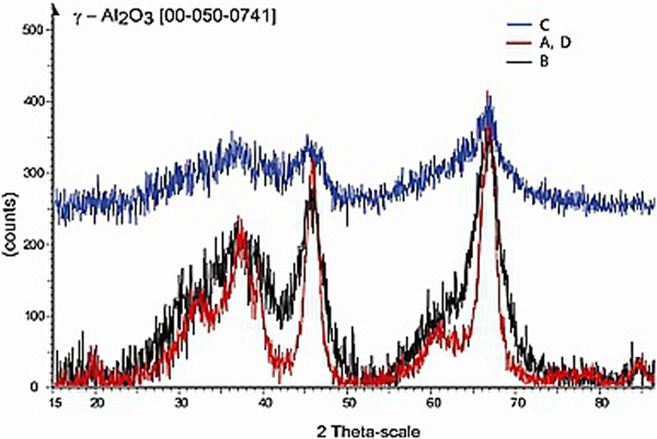
Phase analysis of nanopowders A–D
In order to estimate the agglomeration tendency of the nanopowders, the zeta potential of algal growth media with the nanopowders added was studied.
The lowest zeta potential was revealed in the case of nanopowder A: −22 mV. Solutions of powders B, C and D were characterised by zeta potentials of −12·9, −16·7 and −10·2 mV respectively. The highest zeta potential, −10 mV, was obtained for the commercial powder E. Accordingly, nanopowder A appeared to be most stable, with the weakest agglomeration tendency.
The pH of the algal growth media with added nanopowders was also measured. The pH of the control solution – without alumina – was 5·84. In culture media with nanopowders added, the pH ranged from 6·5 for nanopowder A to 7·6 for nanopowder C (Fig. 4). After 21 days of the experiment, the pH value of culture media ranged from about 6·4 to 6·8.
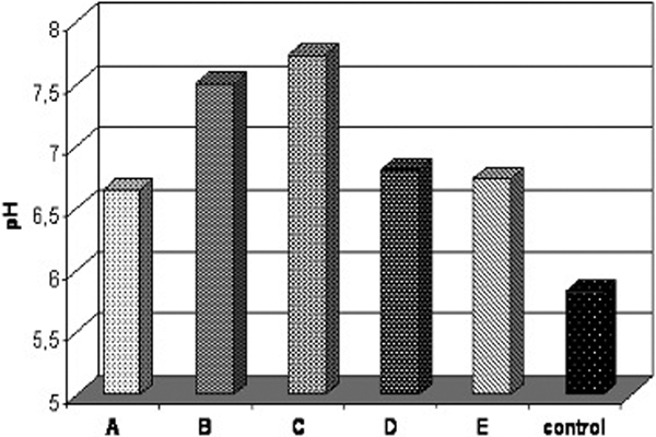
pH of nanopowder containing growth media
In order to estimate Al2O3 solubility in the algal culture media, the conductivity of the solutions containing alumina was measured. The conductivity of the control sample, with no nanopowder, was found to be 258 μS. Immediately after a nanopowder was added, the culture media revealed a conductivity ranging from 263 μS for nanopowder B to more than 300 μS for nanopowder A. After 3, 6 and 8 days of the experiment, a progressive growth in conductivity was observed, probably connected with an increase in Al3+ concentration in the growth medium (Fig. 5).
The influence of different alumina nanopowders on the growth of the green alga S. quadricauda was studied. Two experimental series were carried out with 500 and 1000 mg L−1 of nanopowder respectively. The solubility of the alumina nanopowders in culture medium was very low. The concentration of Al3+ ions in B, C, D and E nanopowders and A nanopowder at the amount of 500 mg L−1 did not exceed 20 mg L−1, while in case of 1000 mg L−1 of the A nanopowder it was 52 mg L−1. After 21 days of the experiment the concentration of Al3+ was 23·7 and 54·5 mg L−1 respectively.
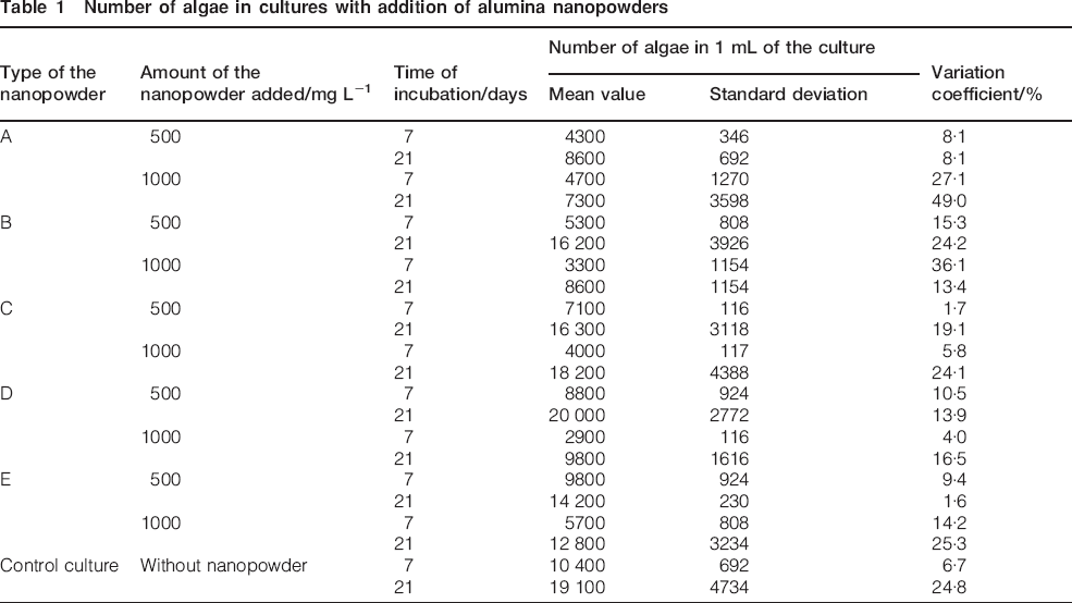
The results of algal counts are presented in Table 1.
Number of algae in cultures with addition of alumina nanopowders
In algal cultures containing 500 mg L−1 of nanopowders, the inhibition effect of nanopowders A–D after 7 days of the experiment ranged from 5% for nanopowder D to 54% for nanopowder A. The commercial powder E revealed no inhibiting activity at this concentration. After 21 days algal growth in the medium containing nanopowder A was inhibited in 55%. The inhibition caused by powders B, C and E was lower than 30%, and for nanopowder D no inhibition was observed (Figs. 6 and 7). Figure 5
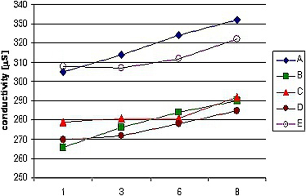
Conductivity of nanopowder containing culture media
Inhibition of algal growth in presence of nanopowders A–E after 7 days of experiment, depending on nanopowder added (500 mg L−1)
Inhibition of algal growth in presence of nanopowders A–E after 21 days of experiment, depending on nanopowder added (500 mg L−1)
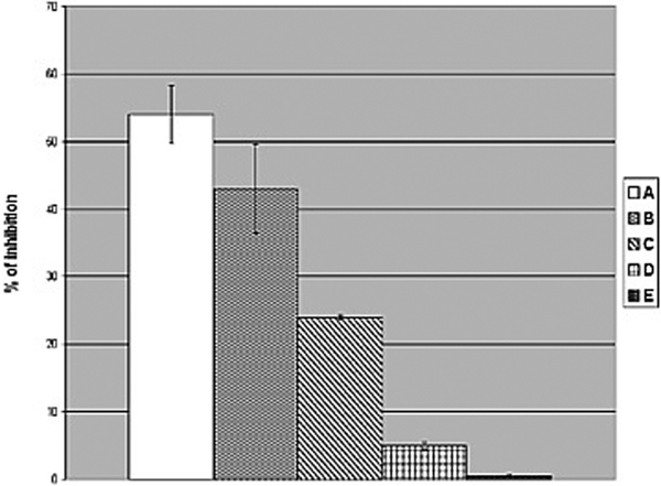
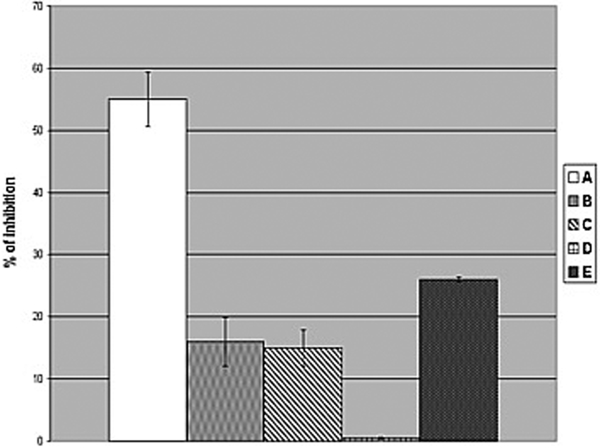
At a nanopowder content of 1000 mg L−1, algal growth was inhibited more strongly, with inhibition after 7 days amounting to about 39–69%. The highest inhibiting activity was observed for nanopowders B and D. After 21 days the inhibiting effect of the nanopowders was less significant, compared with the control culture from 25 to 62%, with the strongest inhibiting activity shown by nanopowder A (Figs. 8 and 9).
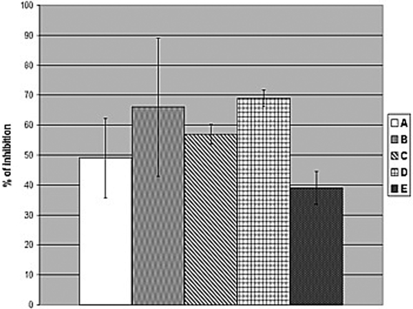
Inhibition of algal growth in presence of nanopowders A–E after 7 days of experiment, depending on nanopowder added (1000 mg L−1)
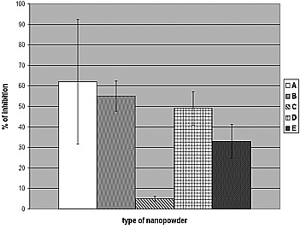
Inhibition of algal growth in presence of nanopowders A–E after 21 days of experiment, depending on nanopowder added (1000 mg L−1)
In case of nanopowders B–E the inhibition level after 21 days was lower than after 7 days of the experiment. This effect was more evident at a nanopowder content of 500 mg L−1. It should be stressed that this effect was not observed for nanopowder A.
Discussion and conclusion
Our research has revealed an inhibiting effect of all the aluminium dioxide nanopowders on the growth of the common European freshwaters green alga S. quadricauda. The negative impact on algal growth was observed for all the nanopowders and depended on their content in the algal growth medium, which is in agreement with literature data concerning metal oxide nanoparticles.10, 16, 17 The maximum inhibition level of powders B and D exceeded 60% after 7 days of the experiment at a nanopowder content of 1000 mg L−1, whereas at 500 mg L−1 it was significantly lower. Nanopowder A revealed a strong inhibiting effect both in a presence of 500 and at 1000 mg L−1 of the nanopowder (the Al3+ concentration of 20 and 52 mg L−1 respectively), with an inhibiting level of ∼50%.
Some differences in inhibiting effect between 7th and 21st day of the experiment were observed mainly in 500 mg L−1 of alumina nanopowder. The impact of nanopowders B–E after 21 days of the experiment was lower compared with that observed after 7 days; however, the nanopowder A revealed an inhibiting effectiveness exceeding 55% at both concentrations.
The solubility of alumina nanopowders in the algal growth medium was very low, so the toxic effect might not be caused mainly by the alumina ions concentration. There are some suggestions that the toxicity effect may result from the entrapping of algal cells by formed metal oxide nanopowder aggregates.18 In case of TiO2, physical disruptions of algal cell membranes as an effect of the nanoparticle structure and surface properties were observed.19
The activity of nanoparticles toward living cells may be determined by their properties and their behaviour in solution. Some literature data suggest that the ecotoxicological effect may depend on the nature of the particle,20 particle size and shape,7, 21 and the surface to volume ratio, allowing for close interaction between nanoparticles and microbial membranes.22 The bioavailability of nanoparticles depends on their aggregation tendencies.12 The toxic effect of nanopowders may also be correlated with their solubility12, 23 and with environmental conditions: the presence of organic matter, pH changes, temperature and ion strength, and light intensity.1, 7, 10
In this study, the grain size and morphology, the specific surface area, the agglomeration level, and the crystallisation degree of the four prepared alumina nanopowders and one commercial powder were determined. Environmental conditions in the presence of Al2O3 powders were characterised by the pH, the conductivity and the zeta potential of the growth medium.
It was found that the grain size fraction of 25–45 nm diameter predominated in powders A–E. In theory, smaller particles should influence cell activity more strongly due to their higher surface to volume ratio, enabling close interaction between the nanoparticles and cell membranes,2, 22 and because of the diameter of pores in the cell walls of algal cells, which determines the size of nanoparticles that may enter the algal cell.10 In this study, no correlation was observed between inhibiting activity and particle size. Powder A, with the largest average grain size, and powder B, with the smallest grains, revealed similar inhibition levels of ∼60%.
The specific surface of the nanopowders had only a slight effect on algal growth: nanopowder A, characterised by the smallest surface area – 58·2 m2 g−1, exhibited the strongest anti-algal activity. However, in the case of the other powders, no correlation with this parameter was discovered. The specific surface areas of nanopowders B and D, which both showed strong inhibiting activity after 7 days of the experiment, differed widely from each other: 316·4 m2 g−1 for powder B compared with 155·9 m2 g−1 for powder D. This finding contrasts with the results described by Navarro et al.10 concerning TiO2 toxicity to the green alga Desmodesmus subspicatus, which depended mainly on the specific surface area of the nanoparticles.
There are some suggestions that the aggregation, adsorption and agglomeration steps of nanoparticles may influence their toxicity due to their local concentration, even without internalisation within organisms.3, 24, 25 The results of this study prove that the degree of agglomeration of a nanopowder may determine its inhibiting properties. Powder A, with the highest degree of agglomeration and agglomerates of ∼1 μm in size, inhibited algal growth the most strongly. Powders B and D, which showed strong inhibiting activity after 7 days, were also characterised by a high degree of agglomeration, with an average agglomerate size of ∼500 nm.
The degree of crystallisation of the nanopowders, connected with their synthesis conditions, appeared to be the decisive factor determining the inhibiting activity of the alumina powders used. The most active nanopowder A showed the highest degree of crystallisation, in contrast to nanopowder C, whose effect on the growth of algae was the weakest.
Moreover, the experiments showed that the inhibiting activity of alumina nanopowders may probably be influenced by the pH of the culture medium, its conductivity and its zeta potential in the presence of nanoparticles.
The addition of nanopowders to an algal growth medium (pH 5·6) resulted in a pH increase: the pH of the medium with a nanopowder ranged from 6·5 in the case of nanopowder A to 7·6 in the case of nanopowder C. There are some suggestions that the charge of nanopowder particles, influencing their activity, is determined by the composition of the organic coatings (citrate, cysteine, carbonate and surfactants).10 The composition of these coatings is determined by the pH of the solution. For example, TiO2 particles are charged positively at pH 6 but negatively at pH>7. Nowack and Bucheli1 also proved that surface charge may influence the activity of nanoparticles. It has been observed that positively charged particles seem to be more toxic than negatively charged ones.26
It was observed that nanopowder A, showing the strongest anti-algal inhibiting activity, was characterised by the lowest zeta potential (−22 mV) and the highest conductivity of the nanopowder-containing growth medium (about 300 μS). The lowest zeta potential, connected with a lower agglomeration tendency, probably resulted in a higher availability to algae. Higher conductivity, caused by the presence of ionic forms as a result of solubilisation, might be responsible for increased destructive impact on algal cells.
The results of the research work allowed to conclude as follows.
Nanopowder A exhibited the most evident inhibiting properties against the green alga S. quadricauda.
The strong inhibiting effect of nanopowder A was accompanied by the highest degree of agglomeration and the highest crystallisation level as well as the smallest specific surface area.
The highest conductivity of the growth medium containing nanopowder A and its comparatively higher solubility than the other nanopowders resulted in increased impact on algal cells. The toxic effect might also be connected with the smallest agglomeration tendency of this nanopowder, as measured by the zeta potential of the algal growth medium.
Such parameters as particle size and specific surface area of alumina nanoparticles did not determine the impact of the prepared nanopowders on algal growth.
It should be noted that the mechanisms of interactions between Al2O3 nanoparticles and algal cells are still unclear. Further research into the activity of alumina nanopowders as well as its influence on the environment is required.