






Abstract
Dispersed, spherical particles of nanosilica with controllable size have been synthesised using a metal alkoxide, i.e. tetraethoxysilane, as starting material, ammonia as base catalyst and non-ionic surfactant as template by sol–gel method. Size of particles and dispersivity were controlled by varying the surfactant chain length and temperature conditions of the reaction mixture. Silica nanoparticles were synthesised using a series of non-ionic surfactants, namely Polyoxyethylene (20) sorbitan monolaurate (Tween 20) and Polyoxyethylene (80) sorbitan monooleate (Tween 80), at different reaction temperatures of 25, 50, 70 and 90°C. The particle size of silica nanoparticles gradually decreased with increasing carbon chain length of the surfactant and at higher temperature particle size became larger. Furthermore, these silica nanoparticles are incorporated into the cementitious system to improve the mechanical properties and reduce calcium leaching in the hydration process. Addition of silica nanoparticles into cement paste improves the microstructure of the paste, and calcium leaching is significantly reduced as silica nanoparticles react with calcium hydroxide, thereby forming a denser calcium–silicate–hydrate gel structure. Synthesised silica nanoparticles and microstructure of cement paste incorporated with silica nanoparticles were analysed using scanning electron microscopy, powder X-ray diffraction, infrared spectroscopy (IR), 29Si MAS NMR and thermogravimetry analysis for morphological and mineralogical attributes.
Introduction
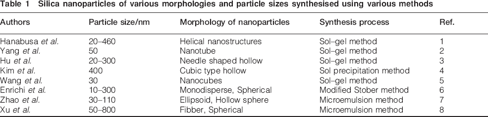
Nanotechnology is gaining widespread attention and being applied in many fields to formulate materials with novel functions due to their unique physical and chemical properties. In the construction sector, nanotechnology is being used in a variety of ways to produce innovative materials. Using nanotechnology as a tool, it is possible to modify the basic structure of the materials at nano and micro levels to improve its bulk properties, such as mechanical performance, volume stability, durability and sustainability. Various methods for the preparation of nanoparticles are employed, such as plasma synthesis, chemical vapour deposition, microemulsion processing, combustion synthesis, sol–gel processing, hydrothermal techniques, etc. Recent efforts for the preparation of nanoparticles are focused on the control size, morphology and surface reactivity of nanoparticles (Table 1). The sol–gel method has been widely used and a method of choice for the preparation of nanoparticles as it has several advantages, such as synthesis may be carried out at low temperature, desired pH to yield high purity and also the reaction kinetics of the process may be controlled by varying the composition of the reaction mixture. The size and shape of silica nanoparticles may be controlled by additives such as electrolytes, surfactants, organic acids, etc. Nanosilica can also be produced in a form of dry powder via pyrolysis of tetraalkoxysilanes or tetrachlorosilane in the presence of water as well as by direct hydrolysis of sodium methasilicate or tetraalkoxysilanes.9, 10 Saito et al. have synthesised nanosilica from per-hydro-poly-silazane with mild conditions using steam as catalyst.11 The density and refractive index of silica prepared from per-hydro-poly-silazane were close to silica glass. Chrusoid and Slusaraski have prepared nanosilica from a stable emulsion of alkoxysilanes by addition of a surfactant and heating the emulsion to remove the residual materials.9 The addition of surfactant is necessary to lower the interfacial energy and minimise the surface energy between two liquids. Kim et al. have suggested the aerosol assisted sol–gel method to produce nanosilica.12 In this method, tetraethoxysilane (TEOS), water and ethanol were allowed to react according sol–gel chemistry. Sufficient hydrolysis time was given, and then the solution was aerosolised with sodium chloride. Sodium chloride was employed both as an agent to accelerate the kinetics of silica gelation and as a templating medium to support the formation and stability of pore structures. Silica nanoparticles can also be obtained under acidic conditions.13 The rates of hydrolysis and condensation of TEOS are not the same under acidic or alkaline conditions, and therefore, the formation mechanism of these two species of silica materials is different. Tapasi and Dolui used sodium silicate with dilute hydrochloric acid as catalyst in poly (vinyl alcohol) to produce poly (vinyl alcohol)/silica nanocomposites.14 This is a sol–gel method in which acid plays a catalytic role in enhancing the sol–gel condensation of silicon alkoxides within poly (vinyl alcohol). Jal and co-workers, who synthesised nanosilica by precipitation method, characterised it by various analytical tools.15 The silica particles were found to have almost spherical shape with a dimension of ∼50 nm. The surface area was found to be 560 m2 g−1, and the density was 2·2 g cm−3. The total silanol density in silica was found to be 7·68 nm−2. The number of reactive silanols that formed hydrogen bond with water molecules was found to be 2·48. The infrared spectral data supported the presence of hydrogen bonded silanol group and the siloxane groups in silica. Nanosilica is used in a variety of products ranging from cosmetic to construction materials. Most of the nanoparticles reported in the literature exhibit various sizes, shapes and orientations and therefore significantly differ in product development. These nanoparticles are either in colloidal form or agglomerated dry powder. Our efforts are to prepare uniform size, dry powder of silica nanoparticles and to study their beneficial role in cementitious materials. Recently, researchers have shown enormous interest to investigate the properties of building materials, such as mechanical, temperature and strain sensing, durability, etc., with nanomaterials.16–22 If dry powder of nanoparticles is added, they may behave significantly different than that of commercially available colloidal or agglomerated silica nanoparticles. During the cement hydration process, these nanoparticles act as reaction centres. During the hydration of cement paste, calcium–silicate–hydrate (C–S–H) is formed as a major constituent (∼65%) along with calcium hydroxide (CH, ∼25%), which leaches out during the hydration process. Our efforts are to control the calcium leaching in the hydration process by the reaction of dry powdered silica nanoparticles with CH, thus imparting higher strength and durability to cementitious materials.
Silica nanoparticles of various morphologies and particle sizes synthesised using various methods
In the present work, we report the preparation of dispersed, stable and dry powder form of silica nanoparticles synthesised by simplified sol–gel method and the effect of addition of these nanoparticles in cement pastes for mineralogical and morphological attributes.
Experimental
Materials and synthesis
Tetraethoxysilane (99·9%, Alfa Aesar, UK), absolute ethanol (99·9%, Merck, India), ammonium hydroxide (25%, Thomas Baker, India) and polyoxyethylene (20) sorbitan monolaurate (Tween 20) and polyoxyethylene (80) sorbitan monooleate (Tween 80) (Loba Chemie, India) were used without any further purification. The chemical structure of the surfactants used in the present studies is shown in Fig. 1. Ordinary Portland cement, 43 grade, conforming to IS 8112 was used as such.

Surfactant used for synthesis of silica nanoparticles
For each experiment, first, ethanol, TEOS, water and surfactant were mixed and stirred for 30 min. Then, ammonia was added quickly to the previously prepared mixture. A white turbidity appears after the addition of ammonia. The reaction was completed in 2 h. The concentrations of ammonia, water and TEOS were fixed at 0·2, 3·2 and 0·2M respectively. The resulting white powder was dried overnight at 100°C and then calcined in air for a period of 3 h at 650°C. The reaction was also performed without surfactants for comparative studies of particle size of silica nanoparticles.
To study the effect of silica nanoparticle addition in cement paste, 5% (w/w) of dry powdered silica nanoparticles was added to cement paste, and comparative morphology and mineralogy were analysed through scanning electron microscope (SEM), X-ray diffraction (XRD) and thermogravimetry analysis (TGA) analysis. For morphological and mineralogical analyses of cement paste with silica nanoparticles, 0·2 g of silica nanoparticles was mixed with 4 g of cement. A water/cement ratio (w/c) of 0·4 was fixed for all the experiments. Acetone was used to discontinue the hydration process of cement paste. Fresh cement paste with varying composition of silica nanoparticles (0·25, 0·5, 1·0, 2·5 and 5·0%) was casted with moulds (25×25×25 mm) to prepare specimens for the measurement of compressive strength. After being demoulded at the age of 24 h, all the specimens were cured in water, and three cubes were tested at a given age (1, 3, 7 and 28 days) using a HEICO universal testing machine.
Characterisation of silica nanoparticles and nanomodified cement paste
The morphological attributes of nanosilica were measured using SEM (LEO-438 VP) at an accelerating voltage of 20 kV. The samples were analysed under high vacuum mode with gold coating so as to improve the image quality. Cement samples were also analysed under variable pressure mode without any gold or carbon coating so as to keep the morphology unaltered. The samples were deposited on a sample holder with a double stick conducting carbon tape. The average particle size was determined based on the measurement of a number of particles from the SEM images. The term particle size used in this paper refers to the average diameter of the silica nanoparticles. Powder XRD data were recorded at room temperature on a Rigaku D-Max 2200 using Cu Kα radiation at a scanning rate of 1° min−1. Various chemical bondings of silica nanoparticles were studied with a Thermo Nicolet NEXUS FTIR. Each IR spectra were collected from 400 to 4000 cm−1. Surface modification of the synthesised silica nanoparticles using nonionic surfactants as organic templates was analysed on a Bruker AV300 NMR spectrometer operating at 59·6 MHz for 29Si. All the NMR spectra were obtained using a contact time of 10 ms and a pulse repletion time of 0·5–1·0 s. All the chemical shifts are reported with respect to the silicon resonance in liquid Me4Si.
Results and discussion
The sol–gel process involves the formation of a colloidal suspension (sol) and gelation of the sol to form a network in a continuous liquid phase (gel). Generally, TEOS is used as a precursor and ammonia as catalyst for synthesising nanosilica.
The chemical reaction of nanosilica synthesis can be summarised as



Images (SEM) of silica nanoparticles synthesised a without surfactant and using surfactants b Tween 20 and c Tween 80 respectively
The particle size of nanosilica also depends on the temperature of the reaction system. At the same concentration of all reagents, the effect of temperature on the particle size and dispersivity of silica nanoparticles was investigated. The SEM images of four samples synthesised at different temperatures 25, 50, 70 and 90°C are illustrated in Fig. 3. Particle size becomes larger and more uniform with the increase in temperature as silica particles are stabilised by electrostatic repulsion. The charge originates from silanol groups, which are relatively acidic and dissociate in the presence of ammonia. Under basic conditions, TEOS undergoes hydrolysis and polycondensation reaction, which result in the formation of dispersed spherical particles of amorphous silica.

Image (SEM) of NS particles (100–250 nm) prepared at reaction temperatures of a 25°C, b 50°C, c 70°C and d 90°C
The XRD patterns of silica nanoparticles shown in Fig. 4 reveal that the silica nanoparticles synthesised by the sol–gel method are amorphous, which is advantageous for cementitious reaction. A representative IR spectrum of silica nanoparticles synthesised using Tween 20 and Tween 80 is shown in Fig. 5. The two strongest IR absorption bands at ∼1100 and 480 cm−1 initiate from the extension and flexural vibrations of Si–O–Si bonds. The IR absorption band at ∼800 cm−1 originated from the vibration of (SiO4) tetrahedrons. The ∼3453 and 1630 cm−1 absorption bands come from the O–H bonding vibration of adsorbed molecular water. Infrared analysis also showed that the band at 1076 cm−1 was slightly shifted towards lower wave number as the particle size is reduced. This observation suggests a change in the local bonding structures of Si and O atoms at smaller particle size.24, 25 Figure 6 shows the 29Si MAS NMR spectra of all silica nanoparticles synthesised with and without surfactants. The 29Si MAS NMR spectra of silica nanoparticles show the T and Q sites. The signals at ˜−113·0 and −103·0 ppm arise from the Si species Q4 [Si(OSi)4] and Q3 [Si(OH)(OSi)3] respectively. A low intensity peak at −94·20 ppm arises from chemical shift correlation and relaxation data to germinal-hydroxyl silanol sites.26

Pattern (XRD) of synthesised silica nanoparticles (A) without surfactant and using (B) Tween 20 and (C) Tween 80 respectively

Spectra (IR) of nanosilica particles synthesised (A) without surfactant and using (B) Tween 20 and (C) Tween 80 surfactants respectively

Si (29) MAS NMR spectra of silica nanoparticles synthesised (A) without surfactant and using (B) Tween 20 and (C) Tween 80 surfactants respectively
Further, these synthesised silica nanoparticles (∼50 nm) are incorporated into the cementitious system to improve the mechanical properties and reduce calcium leaching in the hydration process. The mechanical properties of cement are enhanced by the addition of silica nanoparticles as compressive strengths after 1, 3, 7 and 28 days are substantially enhanced (Table 2). The nanoparticles have high surface energy, and atoms at the surface have a high activity, leading the atoms to react rapidly. The compressive strength of cement paste containing 5% silica nanoparticles is 63% higher at 1 day and 34% at 28 days than that of control cement paste. The standard deviation for the compressive strength of cement paste is ±3·8. The difference in the strength development of the paste is attributed to the pozzolanic reaction of silica nanoparticles with CH and forming a denser C–S–H structure. To verify this mechanism of increased compressive strength by the formation of a denser structure of C–S–H, SEM, XRD and TGA studies were carried out for the morphological and mineralogical attributes of cement pastes.
Mix proportions and compressive strengths of plain cement and nanosilica incorporated pastes
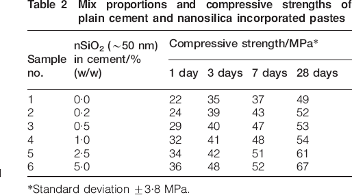
*Standard deviation ±3·8 MPa.
The two silicate phases of cement, i.e. tricalcium silicate and dicalcium silicate, give calcium–silicate–hydrate (C–S–H) and CH as hydration products. The C–S–H gel being the main component of cement hydration is responsible for the strength and microstructure of the cement paste. The amount of CH formation in cement paste was evaluated by TGA during the hydration process. This was characterised by CH residue determination in the cement paste admixture. Further, silica fume was also added to cement paste for comparison. Hydrated cement shows a reduction in weight up to 200°C due to surface water desorption as well as loss of water from C–S–H gel layer and from the dehydration of ettringite. Further, the weight loss of ∼400–500°C is due to the thermal decomposition of CH followed by the decomposition of carbonated phases and calcite. The amount of CH in the specimen is calculated directly from the TG curves using the following equation

The SEM images of prepared nanosilica and commercially available silica fume are shown in Fig. 7. The TGA curves of pure, silica nanoparticles and silica fume incorporated cement pastes are shown in Fig. 7 at 28 days of hydration. The CH content in various cement pastes during the hydration process is shown in Fig. 8a and b. At the early stage of hydration, plain cement paste has 4·4% of CH, whereas silica fume incorporated cement paste has 2·3% and silica nanoparticle incorporated paste has only 0·5% of CH content. During the hydration, CH is continuously formed, and at 3, 7 and 28 days, it amounts to 7·7, 12·8 and 20·3% respectively in plain cement paste, whereas the CH content in silica fume incorporated cement paste is up to 16·3% at 28 days. Silica nanoparticles have a much significant effect as compared to plain and silica fume incorporated cement pastes, and at 28 days of hydration, only 8·5%CH content was observed (Table 3). The standard deviation for the CH content in cement paste, silica fume and silica nanoparticle incorporated cement paste is ±0·28. The SEM images of plain cement paste, silica fume and with silica nanoparticles (5%) at 7 and 28 days are shown in Fig. 9. Depending on the time of hydration and the Portland cement composition, several crystalline phases can be observed in the hydrated cement paste. Secondary electron images show the hexagonal plates of CH, the needle-like habit of ettringite and the sheet-like, flaky habit of calcium–silicate–hydrate. The CH has a distinguishing hexagonal shape. The dimensions, of course, depend on the concentration in aqueous solution and the age of the crystal. The dominating phase in forming the shape in the contact surface is C–S–H probably due to its significantly higher strength. It was observed that in the microstructure of the plain cement paste and the silica fume incorporated cement paste, the C–S–H gel existed along with needle and plate shaped hydrates of CH. The deposited CH around the C–S–H gel is uniformly distributed among the entire cement phases (Fig. 9). The microstructure of cement paste with the addition of silica nanoparticles revealed that the formation of hydration products was denser, becomes significantly different and shows the absence of needle and hexagonal plate shaped crystals of CH. The XRD profiles of plain cement, silica nanoparticles and silica fume incorporated pastes at 7 and 28 days of hydration are shown in Fig. 10a and b. It is evident from the XRD profiles that the CH peak almost disappeared with the addition of silica nanoparticles, while the same is significantly present in plain and silica fume incorporated cement pastes. It is therefore inferred from Fig. 10 that silica nanoparticles react with CH produced during the hydration process. Therefore, the pozzolanic reactivity of silica nanoparticles at the early stage of hydration is significantly high and improves the microstructure of cementitious system, thereby enhancing the durability and mechanical properties of cementitious materials.

Images (SEM) of a synthesised silica nanoparticles and b silica fume

TGA curves of cement pastes at a 1 day of hydration and b 28 days of hydration

Images (SEM) of plain cement paste (PC7&PC28), silica fume incorporated cement paste (SF7&SF28) and nanosilica incorporated cement paste (NS7&NS28) at 7 and 28 days of hydration respectively

X-ray diffraction pattern of cement pastes at a 1 day of hydration and b 28 days of hydration
Calcium hydroxide content in cement pastes
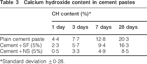
*Standard deviation ±0·28.
Conclusion
Spherical and amorphous silica nanoparticles can be prepared by the hydrolysis reaction of TEOS in ethanol using water and ammonia using the sol–gel method. The particle size of nanosilica can be controlled by adding Tween 20 and Tween 80 surfactants. The particle size of nanosilica powder also depends on the temperature of the reaction system. The particle size increases with the increase in temperature of the reaction system. It was observed from SEM, XRD and TGA studies that addition of silica nanoparticles to cement reduced CH leaching by reacting at the early stage of hydration. It was found that the CH content in silica nanoparticle incorporated cement paste reduced by ∼89% at 1 day and up to ∼60% at 28 days. Therefore, the addition of a small quantity of silica nanoparticles significantly improves the morphology and mineralogy of cementitious materials.
Footnotes
Acknowledgements
The authors are thankful to the Uttarakhand State Council for Science and Technology (UCOST), Dehradun, India, for financial support. One of the authors (S. Ahalawat) is grateful to UCOST, Dehradun, for fellowship assistance.