
Abstract
Plate-like potassium magnesium titanate (KMTO) powder prepared by molten salt growth method and the KMTO porous ceramic synthesised by polymeric sponge replication method were used as sorbents to remove nickel ions from wastewater. Both powder and porous ceramic were characterised by X-ray diffraction, scanning electron microscopy, transmission electron microscopy and X-ray photoelectron spectroscopy. The maximum adsorption capacities of powder and porous ceramic were 96 and 24 mg g− 1 respectively at a pH value of 6 (25°C). However, the removal efficiencies of both could reach up to 99%. Moreover, the adsorption kinetics for the KMTO powder and the porous ceramics followed the pseudo-second-order kinetic model, and the equilibrium data for the KMTO powder fitted the Langmuir isothermal model well, while the porous ceramics fitted with the Freundlich model. The mechanism of the adsorption by the KMTO powder and the porous ceramic was ion exchange. It was also shown that the nickel saturated KMTO powder and the porous ceramics were stable in leaching tests.
Introduction
Heavy metal ion pollutants have long been a concern: among the potential contaminants, nickel is one of the most widespread toxic pollutants in the environment. Nickel salts are commonly used in electroplating, electrolysis and pesticides. 1 Prolonged and accumulated nickel inside the human body can cause adverse health impacts, such as anaemia, diarrhoea, encephalopathy, hepatitis and central nervous system dysfunction.
Although various methods have been used to treat heavy metal ions in wastewater, including chemical precipitation, ion exchange, reverse osmosis, electrochemical treatment and adsorption,2–8 the removal of nickel is not as satisfactory as that of other heavy metal ions, because the complex formed by nickel in aqueous solution after adsorbent addition is unstable. 9 Some types of materials are used to treat nickel ions, such as natural organic materials, mesoporous silica and other minerals.10–14 Oven dried biomass of Eichhornia crassipes, Valisneria spiralis and Pistia stratiotes 10 shows good removal efficiency in 15 to 30 mg L− 1 nickel solutions. The adsorption capacity of a macroporous chitosan membrane 9 for nickel ions in wastewater is 5·21 mg g− 1. The adsorption capacity of chitosan–MAA nanoparticles 11 for nickel ions is 0·87 mg g− 1. For zeolite 4A prepared by coal fly ash, 12 the adsorption capacity of nickel ions is 8·96 mg g− 1. Amino functionalised mesoporous silica, NH2-MCM-41, has exhibited good removal efficiency for various heavy metal ions; however, its adsorption capacity for nickel ions 13 is only 12·36 mg g− 1. The adsorption capacity of magnetite iron oxide nanorods for nickel ions 14 is 95·42 mg g− 1.
Therefore, research is still needed to explore new potential materials. Nanometre sized materials are widely studied, including nanoparticles and nanotubes. Titanate nanoparticles and nanotubes are convenient to prepare and cost effective but are apt to cause secondary contamination. Layered titanates have recently been paid much attention by researchers. Akieh et al. 15 reported that layered ion doped titanate NaFeTiO4 can exchange ions with ionic nickel, and show high removal efficiency. Aguilar-Gonzalez et al. 16 studied the removal behaviour of SiO2 doped potassium titanate pellets for lead and nickel. However, reports on the nickel ion removal efficiency of plate-like titanates are sparse.
In this work, the experiment part consisted of two stages. The first stage synthesised and characterised potassium magnesium titanate, K0·8Mg0·4Ti1·6O4 (KMTO), powder and porous ceramic with different temperatures. The second stage used the as synthesised powder and 1000°C ceramic as adsorbents for low concentration nickel ions ( < 100 mg L− 1) in wastewater and further studied the adsorption process.
Experimental
Materials and chemicals
Titanium dioxide, magnesium hydroxide, anhydrous potassium carbonate, potassium chloride and nickel nitrate were purchased from Xilong Chemical Co. Ltd (China): 60 PPI commercial polyurethane sponges and commercial silica sol (9·5 ≤ pH ≤ 10) were bought from the No. 6 Plastic Factory (China) and Shanghai Second Reagent Plant (China) respectively. Sodium carboxymethyl cellulose (CMC), polyacrylamide and sodium benzenesulphonate were bought from Tianjin Kermel Agents (China). Chloride acid and sodium hydroxide were used to adjust the pH value of the aqueous solution. All the chemicals used were of analytical grade and with no further purification.
Preparation of KMTO powder
The KMTO powder, with its plate-like morphology, was prepared by molten salt growth method as previously reported. 17 The required quantities of titanium dioxide, magnesium hydroxide, anhydrous potassium carbonate and potassium chloride were weighed and then milled with ethanol in an aluminium oxide jar for 5 h. The powder slurry was dried at 80°C and calcined at 1050°C for 2 h. The powder was rinsed several times with deionised water and ethanol to remove any extra solvent and then dried.
Preparation of KMTO porous ceramic
The porous KMTO ceramic was prepared by polymeric sponge replication method. 18 Commercial silica sol and potassium dihydrogen phosphate were used as organic and inorganic binders respectively. Sodium CMC was used as a thickening agent. Polyacrylamide and sodium benzenesulphonate were used as dispersing agent and emulsifying agent respectively. A two-step soaking method was used, and the concentrations of the first and second slurries were 65% and 58% respectively. 18 Once the slurry had been stirred until homogeneous, the sponge was immersed in it and soaked for 30 min. The saturated sponge was then rolled to remove any excess slurry. The rolled sponge was coated repeatedly by centrifugal process in the slurry and rerolled before being dried at 50°C for 12 h. The dried sponges were sintered at three different temperatures (900°C, 1000°C and 1100°C) for 2 h.
Material characterisation
The prepared samples were characterised by X-ray diffraction (XRD) (D/max-γ, Japan), X-ray photoelectron spectroscopy (XPS) (K-Alpha 1063, Thermo Fisher), field emission scanning electron microscopy (FE-SEM) (NOVA NANOSEM 230) and transmission electron microscopy (TEM) (JEOL JEM-2100F). The density and open porosity of struts were measured using mercury porosity (model Poresizer 9320). Nickel ion concentrations were measured using atomic absorption spectroscopy (Zeenite 700P, Analytical Jena).
Batch adsorption experiments
Batch adsorption experiments were generally carried out by stirring calibrated Erlenmeyer flasks containing 0·1 g of adsorbent and 100 mL of metal ion solutions with a magnetic stirrer. The effect of pH was studied at nickel ion concentrations of 100 and 25 mg L− 1 for the KMTO powder and porous ceramic over the range 2 ≤ pH ≤ 7 at 25°C. The pH values of the solutions were adjusted using 0·01 M hydrochloric acid and/or sodium hydroxide solution. To study the effect of the contact time, 0·1 g of adsorbent was dispersed in 100 mL of 100 and 25 mg L− 1 nickel ion solutions at pH 6 for the KMTO powder and porous ceramic respectively. After stirring, the solution was filtered. Each experiment was run three times, and the data were averaged. The minimum detection limit of the atomic absorption instrument was 1 ppb for nickel ions.
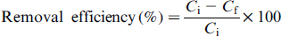
The amount of nickel ions adsorbed was calculated using equation (1):
Results
Phase constitutions
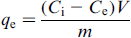
Figure 1a shows the XRD patterns of the KMTO powder before and after adsorption at pH 6. All major diffraction peaks could be attributed to KMTO before adsorption (JCPDS file no. 35-0046). The weak peaks of K2Ti4O9 already existed before adsorption, but became stronger and represented the dominant phase after adsorption (JCPDS file no. 32-0861). The XRD patterns of the different temperature sintered ceramic before and after adsorption at pH 6 were shown in Fig. 1b–d. For the ceramic sintered at 900°C, the XRD pattern agreed with that of K1·54Mg0·77Ti7·23O16 phase (JCPDS file no. 84-0974), and a small amount of KMTO was present. After adsorption, all major diffraction peaks matched K2MgTi7O16 phase (JCPDS file no. 18-1032). For the ceramic sintered at 1000°C, the diffraction peaks were attributed to K1·54Mg0·77Ti7·23O16 phase (JCPDS file no. 84-0974). After adsorption, the XRD pattern matched that of K2MgTi7O16. For the ceramic sintered at 1100°C, both before and after adsorption, all major diffraction peaks could be attributed to K2MgTi7O16 phase.
XRD of a KMTO powder before and after adsorption, b 900°C ceramic before and after adsorption, c 1000°C ceramic before and after adsorption and d 1100°C ceramic before and after adsorption
Powder morphology
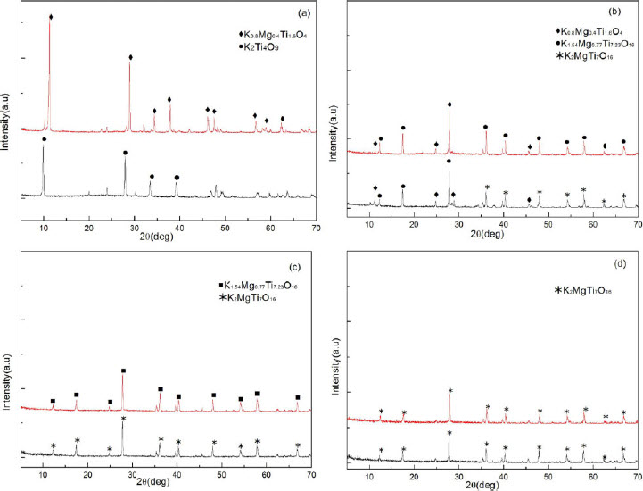
Figure 2a shows the micrograph of the KMTO powder before adsorption. The KMTO powders had a satisfactory plate-like shape and had grain diameters of between 2 and 10 μm. Figure 2b shows the micrograph of the KMTO powder after adsorption. The surface of the KMTO powder before adsorption was smooth, and after adsorption, it was very rough and cracked. A typical diffraction pattern and high resolution TEM image of both powders could be seen in Fig. 3a–d. KMTO before adsorption was identified at measured lattice spacings of 0·37 and 0·23 nm, corresponding to (110) and (111) planes of KMTO respectively. After adsorption, lattice spacings of 0·87 and 0·57 nm were measured, corresponding to (200) and (002) planes of K2Ti4O9 phase.
SEM micrograph of KMTO powder a before adsorption and b after adsorption
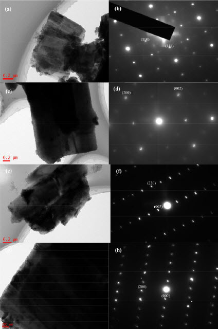
a TEM micrographs of KMTO powder; b diffraction pattern of KMTO powder; c TEM micrographs of KMTO powder after adsorption; d diffraction pattern of KMTO powder after adsorption; e TEM micrographs of KMTO ceramic; f diffraction pattern of KMTO ceramic; g TEM micrographs of KMTO ceramic after adsorption; h diffraction pattern of KMTO ceramic after adsorption
Microstructure of porous ceramic
The microstructures of porous ceramics sintered at different temperatures are shown in Fig. 4. The measured densities were 1·15, 1·27 and 1·43 g cm− 3, and the average porosities were 44·5%, 48% and 34% for ceramic prepared at 900 to 1100°C respectively (consistent with Fig. 4a–f).The average pore size was ∼0·4 μm. The TEM shows more detailed structures of the ceramic prepared at 1000°C before and after adsorption in Fig. 3e–h. For the porous ceramic, before adsorption, lattice spacings of the narrow strip products were determined to be 0·36 and 0·15 nm, which corresponded to (220) and (002) planes of the K1·54Mg0·77Ti7·23O16 phase. The preferential growth direction is [001]. 19 The lattice interspacings of the ceramic after adsorption, the tunnel shape product, were 0·51 and 0·15 nm, corresponding to (200) and (002) planes of the K2MgTi7O16 phase.
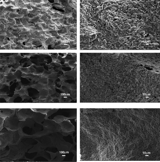
SEM micrographs of 900°C sintered ceramic (a, b), 1000°C ceramic (c, d) and 1100°C ceramic (e, f)
Ion change in adsorption
Figure 5 shows the variation of potassium, magnesium and nickel ion concentrations during adsorption. For the KMTO powder, the nickel ion concentration decreased sharply in the beginning, while the potassium ion concentration increased rapidly. Moreover, there existed small amount of magnesium ions and the concentration only increased in the first hour and then kept stable during subsequent adsorption. For the porous ceramic sintered at 1000°C (shown in Fig. 5c), the potassium ion concentration increased sharply at first and then remained stable. The nickel ion concentration continually decreased, while the magnesium ion concentration increased until adsorption equilibrium was reached. As for ceramic sintered at 900°C (Fig. 5b), the nickel ion concentration initially decreased, but then increased, which was due to the collapsed ceramic. As for ceramic sintered at 1100°C (Fig. 5d), the nickel ion concentration did not change, because the 1100°C ceramic K2MgTi7O16 was tunnel shape and potassium ions were locked and lost the ion exchangeability. By contrast, the ceramic sintered at 1000°C is most suitable as adsorbent,which is used in the following adsorption tests of ceramic.
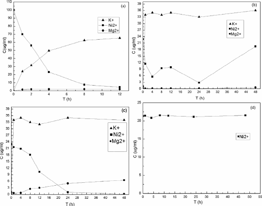
Ion concentration variation with time: a KMTO powder; b 900°C ceramic; c 1000°C ceramic; d 1100°C ceramic
Adsorption kinetics
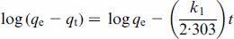
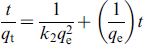
The adsorption process of nickel ions on the KMTO powder was analysed using the Lagergren pseudo-first- or pseudo-second-order kinetic models, which are given by equations (3) and (4) respectively.
20
Kinetic parameters for adsorption of nickel ions by KMTO
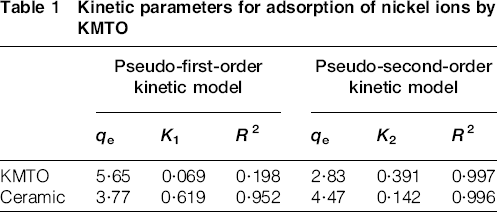
It was found that the pseudo-second-order model was more suitable for modelling the adsorption of nickel ion by the KMTO powder and ceramic than the pseudo-first-order model. The pseudo-second-order model assumes that the rate limiting step is a chemical sorption between the solution and the adsorbent. 21
XPS analyses
High resolution XPS spectra were also recorded from the surface of both KMTO and those porous ceramics sintered at 1000°C to investigate the chemical state and surface stoichiometry after adsorption (Fig. 6). For Ni2p in K2Ti4O9, which was the identified product of KMTO after adsorption, previous studies have reported that the Ni2p3/2 peaked at 855·81 eV and Ni2p3/2 peaked at 861·17 eV, which could be attributed to the presence of nickel ions. 22 Because the crystal morphology is platy, nickel ions were located between the TiO2 layers. For Ti2p in K2Ti4O9, Ti 2p3/2 peaked at 459 eV and Ti 2p1/2 peaked at 464·8 eV, which could be attributed to TiO2. 23 The Ti2p peak line of the porous ceramics after adsorption was almost the same as that of the KMTO powder. The Ni2p line of the porous ceramics after adsorption was composed of one double peak and two single peaks, indicating that ionic Ni was still present. 22 Because the adsorption product K2MgTi7O16 was tunnel shape, nickel ions were located in the tunnel.
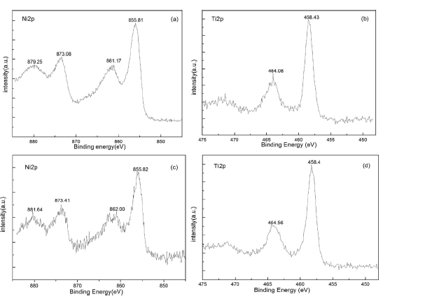
High resolution XPS spectra of KMTO after adsorption (a, b) and ceramic after adsorption (c, d)
Optimisation of adsorption parameters
Effect of contact time
Figure 7a shows the effect of contact time on removal efficiency at a pH value of 6·0. The initial metal ion concentration for the KMTO powder was 100 mg L− 1, while it was 25 mg L− 1 for porous ceramics. The stirring time for the KMTO powder was 12 h, and 24 h for the porous ceramics. The removal efficiency of the powder initially reached 99·3% in the first 4 h, and then the process slowed; therefore, the optimal stirring time was ∼4 h. At the same time, the removal efficiency of the porous ceramic sintered at 1000°C reached 99·5% at 24 h, so its optimal time was 24 h.
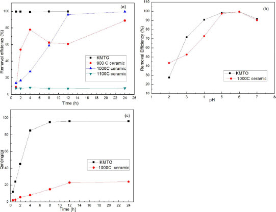
a effect of time on adsorption; b effect of pH on adsorption; c adsorption capacity with contact time
Effect of pH value
The pH value is an important factor for the adsorption of ions, because it affects the distribution of active sites on the titanate surface. 24 The pH values of sample solutions were adjusted to between 2 and 7. A pH of 1 was too acidic to be used in the industry, while a pH of 8 induces precipitation of nickel ions. The results are shown in Fig. 7b. The removal efficiencies of the powder and the porous ceramics increased with increasing pH. Both the powder and the porous ceramics reached an efficiency of up to 99% at a pH value of 6. When the solution pH is 2–4, during the ion exchange process, H+ is more active and easy to exchange with K+ and Mg2+ than Ni2+, which decrease the removal efficiency of Ni2+ ions.
Adsorption capacity and leaching stability
The adsorption capacities of KMTO powder and the porous ceramics at room temperature were determined to be 96 and 24 mg g− 1 (Fig. 7c) respectively. Compared with different adsorbents towards nickel ion removal mentioned before in the paper, it is evident that the adsorption capacity of the KMTO powder is relatively high. The adsorption capacity of the porous ceramic is lower than that of the KMTO powder, but still higher than that of most other adsorbents reported before.10–14 Moreover, the porous ceramic has the advantages of low cost, high removal efficiency and easy to operate.
To examine the stability of the saturated KMTO powder and porous ceramic, leaching tests were performed. Samples of dried saturated adsorbents were dissolved in 100 mL of distilled water at 96°C over a 5 h period and analysed by atomic absorption spectroscopy to determine the nickel ion content. The results showed that there was only 0·3 mg L− 1 nickel ion for both powder and ceramic. Therefore, the KMTO powder and ceramic were deemed to be stable and safe adsorbent materials.
Adsorption isotherms
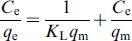
Heavy metal ion adsorption isotherm data starting at different initial KMTO concentrations were investigated following the models of Langmuir and Freundlich. The Langmuir model is given
25
by:
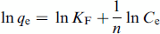
The logarithmic Freundlich equation is given
25
by:
The values of qm, KL, KF and 1/n, and the correlation coefficient (r) for Langmuir and Freundlich models for the KMTO powder and ceramic are given in Table 2.
Parameters of Langmuir and Freundlich isotherm equations
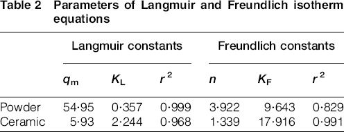
The values of the correlation coefficients for the KMTO powder demonstrated an almost perfect agreement with the Langmuir model, while the values for porous ceramic fitted well with the Freundlich model. This suggested that the adsorption of nickel by the KMTO powder was of monolayer type, and its adsorption by porous ceramic was multilayer.
Adsorption mechanism
For the K0·8Mg0·4Ti1·6O4 powder, as both the layered structure and plated shape (Fig. 1a) can be preserved, the adsorption process can be regarded as a typical ion exchange process.26–28 Mg2+ ions occupied the octahedral sites in the host layers; therefore, substitution for Ti4+ ions in the nominal (TiO6)n framework gave a negative charge that was balanced by interlayer K+ ions. As illustrated in the ‘Effect of pH value’ section, the maximum adsorption capacity was reached at pH 6 with weak acidic solution. Among the process, K+ ions were first replaced by H3O+, and as a result, the interlayer space swells along the b axis. 29 Ni2+ ions had been intercalated among (TiO6)n octahedral slabs during ion exchange with H3O+ and Mg2+ ions, which was also confirmed by XPS results in the ‘XPS analyses’ section. Finally, the adsorption product was K2Ti4O9, with Ni2+ ions located in the layers formed by (TiO6)n octahedral slabs.
The porous ceramics calcined by the KMTO powder at different temperatures formed different phases. For the ceramic calcined at 1000°C, the newly formed K1·54Mg0·77Ti7·23O16 ceramic had an unclosed tunnel shape, which has similar structure with wire shaped KTi8O16·5. 19 Because KTi8O16·5 has an incommensurate structure along the [001] crystallographic axis, 30 this phase was not stable. The edge sharing Mg–TiO2 octahedrons could not close to form a complete tunnel, so the K+ ions have freedom to dissociate to exchange with Ni2+ ions during adsorption. The exchange process is based on the same mechanism with powder. Both K+ and Mg2+ ions exchanged with Ni2+ ions, and finally reached a product of K2MgTi7O16 ceramic, which has closed tunnel with Ni2+ ions locked in. By contrast, K2MgTi7O16 phase formed at 1100°C had no ion exchange property (Fig. 5d).
Conclusions
The plate-like KMTO powder was fabricated using the molten salt method, and the porous KMTO ceramic was prepared by polymeric sponge replication method. The phase structure and porosity of the porous ceramics were affected by sintering temperature. After adsorption, the products were stable in leaching tests. Nickel ion adsorption activities of KMTO powder and ceramic were investigated under different experimental conditions. The removal efficiency of both powder and ceramic reached 99% at a pH value of 6 at room temperature. The pseudo-second-order model provided a good fit to both adsorption kinetics (powder and ceramic), which therefore assumed chemical sorption. The KMTO powder demonstrated an almost perfect agreement with the Langmuir model, while the Freundlich model provided a good fit for the porous ceramic. The adsorption of the KMTO powder and the porous ceramic was based on ion exchange.
Footnotes
Acknowledgement
We thank the National Natural Science Foundation of China for their financial support (grant nos. 51272289 and 51301203).