
Abstract
Two collagen based nanocomposites were synthesised by initiating the reconstitution of collagen solutions in the absence or presence of alginate with simultaneous hydroxyapatite synthesis. The structural characteristics and thermal behaviours of both collagen-hydroxyapatite (M1) and collagen/alginate-hydroxyapatite (M2) were evaluated in comparison with rabbit bone (RB), which confirmed the bone-like features of synthetic nanocomposites. However, differences in microstructure, phase and composition were found among the respective samples calcined at 600 and 1000°C, as revealed by X-ray diffraction and scanning electron microscopy. Especially, in contrast to the presence of beta tricalcium phosphate in M1, pure HA was obtained in both M2 and RB after calcination at 1000°C, confirming that the introduction of alginate with Ca capturing capability is beneficial to achieve nanocomposites with better analogue to natural bone.
Introduction
Vertebrate bone is a naturally occurring composite formed mainly with nanoscale hydroxyapatite (HA) and fibrous collagen with a small amount of non-collagenous proteins and polysaccharides, as well as with binding water. The weight percentage for each component is ∼65:25:10. Bones are constantly being remodelled in response to the applied stresses in the physiological environment. 1 The ongoing bone resorption and formation in a dynamic balance are achieved by normal functions of osteoclasts and osteoblasts. Generally, bone defects caused by trauma or tumours are far beyond the self-regenerative ability of bone. Autologous bone grafting is considered as the gold standard of clinical bone substitution, but autologous bone grafts bear several shortcomings such as limited amount of available bone and additional surgery for the graft extraction. Thus, artificial bone substitutes are still required to meet the demands of restoring the bone defects in many cases. Because the biological and other properties of bone depend on its compositions and microstructures, various artificial composites3 4 –5 or hybrid materials6 7 –8 based on polymers and HA have been extensively studied. Among these synthetic materials, the biomimetic composite of collagen and HA is one of the most promising bone substitute.7–10
Type I collagen is the primary constituent of extracellular matrices and accounts for up to 90 wt-% of the organics in bone. Owing to its excellent biocompatibility and biodegradability, collagen has been widely used for various tissue engineering purposes. In the formation of bone, non-collagenous proteins such as glycosaminoglycans play an important role in directing the nucleation and growth of HA. Several in vitro collagen mineralisation studies also demonstrated that the introduction of non-collagenous proteins 11 or their functional analogs such as natural or synthetic polyanionic polymers12–14 resulted in mineralisation of collagen much closer to natural bone or achieving collagen based bone nanocomposites with better properties. Alginate is a natural linear anionic polysaccharide rich in brown algae and has been used for various biomedical purposes due to its good biocompatibility and elasticity.12,14,16 The complexing capability of alginate with metallic ions such as Ca2+ is very attractive for developing collagen based nanocomposites.
In this study, two kinds of collagen based nanocomposites were prepared by collagen self-assembly with simultaneous HA synthesis. One is collagen-hydroxyapatite (Col-HA) and the other is collagen/alginate-hydroxyapatite (Col/Alg-HA) nanocomposite. Scanning electron microscopy (SEM), X-ray diffraction (XRD), Fourier transform infrared spectroscopy (FTIR) and thermal analyses were employed to characterise the physicochemical properties of the as received synthetic nanocomposites in comparison with natural bone. Their calcined products at 600 and 1000°C were further analysed. The present study not only validated the bone-like properties of synthetic collagen based nanocomposites but also revealed the delicate difference in the composition of Col-HA and Col/Alg-HA nanocomposites. The relevant results provide valuable data for developing bone-like bone grafting materials in the future.
Experimental
Chemicals
Collagen type I used in this study was extracted from calf skin by pepsin digestion in authors’ laboratory as previously described. 15 The acidic collagen stock solution (8 mg mL− 1, pH 2.5) was obtained by diluting the extracted product with 0.3 M acetic acid aqueous solution at 4°C. Sodium alginate (molecular weights of 120–190 kDa, viscosities of 20–40 Hz), CaCl2 and NaH2PO4.2H2O were purchased from Sigma-Aldrich Co. LLC (MO, USA). NaOH was purchased from Kelong Chemical Co. (Chengdu, China). All chemicals were of analytical grade and used without further purification. Deionised water (18.3 MΩ cm) was used throughout the experiments. Fresh rabbit femur bones were provided by the National Engineering Research Center for Biomaterials in China.
Preparation of collagen based nanocomposites
The previous processes were applied to prepare collagen based nanocomposites with some modificaitons. 8 Briefly, phosphate containing aqueous solution was added to acidic collagen solution with vigorous stirring at 4°C, and the resultant solution was neutralised to pH 7.2 by slowly adding NaOH solution. Sodium alginate aqueous solution (2 wt-%) was then mixed with the neutral solution to obtain a mixture with Col/Alg = 2:1. Subsequently, CaCl2 aqueous solution was introduced drop by drop into the Col/Alg mixtures at the Ca/P ratio of 1.67, and the pH value was adjusted to 9.0 by adding NaOH solution. After stirring for 2 h, the obtained homogeneous feedstock solutions were degassed by low temperature centrifuged at 1000 rev min− 1 for 5 min. Finally, the resultant hybrid colloids were incubated at 35°C to give rise to a gel initiated by collagen self-assembly (fibrillogenesis). The obtained gels were washed thoroughly with deionised water to remove the reactive residues. The dense nanocomposites were obtained by dehydration of the washed gels under a laminar flow of air at room temperature. The as received nanocomposites in the absence or presence of alginate were designated as M1 (Col-HA) and M2 (Col/Alg-HA) respectively. Meanwhile, the fresh rabbit femur parts were also dehydrated and the as received natural bone samples were designated as rabbit bone (RB).
Calcination
RB, M1 and M2 samples were calcined in a horizontal furnace with an alumina tube. They were performed in air atmosphere at the heating rate of 10°C min− 1 from 25 to 600°C and from 25 to 1000°C. The samples were heated to the designed temperature and then removed from the furnace after cooling down to room temperature. Calcinations at 600 and 1000°C yield the decomposition of organics and crystalline product without decomposition of stoichimetric HA respectively.
Characterisation
SEM was used to examine the morphological microstructures of samples before and after calcination (Hitachi S-4800, Japan). A thin layer of gold was coated on all the samples for SEM observations. Differential scanning calorimetric (DSC) and thermogravimetry analyses (TGA) were carried out in a thermal analyser (STA 449C, Netzsch, Germany) with an empty aluminium pan as a reference. For DSC and TGA measurements, the heating rate was 10°C min− 1 from 25 to 800°C in air atmosphere. The sample amounts for TGA and DSC experiments were 3–4 mg. Derivative thermogravimetric analysis (DTG) was obtained according to TGA. FTIR was adopted to identify the chemical species of the as received samples and calcined samples at 600°C. The experiment was performed on a Nicolet 6700 system with 4 cm− 1 resolution and 20 times scanning. XRD was applied to monitor the phase composition features of as received and calcined samples at 600 and 1000°C. The results were collected using a DX-1000 X-ray diffractometer (Cu Kα radiation, λ = 0.1542 nm) with 40 kV working voltage and 25 mA current. Phase identification was performed with reference to the standard data of JCPDS Card No. 09-0432 for HA and JCPDS Card No. 09-0169 for beta tricalcium phosphate (β-TCP).
Results and discussion
As received samples
SEM
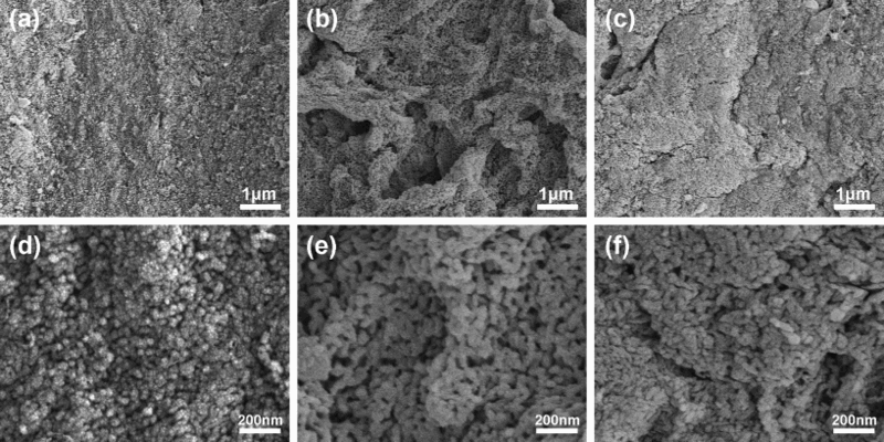
The microstructures of as received samples are displayed in Fig. 1. The cross-sectional SEM image (Fig. 1a) of RB shows a typical femur structure distributed with a portion of microsized and nanosized pores. Mineralised collagen as fibrous substances or bundles can be discernible in RB. According to the magnified image (Fig. 1d), the diameters of mineralised fibres are 40–200 nm. In contrast, the presence of nanosized particles is prevalent in two synthetic nanocomposites in which the delicate microstructure of RB is absent. In fact, both synthetic samples are dense materials, but their microstructural feature is different. In Col-HA sample (M1), there exist many ∼10 nm sized particles. The organisation of these nanoparticles into fibre-like objects is discernible (arrows in Fig. 1e), suggesting the presence of mineralised collagen. In the case of Col/Alg-HA sample (M2), fibrous object is arrowed in Fig. 1c. However, the size and morphology of nanoparticles in M2 are different as compared with M1. In most cases, bundled particles can be observed, and they are ∼20–40 nm in diameter and 100–200 nm in length (Fig. 1f). It is plausible that the microstructural differences among the examined samples are due to component dependent modulating effects on collagen fibril formation and HA synthesis. 12
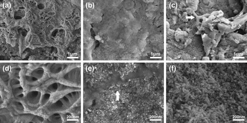
SEM images of cross-sections of a,d RB, b,e M1 and c,f M2
FTIR and XRD
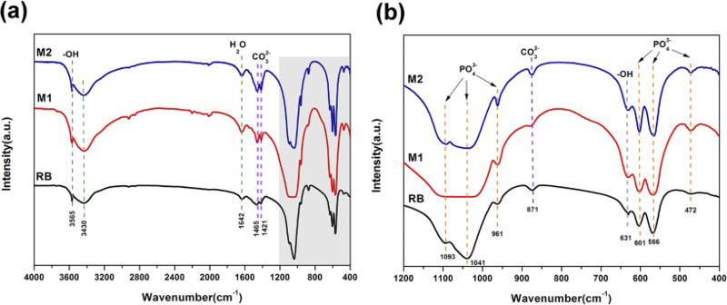
The FTIR spectra of RB, M1 and M2 are given in Fig. 2a. Each spectrum shows similar characteristic peaks of HA (at 561, 601 and 1039 cm− 1 due to phosphate vibrations) and collagen (C = O stretching vibration at ∼1645 cm− 1, N–H in plane bending at 1547 cm− 1, C–N absorptions at 1241 cm− 1, namely, amide I, II and III bands; C–H and N–H stretching modes at ∼2931 cm− 1).16,17 The decreased intensity of characteristic adsorptions of amides II and III in M2 compared with those in M1 represents the molecular interactions that occurred between collagen and alginate. 12 Additionally, the typical bands of carbonate substituting for phosphate sites in the apatite lattice are also present, as indicated by the peak at 871 cm− 1 and double bands at 1413 and 1455 cm− 1. 17 The presence of carbonate characteristic peaks and absence of peaks at 631 and 3565 cm− 1 due to hydroxyl in apatite in all samples confirm that HA in synthetic samples is poorly crystallised apatite.
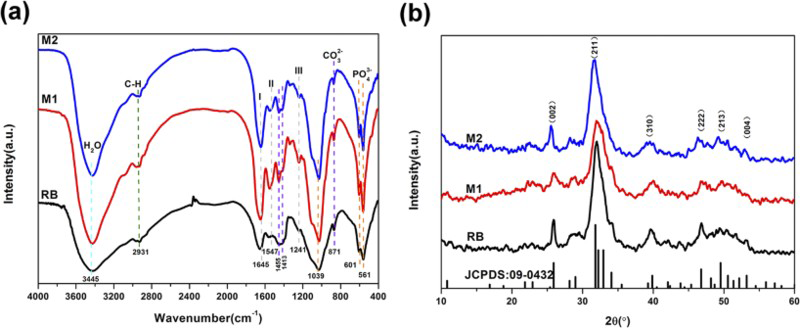
FTIR spectra (a) and XRD patterns (b) of RB, M1 and M2, confirmative of bone-like characteristics of M1 and M2
The XRD patterns of as received samples are given in Fig. 2b, together with the standard data of JCPDS Card No. 09-0432 for HA. The strong reflections at 26.0° and centred at 32.0°, and minor reflections at 40.0, 46.8, 49.4 and 53.0° (2θ) correspond to the (002), (211), (310), (222), (213) and (004) planes of HA respectively.16,17 The broad nature of these reflections indicates again that the inorganic phase is poorly crystallised HA. The similarity in XRD patterns and FTIR spectral traces of synthetic collagen based nanocomposites and natural bone confirms that the present nanocomposites are bone-like materials.
Thermal analysis
A multistage weight loss has been recorded for all the as received samples in Fig. 3a. In the case of RB sample, there are two stages of weight loss. The first stage yields a value of 10.0% weight loss before 200°C due to a complete evaporation of loosely bonded water in bone; the higher weight loss (∼27.0%) occurs between 200 and 600°C, mainly arising from the degradation and combustion of collagen.18–21 In contrast, three stages of weight loss appear in Col-HA (M1) sample. Before 200°C, the 7.5% weight loss is caused by dehydration. Between 200 and 400°C, 13.5% weight loss is given by collagen degradation. Above 400°C, the weight loss is due to the combustion of degraded collagen.20,21 For Col/Alg-HA (M2), there are four weight loss stages: before 180°C, the loss of water is ∼8.5%; between 180 and 300°C, the 9.0% weight loss is deemed to alginate decomposition combined with collagen degradation; ∼10.5% of weight loss between 300 and 400°C is due to collagen continuous degradation; above 400°C, 14% weight loss is the result of the combustion of remaining organic matters that derived from degraded alginate and collagen.20–22 The accurate temperature values associated with multistage weight loss are measured from DTG (Fig. 3b). As a result, the final HA contents at 800°C were 63% in RB, 64% in M1 and 58% in M2. The respective weight loss curve of as received samples shows a component dependent decomposition behaviour.
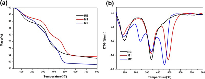
TGA (a) and DTG (b) curves of RB, M1 and M2, indicative of varying weight loss behaviours
DSC recorded heat flow changes in thermal decomposition of all samples, and curves are given in Fig. 4. The exothermic peak of RB occurs at 351°C with a left shoulder, and this value is consistent with reported results.18,20,21 In the case of M1, distinct exothermic peaks are observed at 350 and 456°C. The exothermic reaction ∼350°C presumably represents the decomposition of collagen molecules, while the intense exothermic reaction at 456°C is due to collagen combustion. 9 M2 shows similar results; however, the maximum exothermic peak decreases to 425°C.
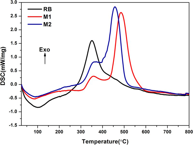
DSC thermograms of RB, M1 and M2, showing respective exothermal peak at 351, 485 and 456°C
600°C calcined samples
SEM
SEM images of RB and synthetic nanocomposites calcined at 600°C are given in Fig. 5. For better comparison, the presence of porous femur parts was intentionally avoided for SEM observation of calcined RB. The most striking feature among all the samples is the appearance of nanoporous structure due to the removal of organic components. In calcified natural bone (RB), spherical nanoparticles are distributed (Fig. 5a and d). These nanospheres are ∼50 nm in diameter and are actually aggregated by very small nanoparticles. In Fig. 5b and d of Col-HA (M1), the nanoparticles and their organised fibre-like objects of the as received M1 (Fig. 1b and d) completely disappear. Instead, a porous network structure is observed, primarily with 70 nm sized particles. The SEM images (Fig. 5c and f) of calcined M2 sample show the presence of bundled nanoparticles which appear in the as received M2 sample (Fig. 1f). In comparison, the calcination of synthetic nanocomposites gives rise to more nanopores in their cross-sectional samples.
SEM images of cross-sections of 600°C calcined a,d RB, b,e M1 and c,f M2
FTIR and XRD
The FTIR spectra of RB, M1 and M2 after being calcined at 600°C are shown in Fig. 6. The characteristic adsorptions of carbonated HA still exist, while the bands of organics in Fig. 2a disappear. The broad adsorption at 3430 cm− 1 and weak adsorption at 1642 cm− 1 arise from absorbed water. New absorptions at 3565 and 631 cm− 1 assigned to hydroxyl groups of HA indicate the enhancement of crystallinity, and this result is further validated by their XRD patterns (Fig. 7). 17 However, it should be noted that HA of samples calcined at 600°C are still poorly crystallised apatite.
FTIR spectra in range of a 4000–400 cm− 1 and b 1200 − 400 cm− 1 of 600°C calcined RB, M1 and M2
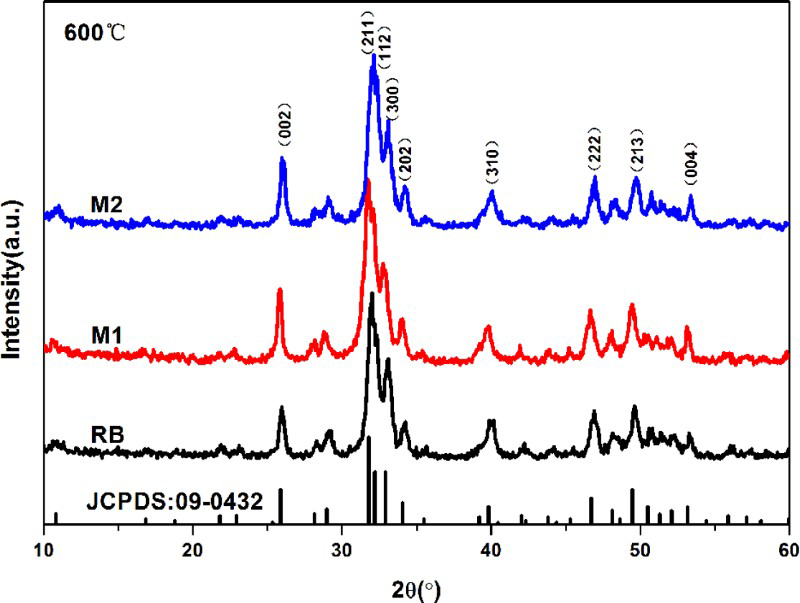
XRD patterns of 600°C calcined RB, M1and M2
1000°C calcined samples
SEM
Significant changes in morphology and crystal size of all the samples took place after being calcined at 1000°C. In calcined RB, well grown hexagonal crystals exist. They are ∼300–700 nm in diameter and 500 nm to 3 μm in length. These crystals of rabbit femur grew with an oriented alignment (Fig. 8a and d). Similar hexagonal crystals are also observed in Col/Alg-HA nanocomposite (M2) (Fig. 8c and f), but these crystals lack the oriented alignment achieved in natural bone. Strangely, such well grown hexagonal crystals are not found in Col-HA nanocomposite (M1). Actually, a porous structure formed in M1 sample, suggesting the continuous decomposition of this sample.
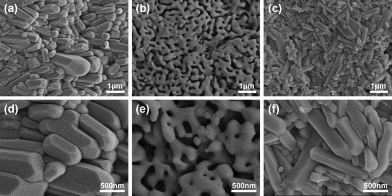
SEM images of cross-sections of 1000°C calcined a,d RB, b,e M1 and c,f M2
XRD
The phases of calcined samples are identified with reference to the standard data of JCPDS Card No. 09-0432 (HA) and JCPDS Card No. 09-0169 (β-TCP, not given in chart). The XRD patterns are shown in Fig. 9. Compared with as received and 600°C calcined samples, much stronger reflections are recorded for 1000°C calcined samples, indicative of the high crystallinity of inorganic phases after 1000°C heat treatment. The XRD patterns of both RB and M2 samples belong to pure HA. However, a mixture of HA and β-TCP is observed in Col-HA (M1). No other phases such as CaO and CaHPO4 were recorded in all the samples. In general, the presence of CaO is due to the Ca/P of initial samples >1.67, while the latter is < 1.50. When the Ca/P value of initial is between 1.50 and 1.67, heat treatment or calcinations especially at high temperature would yield biphasic calcium phosphate materials of HA and TCP.23,24 Accordingly, the present XRD results actually indicate that RB and synthetic M2 sample have stoichimetric HA. This difference between Col-HA and Col/Alg-HA nanocomposites demonstrates that the introduction of Ca capturing alginate into collagen reconstitution with simultaneous HA synthesis could better realise the experimental design from the feedstock solutions.
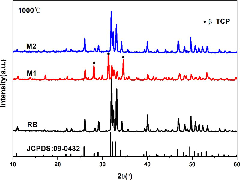
XRD patterns of 1000°C calcined RB, M1 and M2
Conclusions
In this study, Col-HA (M1) and Col/Alg-HA (M2) nanocomposites were prepared by virtue of collagen reconstitution with simultaneous HA synthesis. The as received nanocomposites and their calcined products were systematically analysed with natural bone (rabbit femur, RB) as a control. XRD results showed that all the as received samples were poorly crystallised apatite, confirmative of achieving bone-like features in synthetic nanocomposites. Although M1, M2 and RB gave the similar XRD reflections and FTIR spectral traces, the difference in the molecular species of synthetic nanocomposites and RB was revealed by TG and DSC analyses in which a different multistage weight loss and heat flux change behaviours were recorded. In addition, the structural analyses of calcined samples further indicated the existence of microstructural and phase composition differences among as received and natural bone samples. In 600°C calcined samples, the removal of organic species led to the formation of nanosized pores, but the crystallinity of samples remained low. After being calcined at 1000°C, well grown hexagonal HA crystals formed in both natural bone and Col/Alg-HA nanocomposites (M2), but the oriented alignment of these HA crystals was only achieved in natural bone sample. Surprisingly, thermal decomposition was recorded in Col-HA nanocomposite (M1), and the thermal decomposition product was a mixture of HA and β-TCP without any other phases such as CaO. The present results indicate that the introduction of Ca capturing alginate is beneficial to achieving stoichimetric HA in synthetic nanocomposites. The relevant experimental data are useful for guiding the preparation of bone grafting nanocomposites.
Footnotes
Acknowledgements
This work is supported by the National Basic Research Program of China (grant no. 2012CB933600) and the National Natural Science Foundation of China (grant no. 30670561).