
Abstract
Inonotus hispidus, Scytalidium ganodermophthorum and two strains of Scytalidium lignicola were tested for their ability to produce yellow extracellular pigment on media plates, sterile wood blocks and non-sterile logs to determine their suitability for use as spalting fungi. All three fungi produced a penetrating yellow pigment in the non-sterile logs after 12 weeks of incubation; however, results from the sterile block tests indicated that the incubation time necessary for I. hispidus to produce sufficient yellow pigment may be as low as 4 weeks of incubation. An incubation period of 4 weeks is the shortest recorded for controlled spalting and will allow for the currently utilised production time for yellow spalted wood of 12 weeks to be substantially decreased using an isolate of I. hispidus as the inoculum.
Keywords
Introduction
The field of controlled spalting, a process by which wood is naturally coloured with extracellular fungal pigments, continues to develop as new inoculation methods are established and new fungi are tested for their pigmenting ability. Spalted wood is of particular interest within value added wood products, as the process can be applied to any wood species and results in a substantial price increase to the raw or final product, depending on the type of spalting.
Of the three types of spalting, bleaching, zone lines and pigmentation, the first two are well understood and have been extensively researched (Robinson 2012). Pigmentation, however, which refers to the non-black colours produced by fungi internally in wood as extracellular pigment, is less well understood. While many fungi, especially airborne moulds, are capable of producing extracellular pigment, very few of these fungi are capable of growing inside wood and releasing pigment broadly through the substrate (Robinson et al. 2011a).
Fungi that have the capacity to grow internally within wood and also produce extracellular pigment are best suited to spalting. Unfortunately, in addition to their infrequent occurrence in North American forests, many of these fungi can be difficult to maintain in laboratory conditions, necessitating the use of malt agar plates amended with prespalted wood chips in order to insure that the fungus continues to produce pigment (Robinson et al. 2012a). These difficulties make finding and testing potential new spalting fungi a time consuming and arduous process, with a substantial failure rate. Hence, potential spalting fungi must be run through a gauntlet of tests, from basic malt agar plate growth through non-sterile log inoculation, to determine their suitability for use.
There are currently two well developed colours for use in spalting: the pink/red of Scytalidium cuboideum (Sacc. & Ellis) Sigler & Kang and the blue/green of Chlorociboria aeruginascens (Nyl.) Kanouse and Chlorociboria aeruginosa (Oeder) Seaver (Robinson and Laks 2010; Robinson et al. 2011a, 2012a), although as with all pigment producing fungi, their effects differ widely based upon wood species and incubation time. The blue/green pigment produced by Chlorociboria species, called xylindein, has been used in wood art for centuries (Blanchette et al. 1992) extensively studied and its structure is well understood (Saikawa et al. 2000). Scytalidium cuboideum is thought to produce several pigments, although only one has been characterised thus far (Golinski et al. 1995). The pigments of S. cuboideum have undergone moderate testing in terms of stability in indoor and UV light, and were found to be significantly more stable in light than their non-fungal biological counterparts (Robinson et al. 2013a).
In contrast to the rarer blue/green pigments, yellow pigments occur with some frequency in naturally spalted wood. The two fungi routinely cited as producing a yellow extracellular pigment within wood are Scytalidium ganodermophthorum Kang, Sigler (Kang et al. 2010) and Inonotus hispidus (Bull.) P. Karst (Ali et al. 1996; Perrin and Towers 1973). The goal of this research was to determine the potential usage of these two fungi as spalting fungi, based upon their ability to (1) be grown in culture media quickly and reliably produce pigment; (2) to produce yellow pigment internally in standard test blocks of two species of hardwoods within 12 weeks; and (3) to determine if internal pigment production of any of the fungi could occur under non-sterile conditions. As the University of Alberta Microfungus Collection and Herbarium (http://www.uamh.devonian.ualberta.ca) notes a production of yellow pigment in Scytalidium lignicola Pesante in addition to its standard blue stain production of melanin (Robinson et al. 2007), two strains of S. lignicola were tested concurrently to see if the species produced similar amounts of yellow pigment to I. hispidus and S. ganodermophthorum. The success of any of the fungi to routinely produce a penetrating yellow pigment through hardwoods would add a third colour option for controlled spalting and increase the marketability of spalted wood through additional colour options.
Methods
Scytalidium ganodermophthorum (UAMH 10320 from South Korea, formerly Xylogone ganodermophthora), Inonotus hispidus (F2037, isolation data unknown, sample obtained from Forintek, Canada) and two isolates of Scytalidium lignicola (UAMH 5122 from British Columbia, Canada and UAMH 1502 from Italy) were grown on 2% malt extract agar plates for 2 weeks, inoculated onto Acer saccharum Marsh (‘sugar maple’, average SGOD = 0·71) and Populus tremuloides Michx. (‘aspen’, average SGOD = 0·41), incubated for various time periods, and then evaluated for per cent surface area coverage of yellow pigment. Sugar maple blocks came from kiln dried lumber from Ontario, Canada. Aspen was from air dried logs harvested in Ontario, Canada.
Stage I: Agar plate growth
Roughly 1×1 cm plugs of actively growing mycelium from each species were placed on 2% malt agar Petri plates (100×15 mm). Five plugs were placed on each plate, with each plug equidistant from the others. Each plate contained only one species and there were three replicate plates of each species used. Plates were incubated at room temperature and humidity, in the dark, for 2 weeks. After incubation, the plates were analysed for colour using a Konica Minolta CR-5 Chroma Meter. A one-way ANOVA followed by Tukey's honestly significant difference was used for yellow colour comparison.
Stage II: Wood blocks
Wood was cut into 14 mm cubes, dried at 40°C for 24 h and then weighed. A total of 240 blocks, 120 blocks per wood species were produced. To prepare for inoculation, the blocks were steam sterilised for 30 min, allowed to cool in a laminar flow hood and then placed into sterilised vermiculite containing glass mason jars as previously described in Robinson et al. (2012b). Three blocks of the same wood species were placed equidistant apart in each jar. An inoculum strip measuring roughly 1×3 cm was then placed between all three blocks, with no side of the agar or mycelium touching any of the wood blocks. Inoculated jars were incubated in a humidity and temperature controlled chamber (27±2°C with 80±5% humidity). Jars were removed from the chamber in groups of four jars per wood species per fungus from the chamber at intervals of 4, 6, 8, 10 and 12 weeks. Following the incubation period, the blocks were taken out of the jars, scrubbed with a dry brush to remove mycelium and vermiculite, and weighed. They were then oven dried for 12 h at 40°C and weighed again.
Colour analysis was performed using an Epson WorkForce 500 scanner as described in Robinson et al. (2009). Blocks were scanned on the external face that contained the most yellow pigment. Once scanned, the blocks were cut in half to expose a radial plane, and then one internal face was scanned again for yellow pigment. A two-way ANOVA with incubation time and fungus species was run using SAS version 9.3 for each fungus species. Means were transformed with arcsine square root to achieve a normal distribution.
Stage III: Non-sterile logs
Sugar maple and aspen logs were split lengthwise and then cut into roughly 20×20 cm sections. The pith and visible heartwood from each section was removed via band saw, leaving the log sections with a roughly 10 cm depth. External bark remained in place.
The log sections were placed in plastic storage bins (49·53×44·45×29·46 cm) and stacked on the end grain, with cut faces against one another and one plate of actively growing mycelium from one of the three fungal species sandwiched between. The bins were surface sterilised with a 10% bleach solution and 85% ethanol solution before use. A total of six bins were used: three for each wood species and a separate bin for each fungus species for a total of 12 log sections of each wood species. Log sections were surrounded by vermiculite wetted to 98% water holding capacity.
The bins were stored with lids in a humidity and temperature controlled room (27±2°C with 80±5% humidity) for 12 weeks. After incubation, the sections were removed, cleaned of vermiculite and mycelium with a dry brush, and then turned on a Delta midi-lathe to determine depth of yellow pigment penetration. Penetration analysis was recorded on a simple yes/no scale based upon whether or not the yellow pigment had fully penetrated the entire log section. The amount of yellow pigment was not quantified; however, secondary pigments produced by the fungi were noted.
Results and discussion
Agar plate growth
CIE L*a*b* colour analysis of the agar plates found colours within the yellow and green spectrum for S. ganodermophthorum (average a* = −5·04, average b* = 27·22), blue spectrum for S. lignicola (average a* = −0·38, average b* = 3·0) and yellow and red spectra for I. hispidus (average a* = 1·16, average b* = 15·7). The intensity of yellow pigment was significantly different between each of the three species (P<0·0001), with S. ganodermophthorum having the highest b* value and I. hispidus having the second highest. Despite culture notes stating that S. cuboideum produced yellow stain in culture, no yellow was detected on any of the S. cuboideum plates for the duration of testing. S. ganodermophthorum plates showed only yellow pigment; however, I. hispidus plates showed yellow pigment within the agar, yellow mycelium and dark brown/yellow zone lines, all of which are common pigment components of I. hispidus (Perrin and Towers 1973; Ali et al. 1996).
Of the tested fungi, all four strains were able to completely colonise the agar plate within 2 weeks. The 2-week standard was developed originally as a method for screening fungi for potential success in block test trials, and continues to be a valuable indicator of how quickly extracellular pigment will penetrate wood (Robinson and Laks 2010; Robinson et al. 2012a). Although S. lignicola did not produce yellow pigment within the agar plates, the fungus was carried through to additional testing with the hope that contact with wood would stimulate the production of yellow pigment.
Wood blocks
There was no significant difference between the two strains of S. lignicola, so the data were combined for further statistical tests. For external yellow stain on sugar maple, both the fungus species (P = 0·01) and weeks incubated (P<0·0001) were significant, as well as the interaction between the two (P<0·0001). S. ganodermophthorum at 10 weeks had the highest amount of external yellow pigment (65%); however, that amount did not differ significantly from I. hispidus at 4 weeks, S. ganodermophthorum at 12 weeks, S. lignicola at 10 weeks or S. lignicola at 8 weeks (Fig. 1). There was no significant amount of internal pigmentation among any of the fungal species at any week. The higher yellow pigment amount produced by I. hispidus early in incubation is most likely due to two factors: the early production of the pigments hispidin and hispolon, which are known to decrease markedly after 25 days incubation (Perrin and Towers 1973; Ali et al. 1996), and the white rot decay and zone line formation that occurred later in the incubation cycle, as part of the natural decay mechanism of this fungus. The reason for the decrease in the pigments is unknown; however, it may be due to the bleaching effect of the decay mechanism of I. hispidus overlaying the pigments. If the pigment loss is indeed due to overlay, then the long-term use of yellow stained wood by I. hispidus should not experience colour loss if the fungus is killed before reaching 25 days old. On sugar maple, the amount of yellow pigment from I. hispidus at 4 weeks rivalled that of the other fungi at longer incubation times, marking I. hispidus as an ideal fungus for controlled spalting.
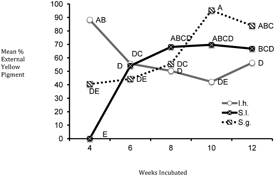
Two-way ANOVA for external yellow pigment on sugar maple blocks: I.h. = Inonotus hispidus, S.l. = Scytalidium lignicola, S.g. = Scytalidium ganodermophthorum; different letters represent significant differences (α = 0·05)
Although I. hispidus proved to produce pigment more quickly than S. lignicola or S. ganodermophthorum, these latter two fungi should not be ruled out for use as spalting fungi. Both managed to produce over 50% coverage of yellow pigment by 8 weeks. Eight to ten weeks incubation is generally used as the maximum time threshold for controlled spalting, with a minimum coverage rate of 30% required in 14 mm block testing (Robinson et al. 2011b, 2012b). Both S. lignicola and S. ganodermophthorum meet these requirements, and are therefore also suitable for use as spalting fungi on sugar maple.
For external yellow pigment on aspen, there was a significant difference between fungi (P<0·0001), weeks of incubation (P = 0·03) and the interaction between the two (P<0·0001). Once again, I. hispidus had significantly more external yellow pigment at 4 weeks (74·3%) than any other fungus at any other time (Fig. 2). The yellow pigment on aspen was much easier to read due to the pale colour of the wood, which likely explains why the dominance of I. hispidus at 4 weeks is so significant. However, unlike with sugar maple, sufficient amounts of yellow pigment coverage were not reached by S. lignicola at any interval, and S. ganodermophthorum just exceeded the 30% threshold at 6, 10 and 12 weeks.
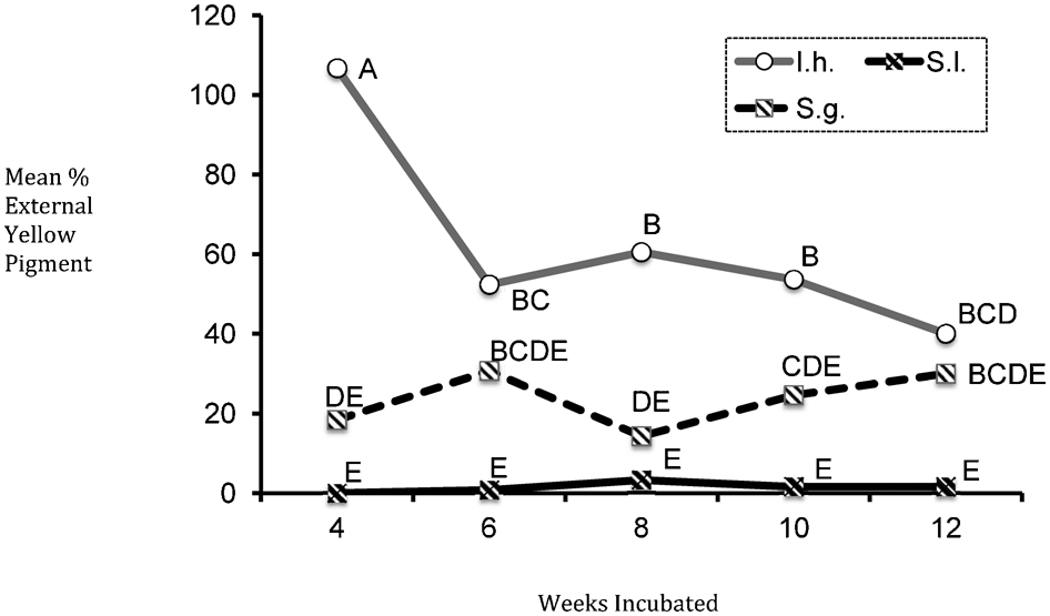
Two-way ANOVA for external yellow pigment on aspen blocks: I.h. = Inonotus hispidus, S.l. = Scytalidium lignicola, S.g. = Scytalidium ganodermophthorum; different letters represent significant differences (α = 0·05)
Internal yellow pigment was significant with fungus species, weeks incubated and the interaction between both (all at P<0·0001). Again, I. hispidus had the most internal yellow pigment at 4 weeks (12%), although this amount did not differ significantly from the yellow pigment on I. hispidus blocks at 10 weeks. The white rot and brown zone lines associated on the external faces of the aspen blocks inoculated with I. hispidus did not penetrate inside the wood, leaving the internal face affected only by the yellow pigments. No significant internal yellow pigment was produced on aspen blocks by S. lignicola at 4, 6, 8 or 10 weeks, or S. ganodermophthorum blocks at any time. Based upon the poor performance of both Scytalidium species on aspen, it does not appear advisable to utilise either for controlled spalting on this wood species. I. hispidus, on the other hand, continued to perform well at 4 weeks externally and unlike with sugar maple, also produced internal yellow stain.
Non-sterile logs
All log sections showed yellow pigment throughout their entire depth, although amounts varied from heavy (Scytalidium species) to sporadic (I. hispidus). In addition to the yellow stain, logs inoculated with S. lignicola showed blue stain (Fig. 3a) and logs with I. hispidus showed white rot and brown zone lines (Fig. 3b and c). Logs inoculated with S. ganodermophthorum showed only yellow stain (Fig. 3d). The same colour variations showed up on the wood block tests as well, although the amount of blue stain and zone lines on the blocks is not included herein. The small amount of yellow stain occurring on logs inoculated with I. hispidus is consistent with the data produced from the block tests. It is likely that more yellow pigment was visible at 4 weeks of incubation and that pigment was either covered or removed by the fungus at a later date during the 12 weeks of incubation.
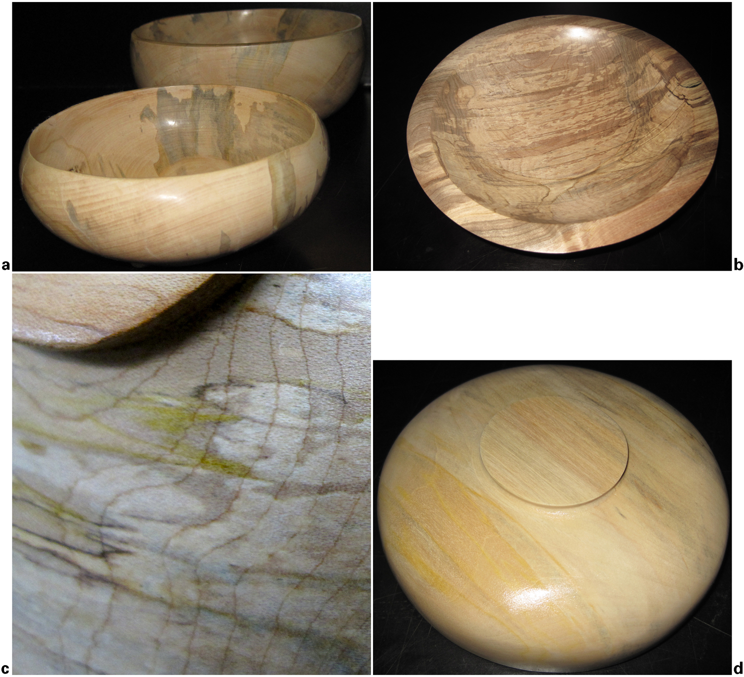
a S. lignicola on aspen; b I. hispidus on sugar maple; c close-up of yellow stain of I. hispidus on sugar maple; d S. ganodermophthorum on aspen
Although no internal yellow pigment was found in the sugar maple block tests and only I. hispidus produced significant amounts in aspen, all of the tested fungi produced internal pigment in the logs. It is not uncommon for fungi to produce more pigment in non-sterile logs than in sterile block tests, and similar results have been seen before in terms of controlled spalting (Robinson et al. 2013b). Many fungi produce extracellular pigments as a response to adverse conditions such as low moisture (Tudor et al. 2012), pH (Tudor et al. 2013), UV light (Perrin and Towers 1973; Ali et al. 1996) or as a method to protect their resources from other fungi (Li 1981; Score et al. 1997). While UV light and low moisture were not conditions prevalent in our incubation chamber, the room itself was not sterilised and no attempt was made to clean the logs before incubation. It is possible that the increased internal pigment was due to interspecific fungal interactions between the inoculated fungus and any fungi already present.
Conclusion
Yellow pigment production by S. ganodermophthorum and I. hispidus was consistent across all three testing stages. S. lignicola failed to produce yellow stain in culture, but did produce the pigment on both sterile block and non-sterile log tests.
Based upon the results of the second and third stage testing, all three fungi are suitable for controlled spalting work. However, the sterile block test results indicate that I. hispidus is superior to the Scytalidium species in terms of early pigment production, and of the three fungi may be the best suited for spalting work that requires a quick production time. In terms of all currently utilised spalting fungi, the 4 week incubation time of I. hispidus is remarkable and is the shortest time thus far recorded for maximum pigmentation, with the closest other incubation time being 6 weeks for S. cuboideum (Robinson et al. 2011a). Fungi capable of producing sufficient amounts of extracellular pigment on wood substrates in a short amount of time are ideal for controlled spalting and offer landowners and small businesses working with native wood and fungal species a quicker turnaround time for their products. The 4 week incubation time of I. hispidus effectively cuts standard incubation times for yellow pigment in half, allowing yellow spalted wood to be produced twice as quickly as other spalting varieties if carefully monitored.