






Abstract
Objective
This biomechanical study aims to elucidate differences in how skulls with trigonocephaly, normal skulls, and postoperative trigonocephalic skulls respond to intracranial pressure and how this affects the orbital distances.
Materials and Methods
For 10 patients with trigonocephaly (8.2 ± 4.5 months), simulation models were produced based on the computed tomographic data of the skulls. These models were categorized as the Trigono group. For each model, a 15-mm Hg pressure was applied to the neurocranium to simulate the intracranial pressure. The interorbital distances expanded in response to the applied pressure. The amount of the change in the orbital distance was calculated using finite element analysis. The same processes were repeated for 10 models simulating normal skulls (the Control group) and postoperative trigonocephalic skulls (the Remodeled group). The changes in the orbital distance were compared among the three groups.
Results
The changes in the orbital distance were significantly smaller for the Trigono group than for the Control group. However, changes were significantly greater for the Remodeled group than for the Control group.
Conclusion
The expansion of interorbital distances in response to the cranial pressure is restricted in skulls with trigonocephaly. This restriction is eliminated by performing remodeling of the skull. These findings explain why spontaneous correction of hypotelorism occurs postoperatively in trigonocephaly.
Trigonocephaly is a relatively uncommon form of craniosynostosis, with an incidence of 0.3 per 1000 live births (Shuper et al., 1985). The cause of trigonocephaly is attributed to premature closure of the metopic suture (Genitori et al., 1991–1992). Trigonocephalic skulls are characterized by an anterior protrusion, a so-called keel-shaped deformity (Havlik et al., 1999; Aryan et al., 2005; Warschausky et al., 2005; Selber et al., 2007). Although the keel-shaped deformity is the most prominent feature of trigonocephalic skulls, trigonocephaly is also often accompanied by hypotelorism (Dominguez et al., 1981; Richardson et al., 2006; van der Meulen et al., 2008). Because this deformity of the orbits impairs the appearance of patients, it is essential to correct hypertrophy to improve their quality of life.
To effectively treat hypotelorism, its etiology in trigonocephalic patients needs to be clarified. Hypotelorism occurs in skulls where the lateral growth of the orbits is hindered.
The skull grows as bone deposits at the sutures. Craniosynostosis develops when this bone deposition is disturbed for some reason. Given that the bone deposition is a metabolic phenomenon, it naturally is influenced by biochemical conditions of the sutures and intrinsic growth potential of the cells. Besides the biomechanical conditions, however, mechanical stresses also affect the speed of bone deposition (Enlow, 1973; Enlow and McNamara, 1973). When mechanical forces work on the skull, strain develops at the sutures, indirectly affecting cellular activities there.
Among the mechanical forces, the authors are interested in the effects caused by the intracranial pressure. The authors hypothesized that in the growth of the skull, the intracranial pressure works to expand the distances of bilateral orbits. The authors further hypothesized that in trigonocephalic skulls, the expansion is deterred by the prematurely fused metopic suture. The present study sought to verify these hypotheses from a biomechanical point of view.
In the present study, computer simulation models were produced for trigonocephalic skulls and normal skulls. Using biomechanical simulation on these models, the influence of the intracranial pressure on the shapes of the skulls was quantitatively evaluated. By comparing these results between the trigonocephalic skulls and the normal skulls, the etiology of the hypotelorism in trigonocephaly was examined. Additionally, by producing operated trigonocephalic skull models and performing biomechanical analysis on these models, how the remodeling of the skull affects interorbital distances was elucidated.
Materials and Methods
Model Production
Model Production for Trigonocephaly
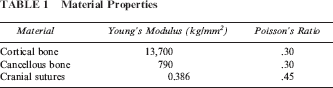
The protocols of the present study were reviewed and approved by the Institutional Review Board of the Department of Plastic Surgery at Keio University. Among the patients who visited the authors’ institutes during the period from 2000 to 2008, there were 10 patients presenting trigonocephaly who constitute the sample for this study. The ages of the patients—none of whom had undergone surgical intervention—were 8.2 ± 4.5 months. For the skull of each patient, computed tomography (CT) images at slice intervals of 0.5-mm thickness were obtained using CB Works (Hitachi Co., Ibaragi, Japan). The CT images of the patients were saved as Digital Imaging and Communications in Medicine files at 300 dots per inch resolution. Then, using the obtained CT data, a three-dimensional computer-aided design model (CAD model) was produced for each patient, simulating his or her skull. First, 40 to 50 specific points were automatically marked on each axial slice of the CT data by means of an originally developed program in a repeatable manner. Thereby, topological characteristics of the bony components of the images were identified. Then the lines between the neighboring points were divided by points at ratios appropriate for the topological complexity of each line. By connecting thus-processed slices, a three-dimensional CAD model was produced. The model production and subsequent analysis were performed using structural analysis software (ANSYS 11.0; ANSYS Co., Canonsburg, PA). In the CAD model, bone thickness of each part of the skull was assigned after careful measurement of the CT images. Material properties shown in Table 1 were allotted to the cortical bone, cancellous bone, and craniofacial sutures. Therefore, the 10 models are homologous in terms of material properties. These material properties were obtained from past literature (Pan et al., 2007). Because all patients of this group lacked the metopic suture, the frontal bones were modeled as a single solid piece in the corresponding models. Each CAD model was divided into 110,000 to 128,000 shell elements (Fig. 1). Each shell element consisted of three layers—two cortical bone layers with one layer of cancellous bone between. Each layer contained 10 nodes. These CAD models were termed the Trigono group.
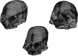
A CAD model of the Trigono group viewed from different angles.
Material Properties
Control Group
Among child patients who visited our institutes for suspected brain injury, 10 who had undergone CT examination were selected. None of these 10 patients presented deformity of the skull. Their ages (8.6 ± 4.3 months) demonstrated no statistically significant differences from the Trigono group patients. To simulate the skulls of these 10 patients, CAD models were produced using the same technique as for the Trigono group. Given that all patients in this group had the metopic suture, the material properties for the cranial suture were allocated to the vertical midline of the frontal bones in the corresponding models. These CAD models were termed the Control group (Fig. 2).
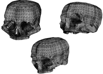
A CAD model of the Control group viewed from different angles.
Remodeled Group
Each model of the Trigono group was remodeled by simulating the operation for trigonocephaly. First, the frontal bone and supraorbital bars were separated and removed from each skull (Fig. 3). The removed entity was divided into six segments—bilateral temporal wings, frontal bones, and supraorbital bars (Fig. 4). These six parts were recombined to present normal morphological features and were fixed to the original skull (Fig. 5). Assuming that the premature fusion of the metopic suture was released and normal flexibility was achieved by the remodeling, material properties for cranial sutures were allocated to the junction that connects the bilateral supraorbital bars (Fig. 4, above right). In contrast, assuming osseous unification occurred between (1) the frontal bone and supraorbital bars, (2) the temporal wings and the original skull, and (3) the temporal wings and supraorbital bars, material properties for bone were allocated to these junctions. Bilateral frontal bones were unconnected. Thus-produced CAD models were defined as the Remodeled group.
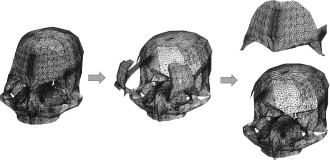
The first step of the simulation surgery. For a Trigono group CAD model (left), the frontal region and supraorbital bar are isolated (center) and removed (right).
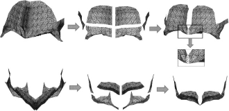
The second step of the simulation surgery. The bone pieces removed from the Trigono group models are divided into pieces and remodeled. The left, center, and right columns indicate the frontal region plus supraorbital bar just after the removal, during processing, and after remodeling, respectively. The upper row views the pieces in the anterior-posterior direction; the lower row views the pieces in the superior-inferior direction. For the junction between bilateral orbital bars, material properties of cranial suture were allocated (close-up rectangle, above right).
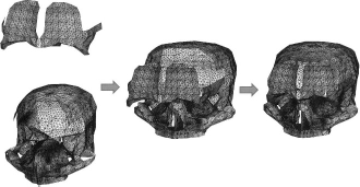
The remodeled bone pieces are returned to the original skulls and fixed.
Load Application and Calculation
After fixing each model by giving zero displacement to the foramen magnum, simulated intracranial pressure was applied (Fig. 6). Because normally the intracranial pressure is less than 20 mm Hg (Rutigliano et al., 2006), a 15-mm Hg pressure was applied to the internal surface of the neurocranium. Each model presented subtle distortion in response to this pressure. The displacement presented by each part of the models was calculated. The finite element method was used for the calculation. In the medical fields, finite element analysis is widely used for biomechanical analyses of such organs as the bone (Remmler et al., 1998a; Remmler et al., 1998b), skin (Mizunuma et al., 2000), vessels (Beller et al., 2007), and blood flow (Shojima et al., 2004). In a series of past studies, the authors conducted analyses on skulls and proved the methodological validity of their analyses (Nagasao et al., 2005; Nagasao et al., 2006; Nagasao et al., 2007a; Nagasao et al., 2007b).
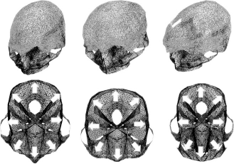
Pressure is applied to the neurocranium of each model. The left, center, and right rows indicate CAD models and their cross-sections belonging to the Trigono group, Control group, and Remodeled group, respectively. The cross-sections in the figures are taken at a vertical height 2 cm superior to the superior orbital rim. A 15-mm Hg pressure was applied to the internal surface of the neurocranium of each model (the netted area in the upper row), simulating the intracranial pressure. The pressure is applied in the outer direction (lower row).
Definition of Parameter to Evaluate Changes of Orbital Distance
For each model, two marking points were set at the inner borders of the bilateral orbital rims. The distance between these two marking points was defined as orbital distance (OD) (Fig. 7). In response to the simulated intracranial pressure, the values of OD altered slightly. The change was defined as ΔOD. Namely, defining the value of the OD before the pressure application as ODpre and the value after the pressure as ODpost, ΔOD is given by

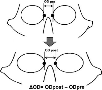
In response to the intracranial pressure, the orbital distance presents subtle change. The amount of change was defined as ΔOD and was calculated by subtracting the orbital distances before and after application of the intracranial pressure.
Evaluation
The differences in the distortion patterns in response to the intracranial pressure were evaluated by the following methods.
Comparison of Contour Maps
The software used in the analysis can demonstrate displacements in the form of scaled contour maps. By referring to the contour maps the models present, morphological transformation patterns of the skulls belonging to the three groups were compared.
Comparison of ΔOD
To evaluate the differing response patterns the ODs presented after application of the intracranial pressure, ΔOD was compared among the three groups. Because ΔOD showed skewed distribution, the Mann-Whitney U test was used for the comparison. Any p values less than .05 were considered statistically significant. All statistical calculations were performed using SPSS Version 10 for Windows (SPSS Inc., Chicago, IL).
Results
General Deformity
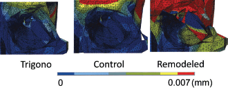
Deformity patterns of the whole skull and of the orbital regions are demonstrated in Figures 8 and 9, respectively. For all three groups, the frontal and superior orbital regions moved laterally in response to the intracranial pressure. The lateral displacement was greater for the frontal regions than for the supraorbital regions (Fig. 8). The lateral deviation of the orbits was greater in the order of the Remodeled group, Control group, and Trigono group (Fig. 9).
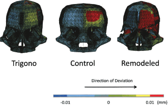
Deformity patterns of the skull models representing the three groups. The color scale indicates the value of horizontal displacement each part of the models presents. Red, yellow, and light green mean positive values, indicating that the areas marked with these colors displace in the left direction in response to the intracranial pressure; blue colors mean negative values, indicating areas that displace in the right direction.
Orbital Distances
For all groups, ΔOD demonstrated positive values, indicating that the intracranial pressure works to expand the interorbital distances (Fig. 10 and Table 2). The ΔOD was significantly smaller for the Trigono group than for the Control group, indicating that the expansion effect is reduced for the Trigono group. The ΔOD was significantly greater for the Remodeled group than for the Control group, indicating that the restriction on the expansion of interorbital distances shown in trigonocephalic skulls are eliminated after remodeling operations.
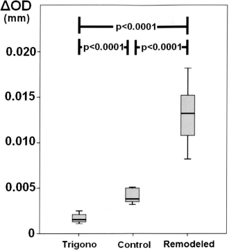
Comparison of ΔOD among the three groups.
Ranges of Change in Orbital Distance (ΔOD)
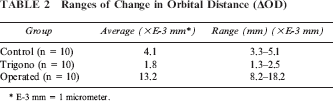
E-3 mm = 1 micrometer.
Discussion
Study Design
Influence of the intracranial pressure on OD was evaluated in the present study. The authors applied pressure on the internal surfaces of skulls, calculated the quantitative change presented by the interorbital distances in response to the pressure, and evaluated the expected growth patterns of the skull by referring to the calculated values. This study design is based on the following considerations.
Enlow states that when mechanical forces work continuously on the skull, the skull grows to conform to the forces (Enlow and McNamara, 1973). For instance, if certain forces keep on compressing the skull from lateral sides throughout the growth process, the skull is expected eventually to present a shape that is long along the anterior-posterior axis and short along the left-right axis.
The mechanical forces that work on the skull during its growth under physiologically normal conditions are grouped into two categories. The first category consists of the forces that work from outside of the skull. Contracture forces of the expression and masticatory muscles belong to this category. The second category—commonly referred to as the intracranial pressure—works from inside the skull. The forces of the first category work temporarily only when the muscles function. On the other hand, intracranial pressure works continuously on the skull. Due to this continuity, the authors perceive the intracranial pressure as one of the most influential mechanical factors on the growth of the skull and orbits.
Extrapolating from Enlow's theory, it can further be hypothesized that the positional relationships bilateral orbits present during growth should be indicated to some extent by quantitative changes of interorbital distances in response to the intracranial pressure. If the interorbital distance in a certain skull stays unchanged or shows little change in response to the intracranial pressure, the skull is expected to present hypotelorism over time. In contrast, if the orbital distance greatly increases on application of intracranial pressure in a certain skull, the skull is likely to develop hypertelorism. Based on these considerations, the authors calculated the incremental change in interorbital distance under application of a simulated intracranial pressure and used it to predict the positional relationships of bilateral orbits.
Clinical Meanings of the Findings
The results of the present study's findings can be summarized in three points. First, for all models, the ΔOD resulted in positive values. Second, ΔOD was significantly reduced for the Trigono group compared with the Control group. Third, ΔOD had a greater value for the Remodeled group than for the other two groups. Clinical meanings of these findings will now be discussed.
The first finding indicates that the intracranial pressure works to expand the OD with all skull types. The authors hypothetically explain this phenomenon as follows. As shown in the upper portion of Figure 11, the roof of the orbit forms a slant whose medial part is lower than the lateral part. When the intracranial pressure works on this structure, part of the pressure pushes it in the lateral direction. Besides this direct effect on the orbital roof, the intracranial pressure also works on the temporal part of the skull (lower portion of Fig. 11), causing lateral movement. Together with this movement, the orbit also moves in the lateral direction.
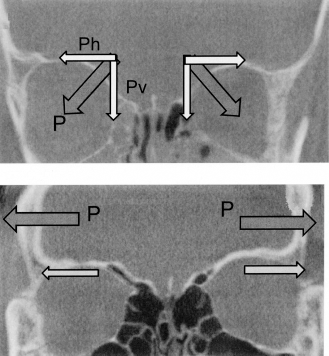
Hypothetical explanation of the effect of the intracranial pressure on the intraorbital distance. Above: Direct effect—When the intracranial pressure works on the roof of the orbit, the force can be divided into horizontal (Ph) and vertical (Pv) components. The horizontal component pushes the orbital roof in the lateral direction, which expands the orbital distance. Below: Indirect effect—The intracranial pressure works to expand the temporal region (P). With this expansion, the orbital roofs are pulled in the lateral direction (small arrows).
The second finding is compatible with the fact that hypotelorism is likely to accompany trigonocephaly. As explained in the previous paragraph, the intracranial pressure works to make bilateral orbits move apart. However, in trigonocephalic skulls, this movement of the bilateral orbits is restricted due to the prematurely closed metopic suture. Consequently, trigonocephalic patients are likely to develop hypotelorism.
The third finding indicates that remodeling of the skull increases the lateral mobility of the bilateral orbits. In the remodeling process of the skull, the constriction by prematurely fused metopic suture is released. This makes it easy for bilateral orbits to grow laterally in response to the intracranial pressure. Some existing studies report that hypotelorism in trigonocephalic skulls corrects itself postoperatively—without expanding the orbital distance by surgical procedures (Marchac et al., 1985; Delashaw et al., 1986; Fearon et al., 1996). Although controversy exists regarding whether this self-correction is extensive enough for the patients to present acceptable appearances (Friede et al., 1990), the third finding provides a hypothetical explanation for this phenomenon.
It should be noted that the third finding is predicated on the junctions of bilateral supraorbital bars being simulated to have material properties of cranial sutures. The authors set this condition because they understand that surgery for trigonocephalic skulls is intended to restore the normality of the skull—both in terms of shape and biomechanical nature. In normal skulls, bilateral orbital parts are connected with sutures. To simulate this condition, the junction between bilateral supraorbital bars was modeled as a suture in the Remodeled group. It should be noted that alteration of this condition can influence the results of the present study. If the junctions are modeled as bone—on the assumption that bilateral supraorbital bars present osseous unification—the increase of the ODs is expected to be smaller than shown in Figure 10. Accordingly, it would be concluded that the likelihood of self-correction of the hypotelorism would decrease. Therefore, the authors believe that osseous unification of bilateral orbital bars after surgery should be avoided to facilitate postoperative self-correction. In performing operations for trigonocephaly, the authors pay attention to avoid osseous unification by keeping a 4- to 5-mm gap between the bilateral bars, and placing soft tissues—such as a galea flap—to fill the gap.
Future Advancement of the Study
Although the authors believe they have provided a rational explanation of why hypotelorism develops in trigonocephalic skulls and how the remodeling operation potentially affects the lateral growth of the orbits, the simulation system used in the present study still has limitations and room for improvement. In the present study, three-dimensional finite element analysis was used. Three-dimensional finite element analysis only allows evaluation of the immediate deformation a skull presents in response to external forces. Although this method enables qualitative evaluation of the growth tendencies of a skull at a given point in time, additional time-related factors need to be taken into consideration to simulate the growth of the skull more accurately because the skull responds differently to external forces as it gradually transforms through growth. Namely, the current system should be advanced to a four-dimensional simulation by taking time-related transformation factors into consideration.
Furthermore, biological characteristics of cranial sutures should be incorporated into the simulation models. For instance, if variation of the speed of bone deposition at the cranial sutures in response to mechanical stresses could be quantified by clinical studies and experiments, the accuracy of the simulation would improve by attributing this relationship to the cranial sutures of the models.
Thus, the authors’ simulation system has room for improvement. However, if these improvements are successfully made, the advanced simulation system will be applicable to prospective analysis of the growth pattern of the skulls. Namely, by using the advanced simulation system, it might become possible to predict how a skull transforms over time. For instance, Maltese et al. (2007) reported an original technique in which they improved the shapes of the frontal and orbital regions of trigonocephalic skulls by expanding the skull using correction springs. Because the forces exerted by the correction springs are known, the shape of the skull after a certain period of time is theoretically predictable in an advanced simulation system where the above-mentioned factors are taken into consideration. To improve the current simulation system to a level applicable for such prediction is not an easy job, and a substantial amount of basic research by multiple institutes is probably needed. To achieve this goal, the authors request that those who are interested in this study contact them for possible joint research.