
Abstract

Introduction
The Achilles tendon is the most commonly ruptured tendon in the human body. 4 This is likely due in part to the increased popularity of endurance sports, such as running and jogging, that lead to chronic overuse of the tendon. 7 Whether treatment by conservative or operative means is superior remains a controversial matter. 8 Operative repair of Achilles tendon ruptures, especially neglected ones, poses a unique set of operative challenges for the surgeon. 1 Many postoperative complications are associated with this surgery, including wound infection, seroma, sural nerve entrapment, fibrotic reaction, and re-rupture of the tendon. 5 Most notable, however, is the high rate of wound complications. 2,3,7
Achilles tendon surgery is associated with a higher rate of postoperative wound problems than most standard operative incisions. 3 Postoperative complication rates are reported to be as high as 17% with most related to wound healing problems. 2,7 Wounds smaller than 1 cm in size can lead to desiccation of the underlying tendon and secondary adhesions because of the relatively poor soft-tissue coverage over the superficial Achilles tendon. 3,4 Paavola et al. 7 reported that in 432 consecutive patients treated operatively for Achilles tendon rupture skin necrosis was the most common postoperative complication. Mitigating factors such as severe swelling and chronic inflammation have been noted to contribute to the poor wound healing in this relatively poorly vascularized area. 3 Other factors inherent to repair of neglected ruptures can potentially lead to wound breakdown, including local skin contracture in the defective zone and augmentation procedures which increase the girth at the distal tendon. 6
Wound necrosis after operative repair can lead to catastrophic sequelae requiring additional surgery and long-term care. 2 Apart from extensive reconstructive procedures to repair the tendon, elaborate local flaps may be necessary to achieve adequate wound closure after wound breakdown. We present a technique using a tissue expander to create a soft-tissue envelope over the distal Achilles tendon. This technique allows low-tension closure at the time of operative repair to avoid wound-healing problems, specifically in neglected ruptures requiring secondary repair or revision surgery.
The technique described is relatively cost-effective (implant cost, $760.00, PMT Corp., Chanhassen, MN) and simple to perform. At our center, the expander has been applied by a plastic surgeon with extensive training in soft-tissue expansion. After observing several of these cases, we believe an orthopaedic surgeon specializing in the foot and ankle can perform the expansion. We have used this technique in three patients and have had one complication unrelated to the expander. There have been no foreign body reactions from this implant. According to one of the author's experience (DE), the expansion rate may need adjustment depending on the patient's skin pliability.
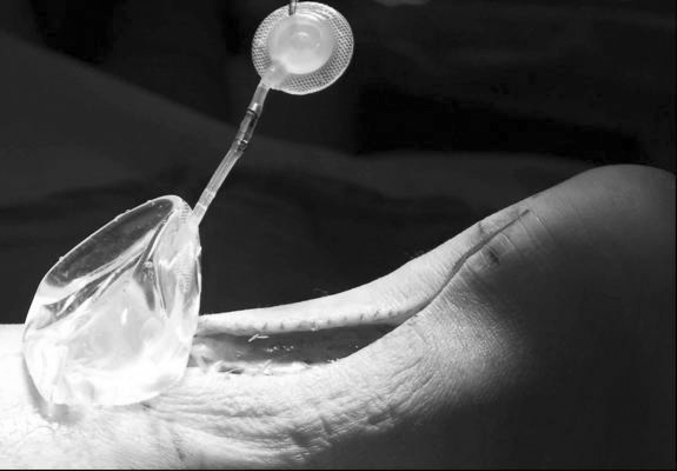
McGhan 70 ml soft-tissue expander.
Inflated soft-tissue expander in place subcutaneously.
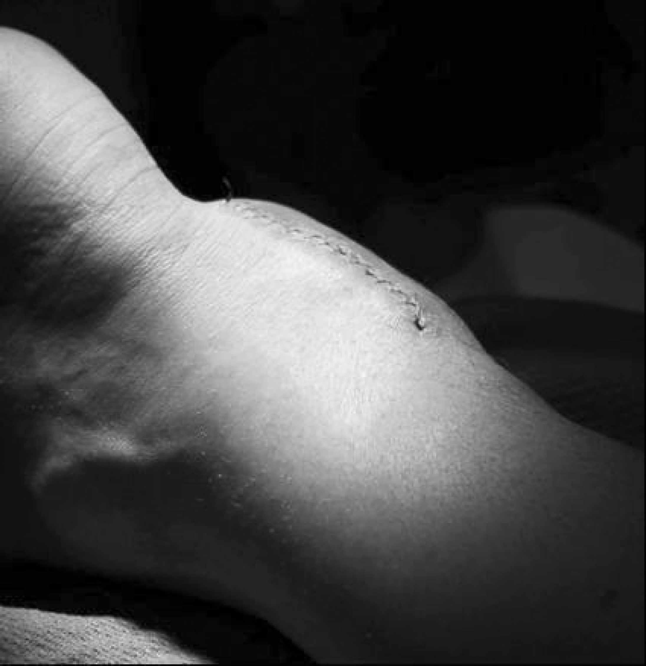
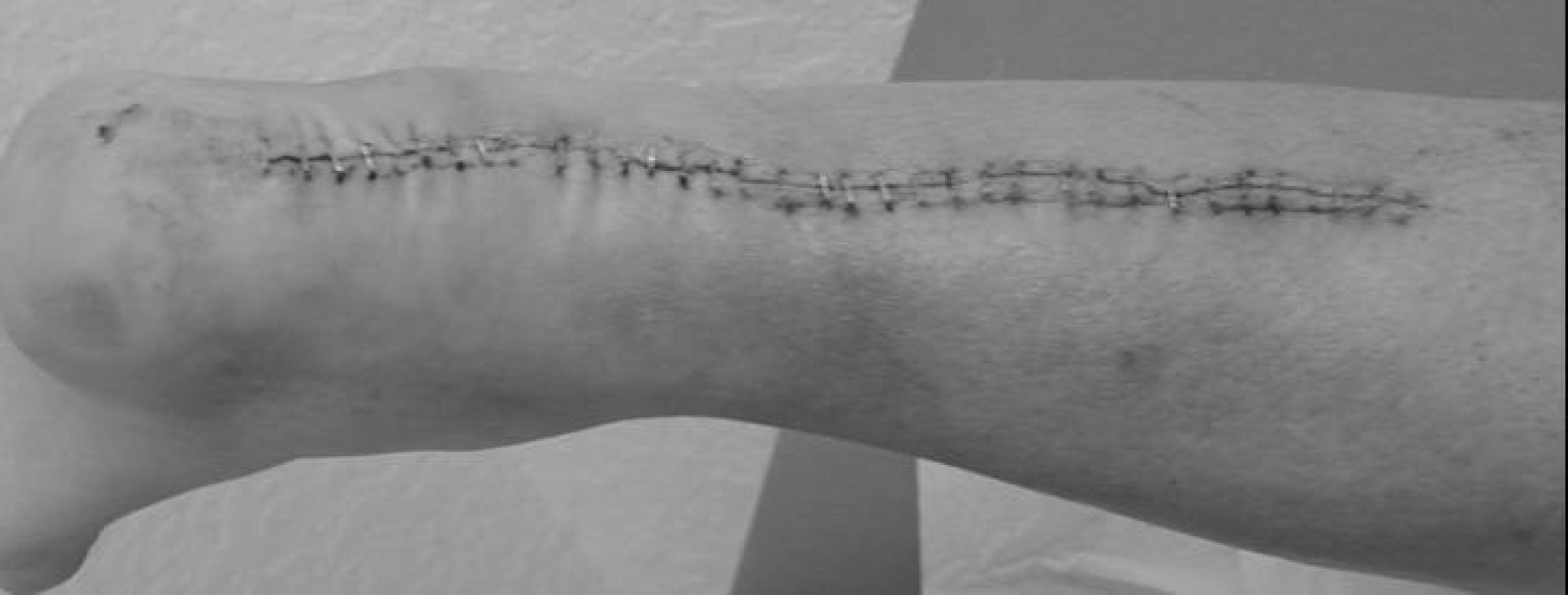
Incision planning for implant removal and subsequent tendon repair.
Operative Technique
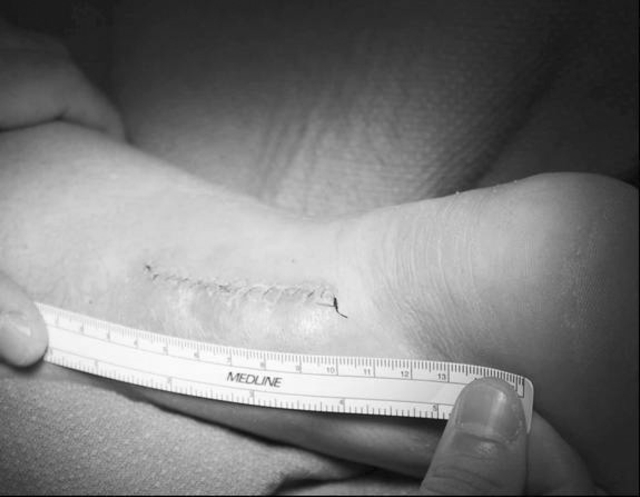
Creation of a soft-tissue pocket through implantation of a soft-tissue expander over the Achilles tendon and subsequent repair of the tendon are done as a staged, two-part procedure approximately 3 to 4 weeks apart. The first stage of treatment involves subcutaneous placement of a 70 ml, rectangular soft-tissue expander (McGhan, Santa Barbara, CA) between the Achilles tendon and the skin (Figure 1). With the patient in the lateral decubitus position, a local block is infiltrated into the operative site. After sterile preparation, a longitudinal incision is made along the medial aspect of the ankle, adjacent to the course of the Achilles tendon, down to the deep subcutaneous level. Superficial subcutaneous elevation is then performed until a pocket, approximately 6 × 4 cm is created. A McGhan tissue expander is inserted into this cavity, and the injection catheter is placed away from the implant. Subcuticular closure is then done with interrupted 4-0 vicryl sutures. The skin is approximated using a running 4-0 nylon stitch. Approximately 10 cc of normal saline is initially injected into the implant through the injection port. The patient is seen 1 week postoperatively for further inflation of the implant. Ten milliliters of normal saline are added to the expander weekly. Patients rarely tolerate more than 30 cc to 40 cc of total volume within the implant, and the expander is removed 3 to 4 weeks postoperatively (Figure 2). At the time of tissue expander removal Achilles tendon repair is done. The previously created incision is accessed and extended as necessary (Figure 3). The expansion balloon is then easily removed from its subcutaneous pocket. Operative repair of the injured tendon is carried out in the surgeon's preferred fashion; in the case of neglected rupture or re-rupture this may include flexor hallucis longus transfer. Careful repair of the paratenon overlying the Achilles tendon is essential at this stage to re-establish the fragile blood supply to this area. Upon completion of the repair, subcutaneous closure is achieved using 4-0 vicryl sutures. Simple approximation of the wound is then possible because of the lack of tension at the expanded skin margins (Figure 4). The operative limb is placed into a short-leg splint in 10 to 15 degrees of plantarflexion after a sterile dressing is placed (Figure 5). Nonweightbearing with immobilization in a short-leg cast is maintained for 3 weeks. Ranges of motion exercises are initiated at 3 to 4 weeks postoperatively. Finally, weightbearing is allowed at 6 weeks after operative repair (Figure 6).
Tension-free skin closure after Achilles tendon repair.
Operative site 1 week after surgery.
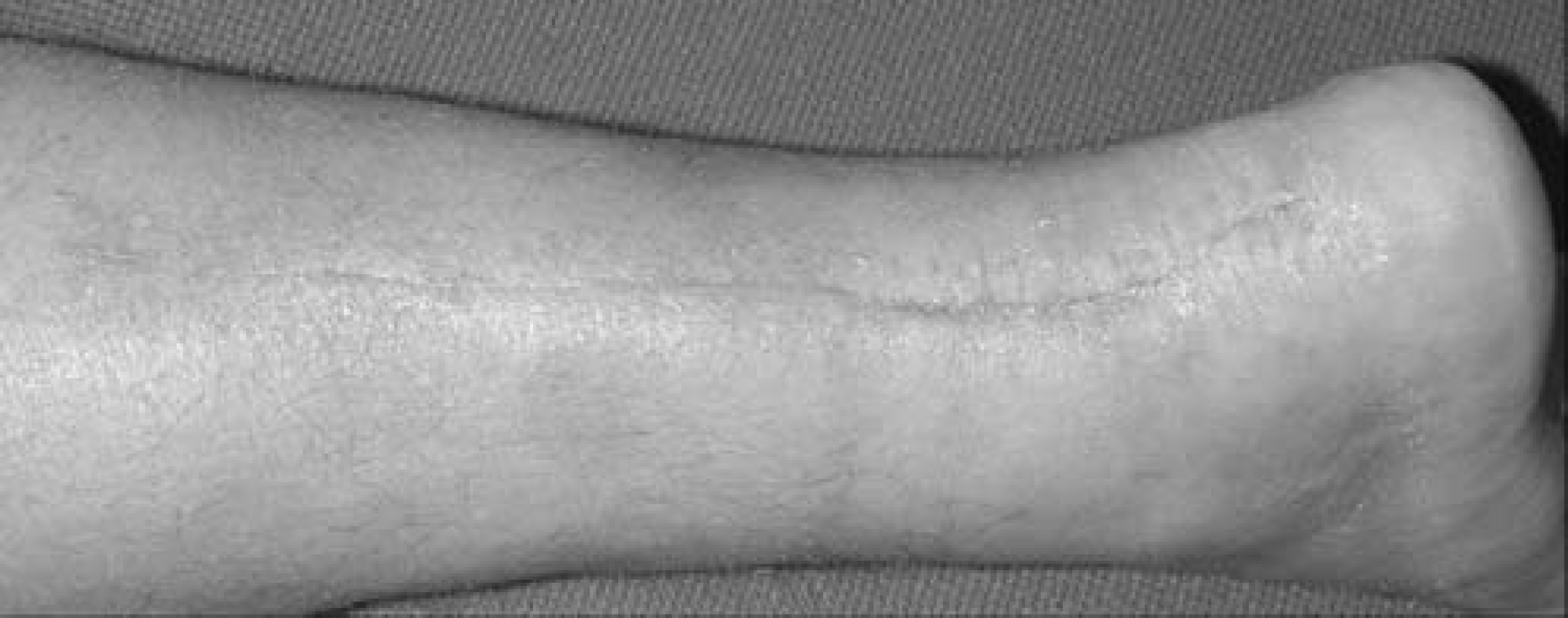
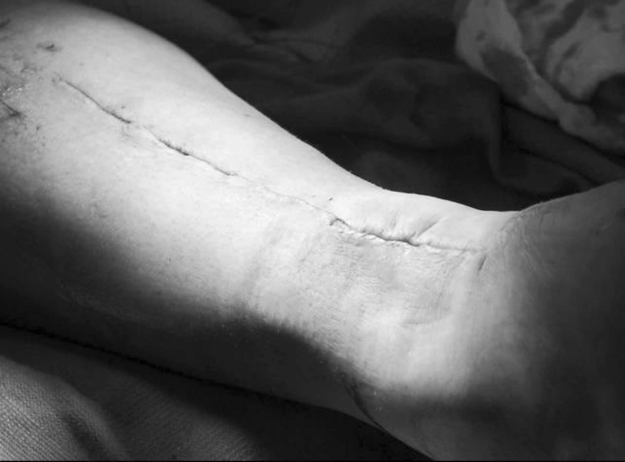
Operative site 8 weeks after surgery.