
Abstract

Introduction
This is a case of a 16-year-old boy presenting with a peroneal spastic flatfoot in which an ipsilateral talar osteochondral lesion was detected on MRI. Peroneal spastic flatfoot is a disorder that usually is caused by tarsal coalition. In some patients, despite a classic clinical presentation, a coalition is not identified. MRI may demonstrate a fibrous coalition or, in some cases, inflammatory changes within the subtalar joint. 5 Talar osteochondral lesions, frequently referred to as osteochondritis dissecans, have been well described and classified. 1–3,7 They appear to have a mixed etiology with some overlap between clearly traumatic osteochondral fractures and atraumatic lesions. Significant trauma is reported in 70% of patients with medial lesions. 2 Osteochondral lesions usually are not considered in the differential diagnosis of a peroneal spastic flatfoot. Remarkably, in our patient the peroneal spasm and flatfoot resolved with the successful treatment of the osteochondral lesion.
Case Report
A 16-year-old boy presented to our clinic with a complaint of left foot pain. The intial clinical impression was one of classic peroneal spastic flatfoot. Plain films were normal with no features of a coalition (Figure 1). CT and MRI did not reveal a coalition but did demonstrate an osteochondral lesion not suspected clinically or on radiographs (Figure 2). The patient was re-evaluated clinically, and it was elected to proceed with operative management. An ankle arthroscopy was done with transcondral drilling of the osteochondral lesion, which was thought to be a stage II lesion. The patient's symptoms persisted, including severe peroneal spasm. A repeat arthroscopy was done 9 months later and confirmed the presence of a large unstable fragment. The fragment was fixed using an open technique with a medial malleolar osteotomy and bioabsorbable screw fixation. At the 6-week followup visit, the patient's peroneal spasm and flatfoot had resolved. At the 6-month followup visit, radiographs revealed that the osteochondral lesion had healed, and the patient had no peroneal spasm. The patient continued to do well 1 year after operative fixation. Informed consent was obtained to publish the results of this case.
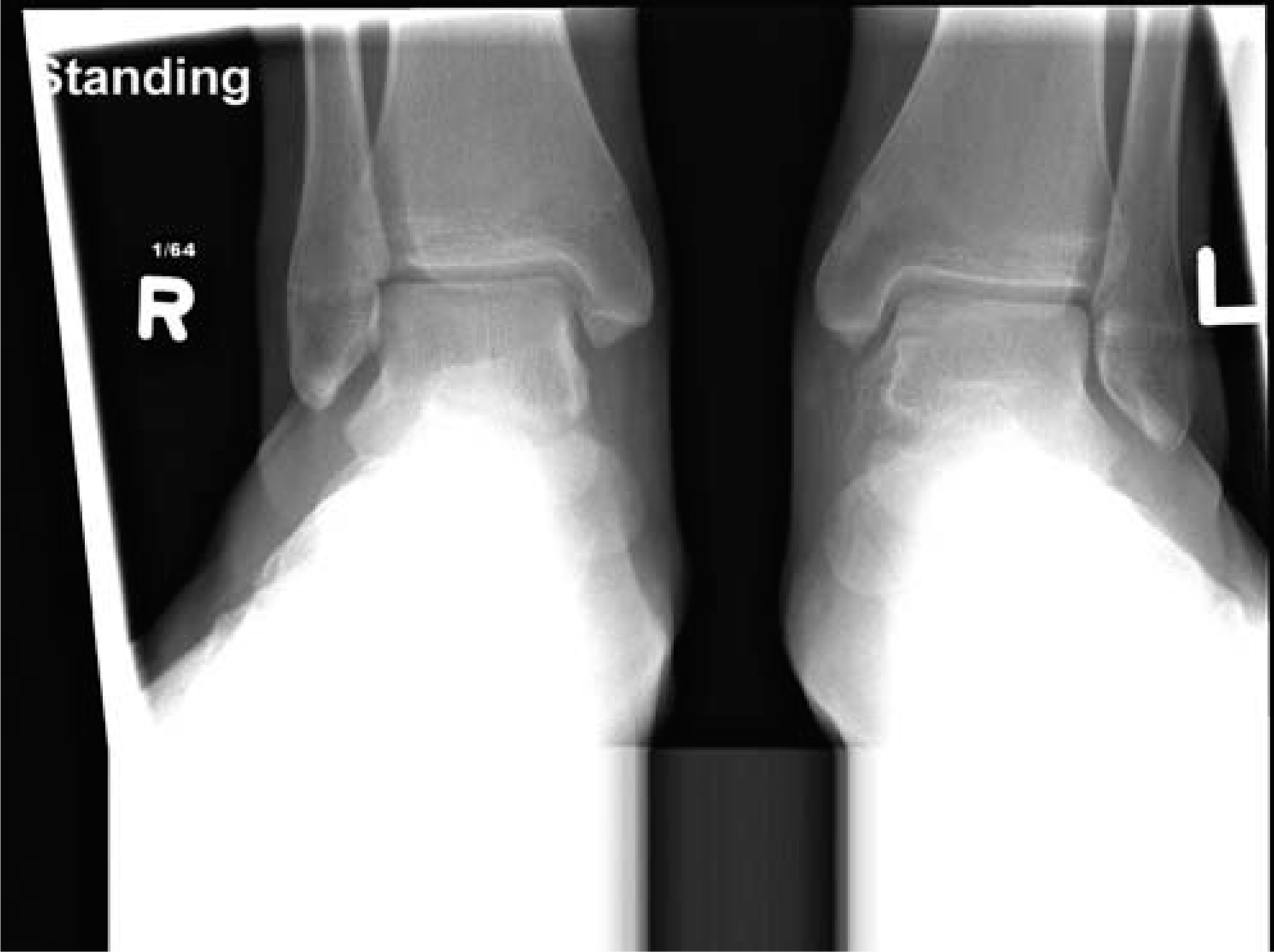
Plain radiograph at presentation.
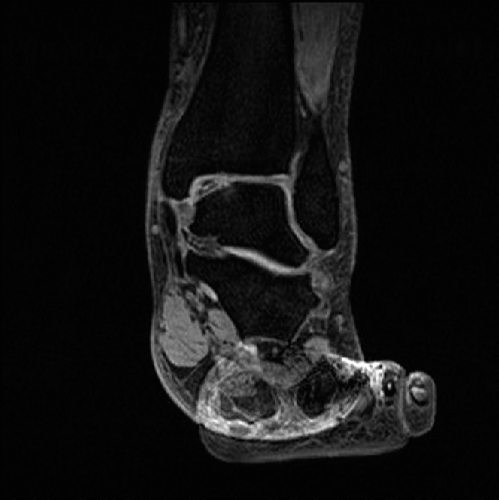
Talar dome lesion demonstrated on MRI.
Discussion
We believe that this patient's peroneal spasm was secondary to the talar lesion. To our knowledge the association between peroneal spasm and osteochondral talar lesions has not been previously reported. An osteochondral lesion occurring in conjunction with a longstanding flatfoot has been described in one patient in a series of patients with osteochondral defects. 5 The patient had been flatfooted since birth and was treated with a medial malleolar osteotomy and screw fixation. The operation was successful in terms of healing; however, the state of the flatfoot was not followed up.
Osteochondral lesions of the talus can be treated several ways depending on the severity of the fracture. There are four fracture stages, as described by Berndt and Harty. 2 The treatment options often are based on the stage of the fracture. For lower stage fractures, arthroscopic drilling can be used to treat the lesion, 2 whereas higher stage fractures may require open approaches. Operative procedures range from excision, curettage, and drilling to bone grafting, osteochondral transplantation, and fixation. 4 The stage of the osteochondral lesion can be difficult to define because radiographs do not necessarily demonstrate the degree of detachment of the talar fragment. MRI can be used to classify a fracture more accurately than radiographs. Arthroscopy also has been described as an extremely valuable diagnostic tool for classifying osteochondral lesions. 4
Adolescent osteochondral lesions are rare, with few published reports in the literature. However, the use of fixation has been previously reported to be successful. 3 Access for fixation frequently is achieved by a medial malleolar osteotomy. The success rate of fixation procedures has been reported to be 73% in 11 adult patients. 2
This patient was initially thought to have a stage II lesion based on radiographic studies and arthroscopic findings. However, the patients’ symptoms persisted despite drilling of the lesion and postoperative cast immobilization. The fixation procedure resulted in a dramatic resolution of both the patient's peroneal spasm and flatfoot posture. At 4-month followup, plain radiographs showed that the loose fragments were properly and successfully fixed into place, with evidence of radiographic healing. As other authors have recognized, 1 the underlying cause of the peroneal spasm can be difficult to define, with mixed results obtained from subtalar exploration. In our patient, operative fixation resulted in positive short-term and long-term outcomes. We believe that the peroneal spasm and flatfoot posture in this patient were secondary to the unstable osteochondral lesion.
Conclusion
The conjunctive occurrence of an osteochondral lesion of the talus and peroneal spastic flatfoot is very rare. In our patient, after treatment with a medial malleolar osteotomy and open reduction internal fixation of the loose fragment, the peroneal spastic flatfoot spontaneously disappeared. The talar dome also healed very well, as shown in followup radiographs (Figure 3). Talar osteochondral lesions should be considered in the investigation of patients presenting with peroneal spasm and no evidence of a coalition.
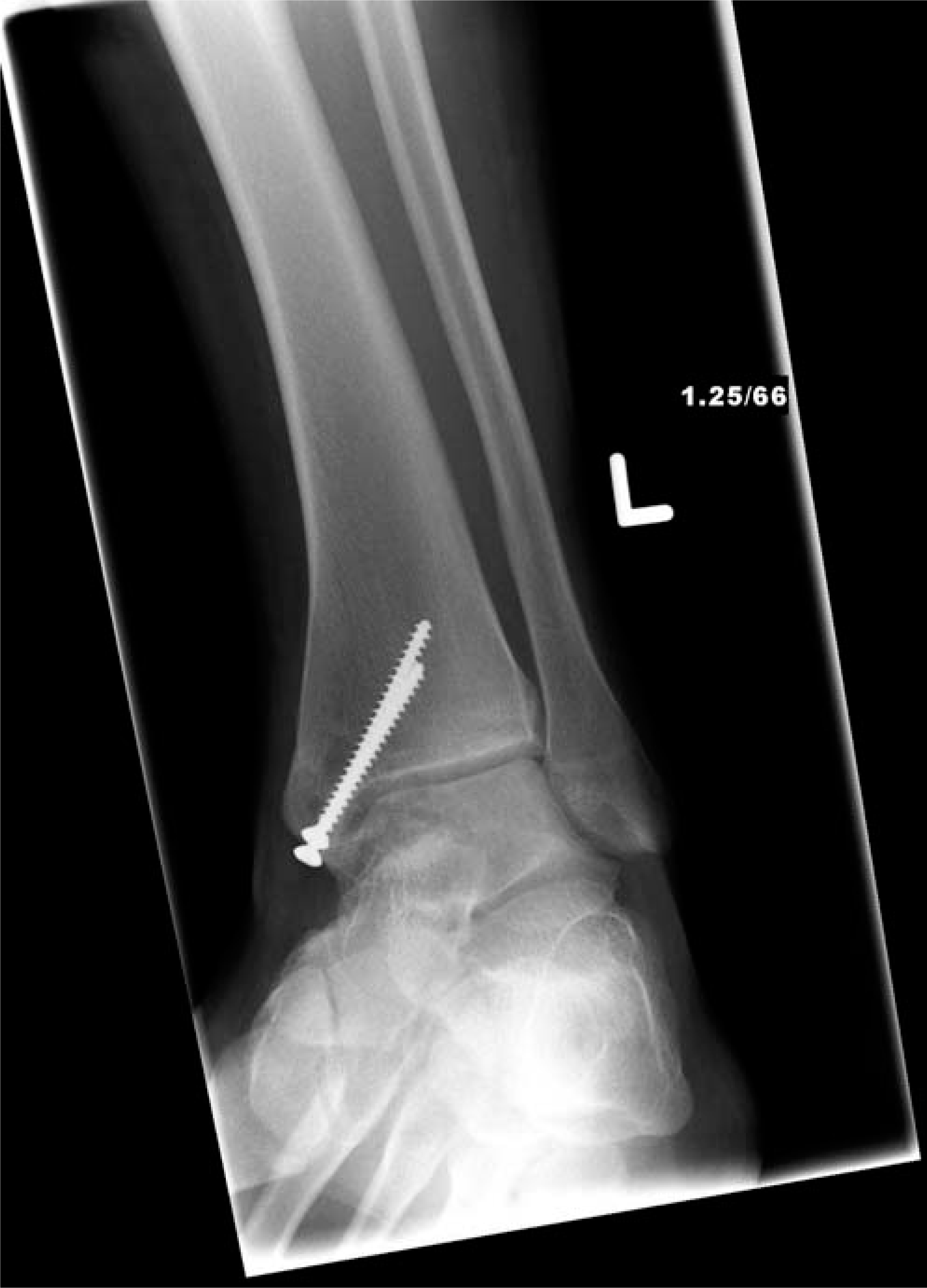
Followup radiograph with radiographic healing of the lesion and clinical resolution of peroneal spasm.