
Abstract

INTRODUCTION
Traumatic pseudoaneurysm of the peroneal artery or one of its branches is a rare phenomenon. Although singular, it has been reported following both non-penetrating trauma as well as iatrogenic trauma: ankle sprain, 3,4,10,11 fracture, 8 ankle arthroscopy, 7 percutaneous tendoachilles lengthening, 5 plantar fasciotomy, 6 femoro-peroneal bypass, 9 removal of hardware from the tibia, 2 and insertion of a catheter into the anterior tibial artery. 12 Due to their uncommon presentation, diagnosis and treatment of these types of pseudoaneurysms are often delayed; since few aneurysms clot spontaneously, medical or surgical intervention is then required. Physicians should be aware of these pseudoaneurysms and understand their natural history. We are presenting the first reported case of a pseudoaneurysm after an ankle sprain with a concomitant subtalar interosseous ligament injury.
CASE REPORT
A 17-year-old male presented to the emergency room with an edematous foot and ankle following a plantarflexion inversion injury while playing basketball. No dislocation was appreciated. Plain radiographs were normal. The injury was treated as an ankle sprain. He was placed in a posterior splint and remained nonweightbearing on crutches. One week post-injury, the medial and lateral aspects of the foot and ankle were ecchymotic and edematous. MRI demonstrated no evidence of fracture. A hematoma measuring approximately 3.2 cm × 1.5 cm overlying the anterolateral aspect of the ankle was thought to be diffuse soft tissue edema involving the subcutaneous tissue. Diffuse edema was also seen in the subtalar joint with partial tear of the interosseous talocalcaneal ligament (Figure 1). The anterior and posterior tibiofibular ligaments, anterior talofibular ligament, and calcaneofibular ligament were all intact. The diagnosis was an inversion ankle sprain with concomitant subtalar joint injury and a partial tear to the interosseous ligament. He was immobilized in a short leg nonweightbearing cast for three weeks. At followup, examination demonstrated decreased soft tissue edema; dorsalis pedis and posterior tibial arterial pulses were palpable. Due to continued pain, he was immobilized in a short-leg walking cast for an additional 3 weeks. At the next examination, there was recurrence of the soft tissue swelling over the anterolateral aspect of the ankle. A pulsatile mass was present. Ultrasound revealed a pseudoaneurysm measuring 3.5 cm × 2.5 cm of the perforating peroneal artery. Interventional radiology attempted localization and thrombin injections. The vessel thrombosed for 24 hours, but the pseudoaneurysm recurred. An angiogram was performed to locate the exact segment of vessel involved (Figure 2). The patient was sent to vascular surgery.
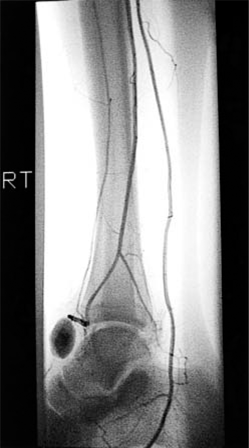
Angiogram showing the pseudoaneurysm.

MRI of injury to the talo-calcaneal interosseous ligament.
Under general anesthesia, an oblique incision was made over the pseudoaneurysm. The pseudoaneurysm was opened and noted to have pulsatile blood flow. The source of bleeding was the perforating peroneal artery. This vessel was ligated and excised. The capsule of the pseudoaneurysm, including the large hematoma surrounding it, was sent to pathology, which confirmed the diagnosis. A compressive dressing was applied for 3 weeks. At 8 weeks followup, his ankle and subtalar joint ranges of motion were normal. He has subsequently returned to full athletic activities.
DISCUSSION
The perforating peroneal artery, a branch of the peroneal artery, pierces the interosseous membrane anteriorly between the distal tibia and fibula. It courses along the lateral talar head to anastomose with the lateral tarsal artery. The anatomical location of the artery makes it vulnerable to inversion injuries of the foot and ankle, which can produce a stretching or shearing force to the outer layers of the luminal wall. In a true aneurysm, the content of the vessel's lumen remains intact, but the artery expands. A pseudoaneurysm is not confined by the vessel wall. This leads to hematoma in the surrounding tissue as well as formation of a fibrous capsule lacking the normal three-layer architecture of a “true aneurysm” and an endothelium lining continuous with the lumen. The natural progression of the arterial pseudoaneurysm is to enlarge. 1 Local symptoms occur from compression of the adjacent tissue. If progression continues, the pseudoaneurysm can become infected, hemorrhage, thrombose, or embolize to distal vessels with substantial patient morbidity.
The diagnosis of a perforating peroneal pseudoaneurysm is often delayed due to vague signs and symptoms of progressive swelling and pain over the lateral malleolus. A palpable pulsatile mass over the lateral ankle may be diagnostic. Often the patient will undergo conservative treatment with immobilization. Needle aspiration will demonstrate arterial blood. Once a pseudoaneurysm is suspected, a duplex ultrasound can be utilized to confirm the diagnosis. Advanced studies with computed tomography, MR arteriography, or angiogram are sometimes utilized to visualize the mass. 1 Once the diagnosis is confirmed, treatment consists of either ultrasound guided compression, interventional radiology or surgical intervention. 1,13 Treatment depends on the patient's age, the aneurysm's size and location, and diagnostic method.
Controversy exists about whether to treat a pseudoaneurysm surgically or non-surgically. Non-invasive measures consist of angiographic-guided thrombin injections or endovascular transcatheter embolization. According to Weaver et al., criteria for non-invasive treatment of pseudoaneurysms are low-velocity injuries, minimal arterial wall disruption (less than 5 mm), intact distal circulation, and no active hemorrhage. 13 Surgical intervention is performed if non-invasive treatment fails. With good collateral circulation, surgical intervention consists of ligation of the non-critical vessel which was the treatment of choice in our case. High velocity injury, limited distal perfusion, or a critical arterial vessel requires repair with lateral suture patch angioplasty, end-to-end anastomosis, interposition graft, or bypass graft. The exact procedure is determined by the extent of arterial damage. 13
CONCLUSION
This is a rare case of pseudoaneurysm of the perforating peroneal artery from an inversion ankle and subtalar joint injury. When presented with unexpected swelling over the anterolateral ankle following ankle or subtalar joint injury, this condition should be included in the differential diagnoses. Early detection and expedient intervention will prevent further morbididty.