
Abstract
Background: In stage II PTTD, flexor digitorum longus (FDL) tendon transfer with an adjunctive bony procedure is the most common method of surgical correction. This paper presents an alternative method of fixation with a biotenodesis interference screw (Arthrex Biotenodesis Screw System) that allows proper tensioning of the FDL tendon transfer. Materials and Methods: We retrospectively reviewed 25 consecutive patients who underwent FDL tendon transfer utilizing a biotenodesis interference screw. Intraoperative stability was noted and any loss of correction was assessed postoperatively. Results: Stable fixation was achieved in 24 of the 25 patients who underwent FDL tendon transfer for PTTD. We were not able to achieve stable fixation in one patient due to improper placement of the bone tunnel. This was recognized intraoperatively and did not affect the final outcome. Conclusion: This method is technically easier to perform than the recommended technique by the manufacturer. It can be performed through a slightly smaller incision without disrupting the normal interconnections between flexor hallucis long (FHL) and FDL tendon at the Knot of Henry.
INTRODUCTION
Adult acquired flatfoot deformity is a common cause of chronic foot and ankle pain which is characterized by flattening of the medial longitudinal arch and dysfunction of the medial soft tissue structures. The posterior tibial tendon (PTT), spring ligament, and portions of the deltoid ligament can become attenuated. The PTT is the strongest inverter of the foot, and pathology of this structure leads to a progressive pes planovalgus deformity. Johnson and Strom described a classification system in 1989 consisting of three stages, and a fourth stage was later described by Myerson. 12,18 In stage II PTTD, patients present with a flexible planovalgus deformity. For patients who have failed conservative care, surgical intervention often consists of transfer of the FDL tendon in conjunction with a calcaneal osteotomy. Myerson et al retrospectively reviewed their results in 129 patients who underwent FDL transfer and medial displacement osteotomy of the calcaneus and found that such treatment in those with stage II PTTD yielded excellent results. 18
The earliest described technique for transfer of the FDL tendon in cases of PTTD involved placement of a drill hole in the navicular from dorsal to plantar and routing the FDL tendon through the hole. 14 The transferred tendon was then sutured to the surrounding periosteum above and below the drill hole. Using the technique described by Mann, the FDL tendon is harvested at the Knot of Henry which requires a longer incision. 7,8,14 Currently, the biotenodesis screw is gaining popularity and is commonly used for fixation of the transferred FDL tendon in such procedures. At our institution a similar approach is taken in those patients with stage II PTTD. We have observed that the recommended technique using a biotenodesis screw for FDL tendon transfer can be somewhat difficult, especially when attempting to place the appropriate amount of tension to the transferred tendon. We present an alternative method of reattaching the transferred FDL tendon utilizing the biotenodesis screw (Arthrex Biotenodesis Screw System).
MATERIALS AND METHODS
This study was reviewed and approved by our Institutional Review Board. Outcome scores were not recorded. Twenty-five consecutive adult patients with stage II PTTD underwent FDL tendon transfer using a biotenodesis screw for fixation and a medial displacement calcaneal osteotomy. Additional procedures were performed as indicated including naviculocuneiform arthrodesis, lengthening of the Achilles tendon or gastrocnemius recession. All 25 patients demonstrated signs and symptoms of stage II PTTD (flexible deformity with inability to do a single heel rise) and had failed non-operative care utilizing orthotaics, braces and physical therapy. Preoperative symptoms exceeded 6 months in all patients. Patient satisfaction was determined by asking the patient if surgery had decreased their pain, and if they would undergo the procedure on the contralateral side. If so, it was considered a satisfactory result.
Operative technique
The patient was placed in the supine position and a thigh tourniquet was used. A curvilinear incision was made starting from the inferior aspect of the medial malleolus extending to the medial cuneiform. The sheath of the PTT was incised and the PTT was transected 2 cm from its insertion into the navicular. It was resected just proximal to medial malleolus. The FDL tendon sheath was identified deep to the resected PT tendon sheath. The FDL tenotomy was made proximal to the Master Knot of Henry under direct visualization (Figure 1).
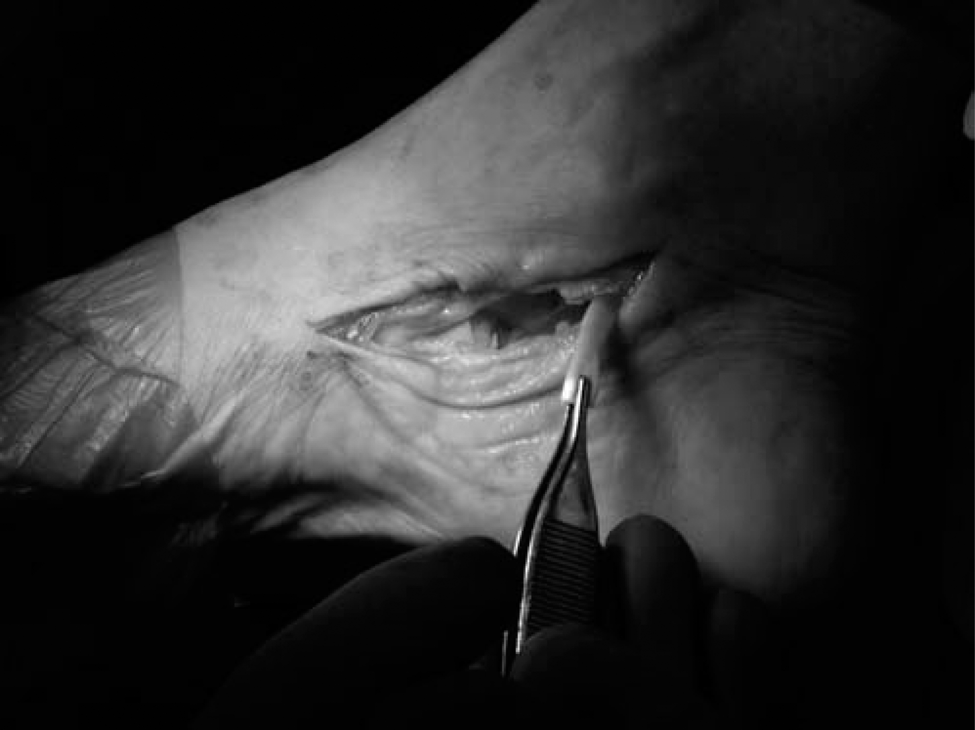
Harvested FDL tendon.
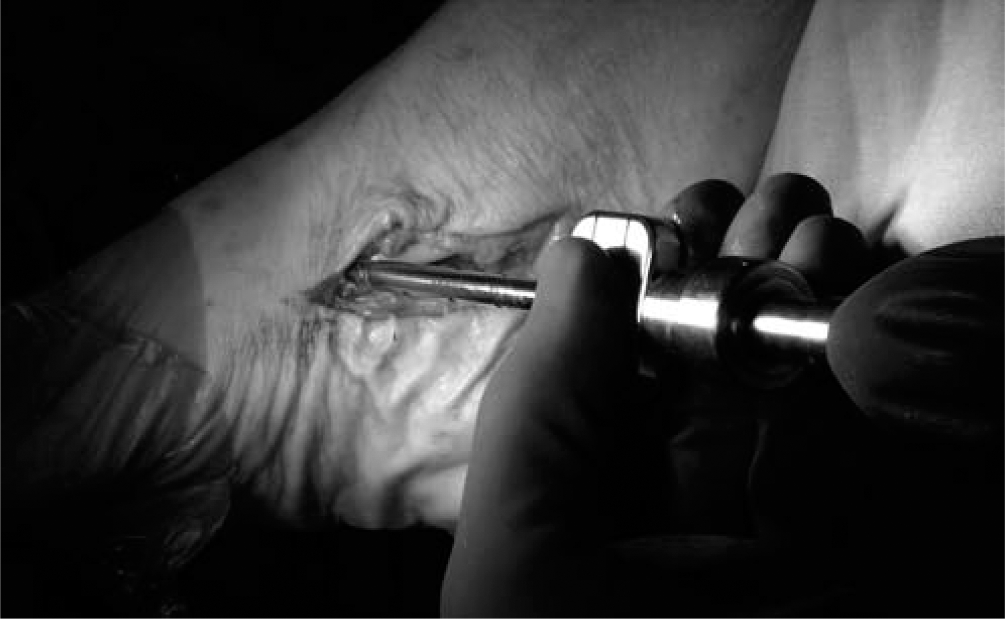
With the ankle plantarflexed and the foot supinated, the location of the transferred FDL insertion into the navicular was marked on the FDL tendon. This was done so that full insertion of the tendon can be confirmed. Typically, 35 mm of the tendon was present distal to where it would insert; ideally, 25 mm of the FDL tendon should be seated in the tunnel and redundant tendon was resected. Arthrex 2–0 FiberWire non-absorbable braided suture (Arthrex, Inc, Naples, FL) was used to tag the end of FDL tendon using a whip stitch. An 18-gauge needle localized the proper insertion point of the screw in the navicular under fluoroscopic guidance (Figure 2).
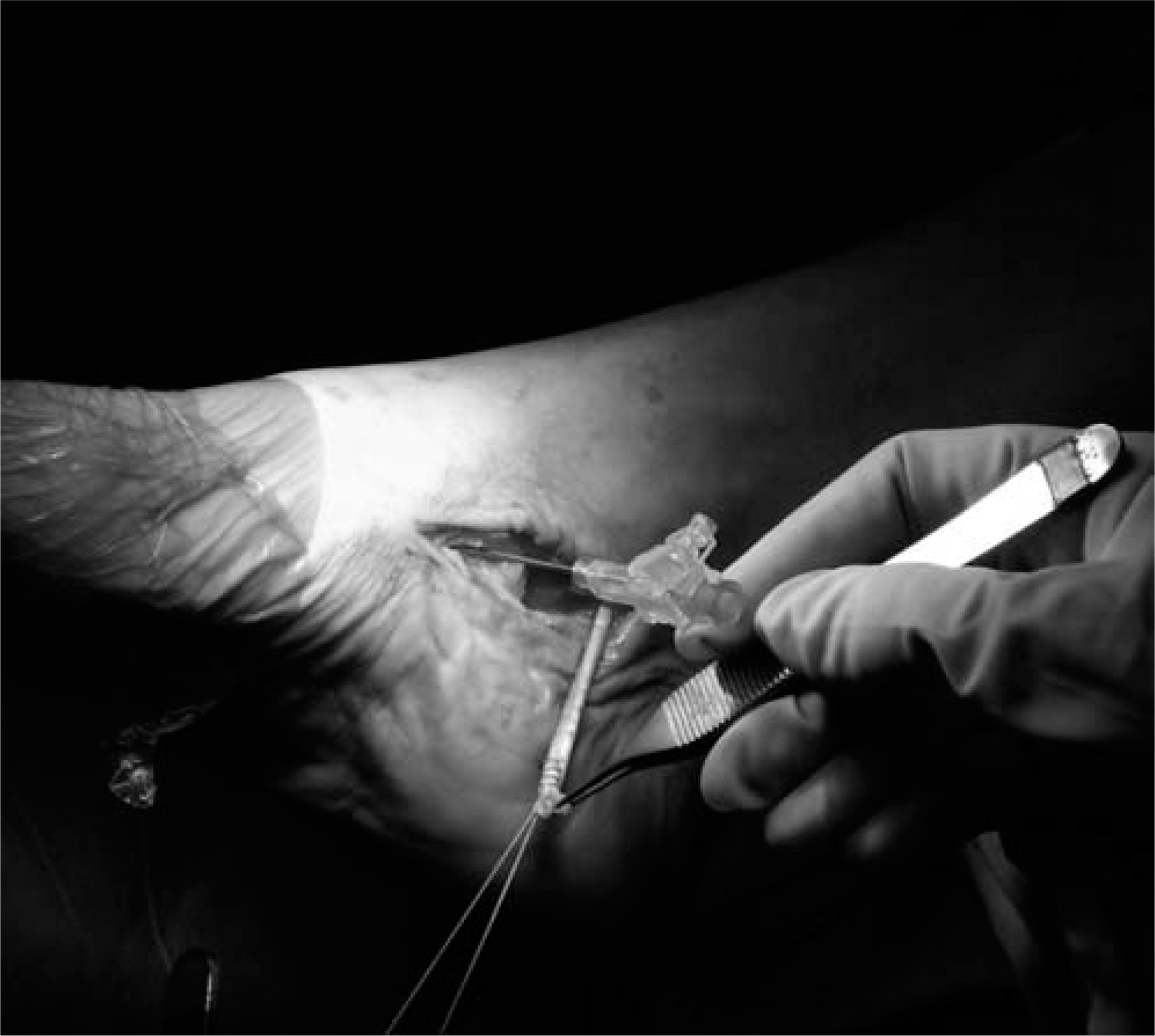
Eighteen-gauge needle used to identify the insertion point in navicular and whip stitch used to tag the end of the FDL tendon.
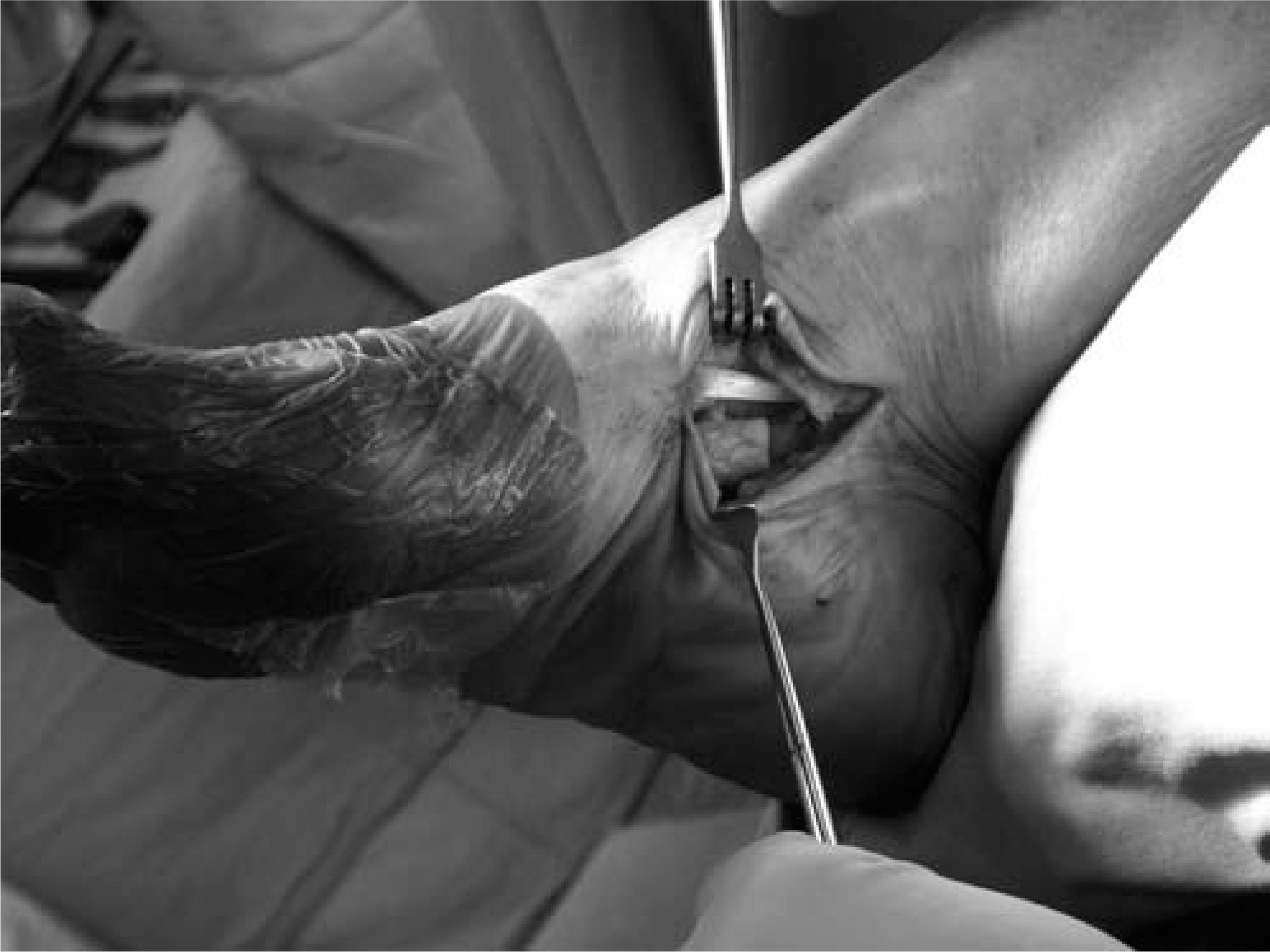
The FDL tendon was sized using the sizer provided by Arthrex. A Beath needle was used as a guide pin, and inserted into the inferomedial navicular and exited on the dorsolateral foot. Proper positioning was confirmed with fluoroscopy. A drill hole was made under direct vision with a cannulated drill, using the Beath needle as a guide, from plantar to dorsal. A drill 0.5 mm larger than the size of the tendon was used. The depth of the bone tunnel was 5 mm longer than length of the tendon that would be secured in the bony tunnel (Figure 3). For instance, if 25 mm of the FDL tendon would be seated in the bone tunnel, a 30-mm tunnel was created. The tendon was usually 4.5 mm in diameter.
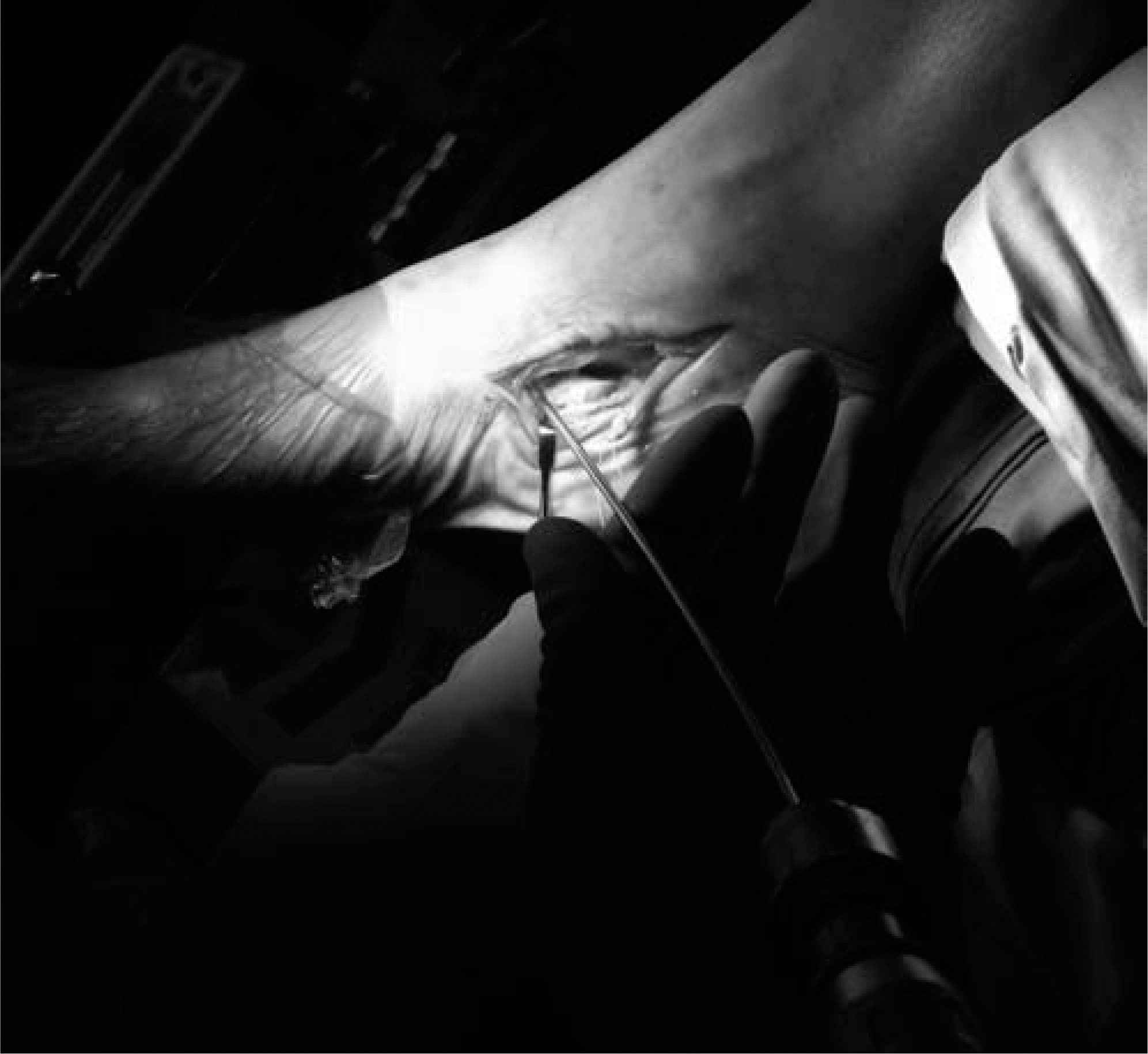
Beath needle driven through navicular from plantar to dorsolateral foot.
The FDL tendon was pulled through the navicular with a Beath needle, while the foot was in maximal supination (Figure 4). A properly sized (usually 4.75 mm × 15 mm) biotenodesis screw was inserted into the navicular over a 0.045 Kirschner wire with the ankle plantarflexed and the foot supinated, while maintaining maximal tension on the suture (Figure 5). The FDL was sutured to the PTT stump with 2–0 FiberWire. Lastly, the 2–0 FiberWire that was used to pull the tendon into the bone tunnel was tied on the dorsolateral foot and allowed to retract into the soft tissue (Figure 6).
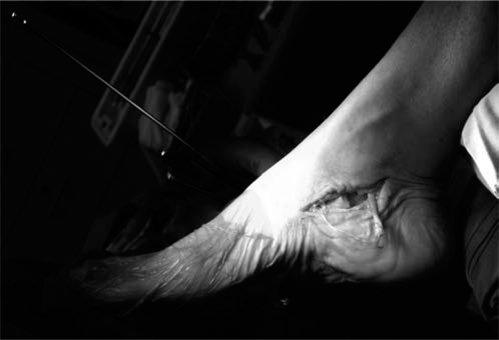
With foot supinated, FDL tendon is pulled through navicular using Beath needle.
Kirschner wire (0.045) placed as a guide and cannulated Arthrex biotenodesis screw is placed in the navicular.
The transferred FDL tendon.
The incision was closed in layers. A non-weightbearing below the knee Jones compression dressing was applied with the ankle in slight plantarflexion and the foot supinated.
Adjunctive procedures such as medial displacement calcaneal osteotomy, Evans opening calcaneal wedge osteotomy and gastrocnemius recession or tendo-achilles lengthening were performed in addition to the FDL tendon transfer procedure.
Rehabilitation
On the first postoperative visit, a below-knee nonweight-bearing fiberglass cast was applied with the ankle slightly plantarflexed and the foot in slight supination. Casting was continued for total of 6 weeks and the patient was then transitioned into a walking boot. At the same time, the patient was started on a range of motion exercises over the next 4 weeks. At 10 weeks, inversion exercises were performed out of the boot walker. Then, formal physical therapy was initiated at 12 weeks. Patients were transitioned to regular shoe gear as tolerated. Most patients received an orthotic device after 3 months.
RESULTS
Proper tension and secure fixation of the transferred FDL tendon was achieved with this technique in 24 of the 25 patients. At a minimum followup of 8 months, no recurrence or clinical failures were noted and 92% of patients reported satisfaction with the procedure performed. The one intraoperative failure occurred in a patient who underwent simultaneous fusions of the naviculocuneiform and first metatarsocuneiform joints with 3.5-mm cortical screws. The orientation of the two 3.5-mm cortical screws in the navicular resulted in suboptimal purchase of the biotenodesis screw. Additional sutures were placed in the transferred tendon and through the PTT remnant to gain additional fixation. This patient was satisfied with the result at 1-year followup. No formal statistical analysis was performed in this study and no patients were lost to followup.
DISCUSSION
The purpose of this study was not to report the results of FDL transfer and calcaneal osteotomy for stage II PTTD, but rather to introduce an alternative method of fixation for the transferred FDL tendon. There is an abundance of studies in the literature supporting the efficacy of traditional methods. The use of a bioabsorbable interference screw system for stabilizing the FDL tendon transfer in treatment of stage II PTTD is being utilized more frequently in some centers. The manufacturer's recommended technique relies on interference fixation of the biotenodesis screw with the FDL tendon. As the screw is inserted into the navicular, it is vital to achieve accurate tension of the transferred tendon with the screw using the suture loop handle provided by the manufacturer. Utilizing that technique, the authors have experienced difficulty with maintaining proper tension of the FDL tendon into the navicular.
Our preferred technique differs in that we do not use the suture loop handle when the FDL tendon is transferred through the navicular. The points of fixation include the interference fit in the osseous tunnel as well tendon-to-tendon fixation at the insertion. These points of fixation allow for bone-to-tendon healing and tendon-to-tendon healing, making the repair less dependent on one point of fixation. We believe this method lessens the risk of loss of proper tension during the healing phase.
The use of an interference screw provides a low profile, intraosseous fixation allowing for less length of graft and an incision that is 1- to 2-cm smaller. The distal extent of our incision is at the level of the naviculocuneiform joint, and we were able to harvest enough tendon in all cases. The procedure was originally described to dissect down to the Master Knot of Henry to harvest a longer length of FDL tendon. 7 With our technique, the FDL tendon can be effectively harvested through a smaller incision with decreased morbidity.
We have successfully employed this technique in 24 of 25 consecutive stage II PTTD patients, achieving patient satisfaction without clinical failure in 92% of patients (n = 23). Our results are comparable to those reported by Myerson and Guyton (94% and 91% patient satisfaction respectively). 8,17 One intraoperative failure occurred due to technical error, but this was recognized immediately. In retrospect, greater care should have been taken to place our biotenodesis drill hole in a more optimal location.
Concerns about the biotenodesis screw include foreign body reaction and screw breakage. We have not observed any osseous changes in the navicular, such as enlargement of the tunnel due to osteolysis. Screw breakage can occur if the size of the bone tunnel is not slightly larger than the biotenodesis screw, especially in patients with excellent bone quality.
CONCLUSION
We have introduced an alternative technique using a biotenodesis screw for FDL transfer for the treatment of stage II PTTD. This new technique provides an alternative method to achieve proper tension when transferring the FDL tendon to the navicular. Early experience with this technique is promising as an alternative to previously described methods.