
Abstract
Background: The purpose of this study was to evaluate the clinical outcomes and objective isokinetic dynamometry on a cohort of patients with chronic insertional Achilles tendinosis, who underwent surgical reconstruction using an FHL tendon autograft transfer through a single incision. Materials and Methods: Forty patients (16 male and 24 female; mean age, 57 years; age range, 39 to 76 years) with persistent chronic Achilles tendinosis were evaluated after surgical reconstruction at an average of 27 months after surgery. At the time of final followup, ankle strength and active range of motion (AROM) were evaluated using Biodex® isokinetic dynamometry. Additionally, patients were assessed with AOFAS Ankle Hindfoot scores, pain on a Visual Analog Scale (VAS) and their self-reported level of satisfaction (Very Good, Good, Fair, Poor). Results: We found no loss of plantarflexion strength or plantarflexion power in the operated ankles; an average of 4-degree loss of AROM was found. The study population scored an average of 96/100 for the total AOFAS-AH score post-repair. The average VAS decreased from 7.5 pre-op to 0.3 post-op. Thirty-eight of 40 patients (95%) were satisfied with their outcome (rated Very Good or Good), two patients rated their outcome as Fair and none as Poor. Conclusion: For individuals with chronic insertional Achilles tendinosis, operative repair using an FHL tendon with the single-incision technique achieved a high percentage of satisfactory results as well as excellent functional and clinical outcomes including significant pain reduction.
Level of Evidence: IV, Retrospective Case Study
Introduction
The Achilles tendon is the largest, strongest and most powerful tendon around the ankle and inserts over approximately a 3- to 4-cm2 area on the calcaneal tuberosity at the posterior heel. The insertion of the tendon is prone to developing tendonitis as a result of overuse injury. Additional intrinsic factors such as a prominent calcaneal tuberosity (Haglund's deformity), muscle weakness, malalignment of the hindfoot, and excess body weight as well as extrinsic factors such as inappropriate footwear, training errors and excessive load on the tendon can cause an overuse syndrome of the Achilles tendon. 12 This condition may result in long-term tendon degeneration and be responsible for a chronic inflammatory process at the insertion sometimes associated with micro-tears. Insertional Achilles tendinosis is a relatively common entity among ankle and hindfoot disorders and is usually treated conservatively. It has a bimodal distribution affecting younger athletic individuals involved in activities requiring forceful push-off or bursts of acceleration; with older more sedentary patients with co-morbidities representing the other, and most common, group. Recalcitrant tendinopathy can become chronic and intensely painful, especially in patients with predisposing factors. Surgical intervention is only indicated in patients unresponsive to conservative treatment.
Various surgical techniques have been described to treat subjects with insertional Achilles tendinosis. The goal of surgery is to provide long-term pain relief without significantly impairing function by sharply debriding the diseased tendon, decompressing the underlying bony prominence and reattaching the tendon at an appropriate length. In older patients, particularly those with degenerative changes replacing more than 50% of the tendon at the insertion, or with co-morbidities which may affect healing, the addition of a tendon transfer to augment the repair has been advocated. 8 A variety of surgical approaches have been described including a transverse incision, 3 a lateral longitudinal incision, 17,24 a medial J-shaped incision, 18,19 or a two-incision approach with medial and lateral incisions. 6,14 McGarvey et al. 16 and later Johnson et al. 11 described the central tendon splitting approach to debride the Achilles tendon, without a tendon augmentation. Wapner et al. 22,23 described using the flexor hallucis longus (FHL) tendon to treat the chronic rupture of the Achilles tendon through a double-incision approach. Another newer surgical alternative is the transfer of FHL tendon through a single incision, as described by Hansen10 and later by Den Hartog. 8 These studies all evaluated results based on clinical results only.
For these patients with more than 50% tendinosis at the insertion, we perform an insertional Achilles tendon debridement, Haglund's exostectomy and FHL tendon transfer through a single central splitting incision, with the FHL tenodesed into a bone tunnel in the calcaneus using a bioabsorbable interference screw.
The purpose of this study was to evaluate the treatment of chronic insertional Achilles tendonitis using a single central tendon splitting incision through which the tendon debridement, Haglund exostectomy and FHL tendon transfer were performed. This is the first study to assess this procedure using both clinical outcome analysis and objective isokinetic dynamometry.
Material & Methods
This study was conducted with the appropriate Institutional Review Board (IRB) approval of the Thomas Jefferson University Hospital. All subjects included in the study agreed to participate and signed the consent form.
Patients
Study subjects were identified from our departmental database on whom the senior author (S.M.R.) had performed surgery for chronic insertional Achilles tendinosis between the years 2001 to 2005, with a final post surgical followup time of at least 18 months.
Inclusion criteria for the surgery was: failed non-operative management for at least 6 months, radiographic evidence of a prominent posterior calcaneal tuberosity (Haglund exostosis) (Figure 1), and degenerative tendinopathy replacing at least 50% of the insertion of the Achilles tendon into the calcaneus on preoperative magnetic resonance imaging (MRI, Figure 2) or ultrasound evaluation (US). Exclusion criteria included: high medical risk and poor healing capacity, including diminished limb vascularity, uncontrolled diabetes, nutritional deficiency, or immunocompromised status. Exclusion criteria for involvement in the isokinetic evaluation aspect of this study included subjects with neurologic deficits, and subjects undergoing revision Achilles tendon surgery, bilateral surgery or significant pathology of the contralateral limb (preventing comparative dynamometry), and known associated ipsilateral limb pathology which might interfere with reproducible testing of the Achilles tendon for the study.
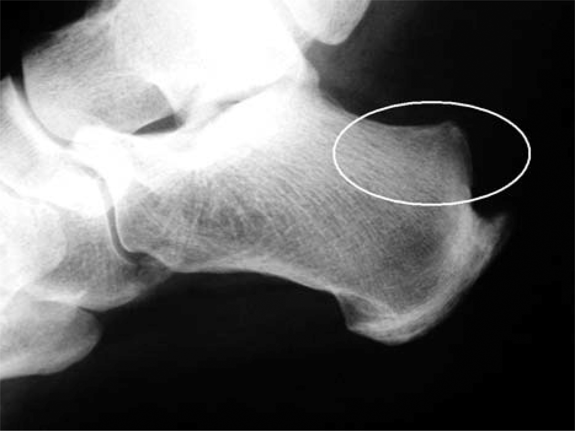
Lateral radiograph of the hindfoot demonstrating the insertional Achilles spur, and Haglund exostosis (encircled).
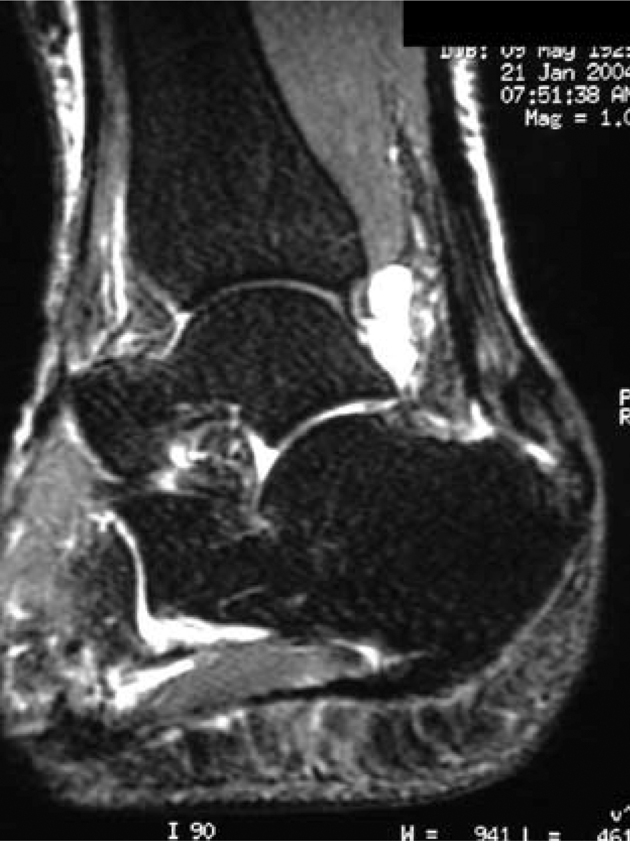
Sagittal plane MRI demonstrating significant insertional Achilles tendinosis with intrasubstance degeneration and longitudinal tears replacing more than 50% of the normal tendon.
A total of 62 consecutive patients (64 ankles, two bilateral) with chronic recalcitrant insertional Achilles tendonitis underwent the surgery during the study period. Twelve patients (14 ankles) did not fulfill the inclusion criteria. Ten patients were not available for followup (six lived out of state, three could not be located, and one had died due to unrelated causes). The remaining 40 patients were included in the study and were evaluated at an average of 27 (range, 18 to 68) months post-surgery. There were 16 male and 24 female with a mean age of 57 (range, 39 to 76) years. The left Achilles tendon was involved in 18 subjects, the right in 22 subjects, (one patient had bilateral surgery; the side which was repaired first was the side evaluated in this study). Average weight of subjects was 97.8 kg (215 lbs), ranging from 61 to 180 kg (134 to 396 lbs). A visual analog pain score (VAS) and American Orthopaedic Foot and Ankle Society–-Ankle Hindfoot (AOFAS-AH) score were obtained preoperatively in all patients.
Operative technique
All procedures were done on an outpatient basis with the patient under general anesthesia with popliteal block augmentation. The operative procedure was done with the patient in a prone position. A thigh tourniquet was used to allow access to the proximal calf and prevent the tourniquet from squeezing the gastrocnemius muscle. After Esmarch exsanguination, the tourniquet was inflated until a compressive dressing was applied at the end of the procedure.
An 8-cm longitudinal midline incision was made over the posterior aspect of the Achilles tendon, extending down to the Haglund's exostosis and the insertion of the spur. The incision was continued through the paratenon, which was reflected full-thickness, together with the skin and subcutaneous tissue, creating full-thickness skin flaps.
A longitudinal central tendon splitting incision was performed down to the insertion into the calcaneus. Intratendinous ossifications were excised, together with the tendinotic portion of the tendon by sharp excision. The diseased tendon has a “fish flesh” appearance, all of which was resected until healthy tendon fibers were visualized. Intrasubstance tears within the tendon were debrided and excised. Most of the diseased tissue was within the central and anterior portion of the tendon through which the splitting incision was made. Additionally the preoperative MRI was useful in guiding the surgeon to the areas of tendon damage and tendinosis requiring resection. The tendon was gently reflected off the insertion to expose the Haglund's deformity and calcaneal spur. A half-inch-wide osteotome was used to perform a posterior calcaneal ostectomy, removing the insertional spur, as well as the Haglund's exostosis completely decompressing the insertion of the Achilles tendon. The posterior calcaneus was smoothed using a rasp to prevent any irritation between the bony edges and the remaining tendon.
In all cases between 50% to 100% of the tendon insertion was detached or excised during the debridement, depending on the pathology determined intraoperatively. The entire tendon was detached in 14 out of 40 (35%) of cases in this study. We do not recommend leaving any diseased tendon undebrided as this could potentially remain as a source of pain. If more than 50% of the tendon had been resected (all of the cases within this study) there was concern that the remaining tendon would be inadequate to sustain the patient's required postoperative function and a FHL tendon transfer augmentation was performed. Dissection was continued to the deep posterior compartment. The deep compartment fascia was identified and split longitudinally, exposing the underlying flexor hallucis longus muscle belly. This lies in the midline immediately posterior to the distal tibia and lateral to the tibial nerve and artery. The FHL muscle belly extends the most distally of the posterior compartment myotendinous complexes making it easy to identify. The tibial nerve and artery run immediately medial to the FHL tendon at this level. An Army-Navy retractor was placed between the tendon and the bundle to protect these structures during harvest of the tendon The tendon was followed around the medial aspect of the ankle (Figure 3). Prior to cutting the tendon, traction should be applied to confirm that the hallux plantarflexes ensuring that the correct structure is being harvested. Once maximum safe harvest length of the FHL tendon was obtained, the tendon was transected, cutting in a medial to lateral direction to avoid potential injury to the adjacent neurovascular structures, (usually at the level of the medial malleolus) and was retracted out of the incision. A Krakow suture was inserted into the distal aspect of the FHL tendon using 0 absorbable suture material. The tendon diameter was then measured using the Arthrex Bio-Tenodesis screw system guide (Arthrex, Naples, FL) and a matching diameter bone tunnel was drilled into the posterior aspect of the calcaneus, just anterior to the Achilles tendon insertion region into the calcaneus. A Beath needle was then used to pull the suture and the FHL tendon into the bone tunnel with the suture pulled through the plantar aspect of the foot. The tendon was held under tension to match the resting tension of the Achilles gastrocsoleus mechanism with the contralateral side with the knees bent at 90 degrees. While held at this tension, the FHL tendon was tenodesed into the bone tunnel using a bioabsorbable interference screw (Arthrex, Naples, FL) 1 mm larger than the bone tunnel, which allowed excellent interference fit in the cancellous bone of the calcaneus, between the FHL tendon and the bone tunnel (Figure 4). The remaining suture was cut at the skin level.
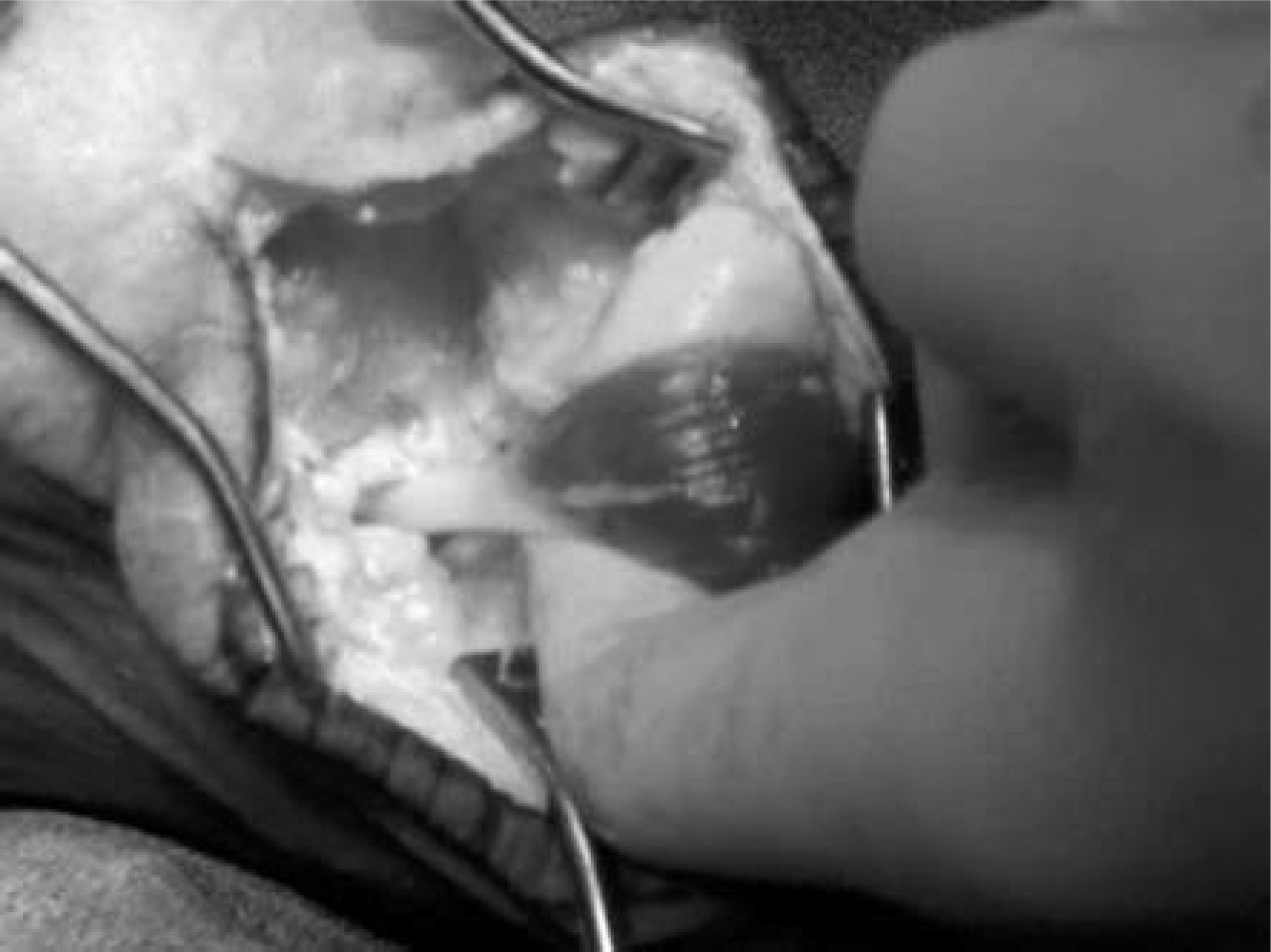
FHL muscle retracted allowing maximal length to be harvested by cutting the tendon at the level of the medial malleolus.
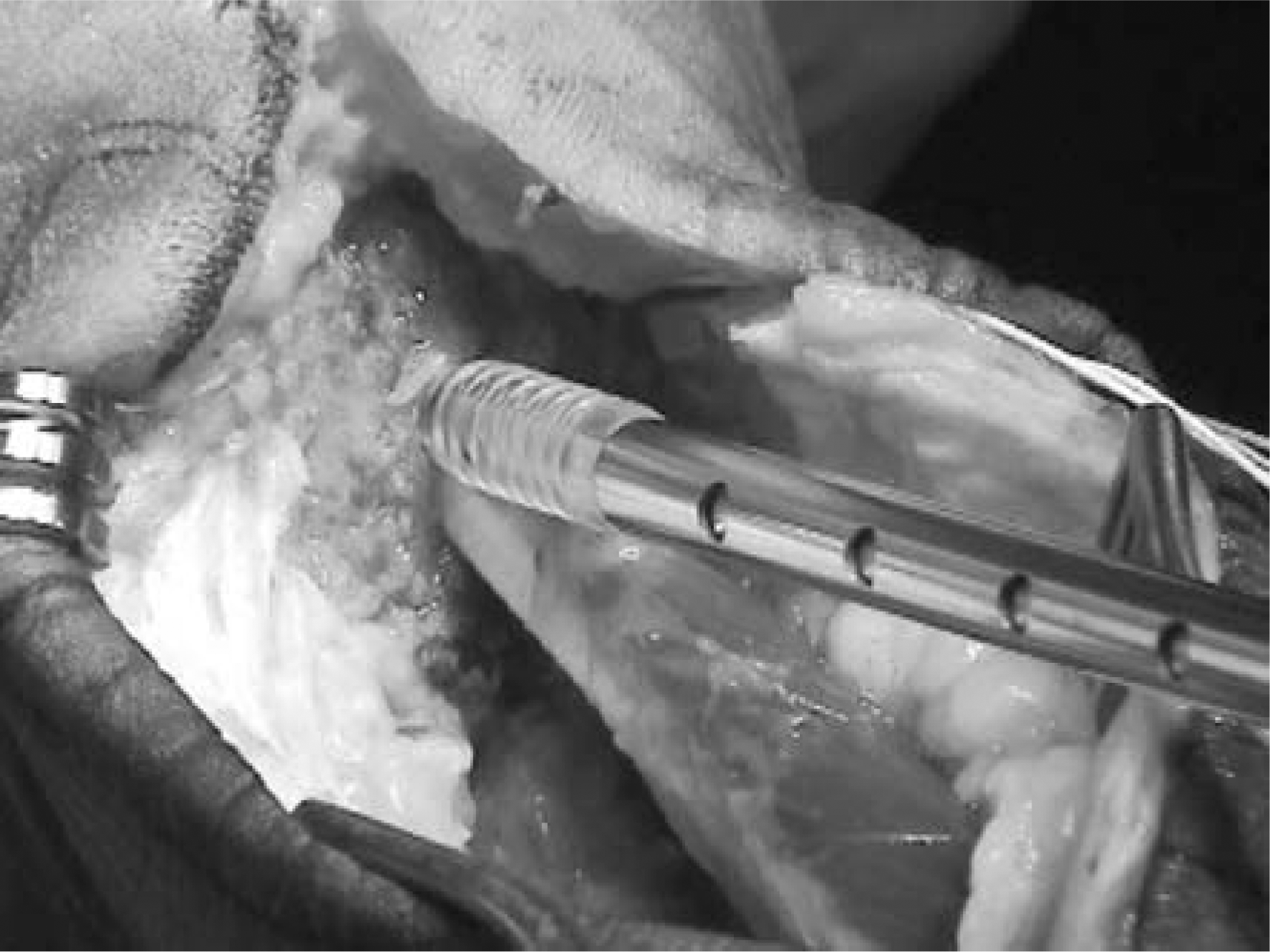
Bioabsorbable interference screw being inserted into the bone tunnel holding the FHL tendon in position with interference fit.
Two 3.5-mm corkscrew suture anchors were inserted into the posterior aspect of the calcaneus, just posterior to the bone tunnel. The attached No. 2-0 braided non-absorbable suture was then used to repair the Achilles tendon onto the calcaneus into the raw bone from which the Haglund deformity had been resected which is slightly proximal to the natural insertion site, accommodating for some of the length of the distal diseased tendon which was excised during the debridement process. The tendon was reattached to create a resting position of the ankle, with the knee flexed, of twenty degrees of equinus. Even though some of the distal tendon insertion had been resected, minimal loss of length was encountered and gentle longitudinal traction on the gastroc-soleus usually allowed the tendon to be advanced to the bony insertion. It is our preference to leave the tendon slightly taut rather than lengthen the gastroc-soleus mechanism; surgical lengthening was not required in any of our cases. Final decompression, tunnel and hardware placement can be checked radiographically (Figure 5). The Achilles tendon longitudinal split was repaired using No. 2-0 absorbable suture in a running continuous non-locked suture. The wounds were then closed in layers using No. 0 absorbable suture to repair the paratenon, and No. 2-0 absorbable subcutaneous sutures and skin staples. A posterior splint was applied to the leg, holding the Achilles tendon at its natural resting tension (approximately 20 degrees equinus) as compared to the contralateral side.
Lateral radiograph confirming the decompression of the insertional spur and the Haglund exostosis; as well as the anchors and bone tunnel (white arrows delineate the edges of the bone tunnel).
Postoperative treatment
Postoperatively the splint was left in place for 2 weeks. After suture removal, patients were placed in Achilles wedge boots (Bledsoe, Grand Prairie, TX). These boots allow insertion of up to 4 (2 are usually used) 10-degree urethane wedges which can be easily removed by the patient at two week intervals allowing the Achilles tendon to slowly be stretched as the ankle is brought into a neutral position. Patients were kept non-weightbearing for the first 6 postoperative weeks. After 6 weeks, patients were re-evaluated clinically. The tendon integrity was tested as was tendon function. Patients were then allowed to start weightbearing on the affected extremity as comfort allowed in a protective Achilles boot brace and were given a prescription to start physical therapy (performed out of their brace) for an Achilles stretching and strengthening program. Twelve weeks after surgery the boot brace was discontinued for ambulation, with the patient continuing the therapy program as needed. Patients were instructed to slowly resume their activity as comfort allowed, but to avoid sudden acceleration, cutting, or jumping activities until at least 6 months after surgery.
Followup evaluation
This clinical outcomes study used a within-subject repeated measures design. Patients were clinically evaluated by a physician (I.E.) other than the treating surgeon in order to minimize observer bias, and then underwent Biodex® System 3 (Biodex Medical Systems, Shirley, NY) isokinetic dynamometer testing at the time of their final evaluation. At followup, data was obtained by the first author who was not involved in any of the surgical procedures or patient management.
Additionally, once followup evaluation of the entire cohort was completed, a thorough review of the patients’ clinical charts which included initial AOFAS-AH score and postoperative VAS pain scores, and postoperative management and complications were noted.
Clinical evaluation included assessment of tendon integrity, incision healing, evidence of infection, and the ability to perform 20 repetitive single-leg heel raise maneuvers. Ankle plantarflexion (PF), dorsiflexion (DF), and AROM were measured using the Biodex® dynamometer. Each patient was seated in the dynamometer with the leg to be tested elevated by a support arm under the thigh. The hip was set at a resting position of 80 degrees of flexion, and the knee was placed in 30 degrees of flexion. The transverse axis of the ankle joint was aligned with the rotational axis of the dynamometer.
Full AROM of the ankle joint was determined when the patient was positioned in the dynamometer. Each patient was instructed to push the ankle as far forward (plantarflexion) and pull the ankle as far back (dorsiflexion) as possible. This procedure was completed on both lower extremities to compare the operated to the non-operated ankle.
Three sets of five consecutive maximal dorsiflexion and plantarflexion concentric contractions were performed by each subject at speeds of 60 degrees/s and 120 degrees/s. Both ankles were tested and deficits were calculated as compared to the unaffected limb. Generally, testing at 60 degrees/s correlates with strength, while testing at 120 degrees/s correlates with power parameters. 26
Each subject also was evaluated using the AOFAS Ankle-Hindfoot scale 13 (maximal 100 points) and the VAS score for pain (10 cm horizontal line ranging from 0 = no pain to 10 = worst imaginable pain), as well as rating their subjective satisfaction as Very Good (VG), Good (G), Fair (F), or Poor (P). A questionnaire was administered to determine satisfaction of the patient with the surgical outcome. Patients also were asked about dysfunction of the hallux after FHL harvest and transfer.
Data analysis
AOFAS-AH scores were computed. Peak plantarflexion (PF) torque, peak dorsiflexion (DF) torque, and total AROM were analyzed for both ankles, for all three trials at each of the two dynamometer speeds (60 degrees/s and 120 degrees/s). Paired t-tests were used to compare peak PF and peak DF torques and total AROM for the involved compared to the uninvolved ankle. A value of p > 0.05 was considered significant.
Results
Demographics
Data were obtained for 40 patients an average of 27 (range 18 to 68) months post surgery. Two patients did not have Biodex® data due to technical issues with the instrumentation at the time of final evaluation. In addition, the patient with bilateral involvement was not included in the Biodex strength and AROM analysis.
Clinical evaluation
Clinical evaluation revealed that 37 patients out of 40 (92.5%) were able to perform 20 repetitive single-leg heel raise maneuvers on the operatively repaired side. The other three patients (7.5%) were able to perform up to 10 repetitive single-leg heel raise maneuvers on their operated side, and a set of 20 repetitive double-leg (both the operated and unoperated side) heel raise maneuvers.
Strength and range of motion
Thirty-seven patients were available for followup independent isokinetic dynamometer (Biodex Medical Systems, Inc., Shirley, NY) evaluation of both the repaired and uninvolved Achilles tendons. Table 1 reports the average AROM and dorsiflexion and plantarflexion torques for both operated and non-operated ankles at 60 degrees/s and 120 degrees/s. No statistically significant differences in dorsiflexion or plantarflexion torque were found between the operated and nonoperated sides. A decrease in active range of motion of 4 degrees (−7%) was seen between the operated and nonoperated sides at 60 degrees/s (p > 0.04). Clinically, there was no discernable difference in dorsiflexion range of motion between the two sides. A number of patients did have tightness of their gastrocsoleus mechanism during the initial post-operative period, but this resolved with stretching and physical therapy in all cases. No patients required heel lift or additional interventions for Achilles tightness.
Average active range of motion (AROM), dorsiflexion (DF), and plantarflexion (PF) torques for operated and non-operated ankles
Means (standard deviation) are presented for both operated and non-operated ankles, with the operated side expressed both as absolute measurement and as a percentage of the non-operated side.
p = significant; n = 37
AOFAS Ankle-Hindfoot score
AOFAS-AH scores were obtained preoperatively and at final followup. Preoperative scores averaged 56.3 (range, 29 to 82) out of 100 possible points. Final followup scores averaged 96.2 (range, 69 to 100) out of 100 points. This is an increase of 39.9 points (p > 0.00001) (Table 2).
Average AOFAS-AH and VAS pain scores pre- versus postoperative results
p = signicant; n = 40
Visual Analog Scale (VAS) score
Visual analog scores for pain were obtained both preoperatively and postoperatively to document pain relief. The average preoperative VAS was 7.5 and the average postoperative pain relief was VAS 0.3. This is a decrease of 7.2 on the pain scale (p > 0.00001) (Table 2).
Subjective satisfaction
When asked if they were satisfied with the results of the procedure and would undergo the procedure again if necessary, 38 (95%) of 40 patients rated their outcome satisfaction as either Very Good (33) or Good (5), two rated their satisfaction as Fair, and none as Poor. All would undergo the procedure again or recommend it to a family member or to a friend if needed.
No patients complained of persistent scar irritation against their shoe counter, although this was a temporary concern in 6 patients when initially wearing a closed back shoe. This was managed with scar massage and a silicone lined Achilles sock while symptomatic, and had resolved in 5 cases by 9 months and the final case by 12 months after surgery. All patients were able to wear standard shoewear without heel lifts or heel counter padding at the time of final evaluation.
With regard to the FHL tendon harvest, none of the patients complained about pain or weakness in the hallux or regretted having the tendon harvested. No patient had noticed any deleterious effect on gait, push off or function.
Complications
A 54-year-old male subject complained of ankle discomfort at the time of final followup and was therefore referred for diagnostic US. A longitudinal sonogram of the posterior ankle demonstrated a hypoechoic line consistent with a longitudinal interstitial split tear at the level of the Achilles insertion on the calcaneus. This patient scored 88 for the postoperative AOFAS-AH and rated the outcome of his surgery as Good. Pain decreased from a preoperative VAS score of 7 to a post-op score of 2. Two other female subjects (62 and 63 years old) referred because of some ankle or hindfoot discomfort had no focal abnormality on US examination. We found no evidence of infection, improper incision healing or nerve injury in any of the 40 patients.
Discussion
Painful chronic insertional Achilles tendinosis is difficult to treat conservatively. Our data show that, after the above-described surgical intervention, patients showed less Achilles tendon pain, the AOFAS-AH score improved, and ankle torque and range of motion were equivalent to the contralateral, undiseased leg. In addition, nearly all patients were satisfied with the results of surgery when compared to their preoperative status.
For the assessment of ankle function we used isokinetic dynamometry, which is a frequently applied method for objectively measuring joint and muscle performance. Studies have shown high reliability, with coefficients above 0.80. 26 Reliability for concentric ankle dorsiflexion peak torque velocities measured on the Biodex device was shown to be excellent, but tended to be lower when measured at higher speeds such as 120 degrees/s as compared to slower speeds of 60 degrees/s. 27 Isokinetic dynamometry, such as the Biodex testing, has been demonstrated to be a reliable procedure for assessing strength, 4,5,26 and for quantification of ankle function. 1,2 Using isokinetic dynamometry, this study demonstrated the ability to restore power and strength to 100% of the contralateral side. That means no loss of isokinetic strength or power as compared to the non-operated leg, and a 4-degree deficit (–7%) in active range of motion after surgical repair. AOFAS-AH scores improved from a preoperative mean value of 56.3 to a long term mean value of 96.2. Post surgical pain was eliminated in most cases, with an overall mean decrease in VAS score from 7.5 to 0.3 (out of 10.0), and subjective satisfaction rates were very high (95%). The results of this study on patients with Achilles tendinosis were similar but slightly better than those reported for similar techniques of FHL-augmented repair.
McGarvey et al. 16 and Johnson et al. 11 described a similar surgical approach and technique, without FHL transfer augmentation. In these patients approximately 50% of the tendon was released and less than 50% excised in all cases. They did demonstrate 68% improvement (53 preoperative to 89 postoperative) in AOFAS-AH, but no clinical strength testing was performed.
Den Hartog reported on a single incision technique using FHL augmentation (attached to the calcaneus using an anchor and suturing to the Achilles reattachment) for chronic Achilles tendonosis. AOFAS scores improved from an average 41.7 preoperatively to 90.1 postoperatively (116%). While no measures of strength and power were collected, the author reported that all patients could toe-walk at final clinical exam. 8
Wapner et al. 22,23 reported using the FHL tendon through a two-incision approach with the second incision made in the medial longitudinal arch where the FHL was harvested at the level of the master knot of Henry 20,21 . Cybex testing showed a 29.5% average decrease in strength at 30 degrees/sec and decreases in torque and work generated by plantarflexion of the ankle of 41.8% and 51%, respectively, as compared to the nonoperated side. 22
Wilcox et al. 25 used Cybex isokinetic testing and AOFAS scores to report on the results of a study of 20 patients with chronic Achilles tendinopathy. Their surgical repair also used the FHL, but with a double incision technique. They report an average loss of range of motion of 2 degrees (range, −13 degrees to +8 degrees), an average gain of 3% in dorsiflexion strength and an average loss of 7% of plantarflexion strength. Postoperative AOFAS scores averaged 86 (range, 43 to 100). Wagner et al. 20,21 reported on 10 patients with complete detachment with proximal V-Y lengthening of the Achilles tendon for chronic insertional Achilles tendinopathy. For this group, they report no mean loss in dorsiflexion or plantarflexion strength as compared to the non-operated side (t-test, p <0.05). In 2005, Martin et al. 15 reported on 44 patients who had FHL augmented Achilles reconstruction. Martin's technique involved complete detachment and reattachment of the Achilles tendon, with resection of the distal 4 to 6 cm of the tendon. Average VAS pain scores post-surgery were 1.5. Nineteen of these patients were evaluated for strength and range of motion. For this subgroup, average post-surgery AOFAS scores were 91.6; plantarflexion range of motion was decreased by 3 degrees and mean peak plantarflexion torque was decreased by 22.8% (30 degrees/s) and 30.4% (60 degrees/s).
Using a z-plasty lengthening instead of FHL augmentation, Costa et al. 7 reported on 21 procedures in 18 patients. Mean concentric peak plantarflexion torques were reported as 42 Nm on the operated side and 47 Nm on the nonoperated side (11% deficit); however, this difference was not statistically significant. Range of motion was reported as a mean increase in dorsiflexion of 5 degrees with a decrease in plantarflexion of 3.5 degrees. Pain at rest was reported as 20 point decrease as measured by VAS.
Considering all surgical options, the single incision technique is the least invasive with minimal risk of potential wound complications and no risk to the medial plantar nerve at the Master knot of Henry when using a proximally harvested FHL autograft for augmentation. Post-surgical ankle strength and power show comparable or better results than those reported with similar or alternative techniques.
While our study population was relatively large as compared to previously published studies for insertional Achilles tendonitis surgery, the cohort still remains small. Additionally, ten patients (20%) were not available for followup as discussed above.
Due to the nature of the design of this clinical follow-up/outcomes study, in which data were collected post-surgery, we did not have preoperative strength and Biodex measured AROM data on our patients. This data might have allowed additional comparisons. However, we believe that the results of this long-term clinical followup study are important. Comparisons of strength and range of motion pre- and postoperatively would not be clinically meaningful. Measuring plantarflexion strength on an individual with persistent chronic Achilles tendinosis would be painful and perhaps meaningless. We believe that the comparison to the unaffected side is more clinically indicative of operative outcome. However, there may have been individuals in the study who had untreated asymptomatic pathology of the contralateral ankle, which may have resulted in diminished strength of the non-operated side. The AOFAS-AH scores and VAS pain scores were available, and pre- to postoperative comparisons were made. Ultrasound and MRI images before surgery confirmed the tendinosis.
Conclusion
Chronic insertional Achilles tendonosis tends to cause more pain than functional difficulty, whereas complete ruptures of the Achilles tend to cause more functional difficulty than pain. 9 After surgical repair, we found no loss of plantarflexion strength or plantarflexion power in the operated ankles; a 4-degree loss of AROM was found. The study population scored an average of 96/100 for the total AOFAS-AH and 0.3/10 for the VAS post-repair. We conclude that for individuals who fail non operative treatment for chronic insertional Achilles tendinosis, operative repair using an FHL tendon autograft with the single-incision technique achieved a high percentage of satisfactory results as well as excellent functional and clinical outcomes including significant pain reduction.