
Abstract
BACKGROUND:
Inconsistencies in the literature concerning the effect of neck pain have led to a lack of understanding concerning the complete pathophysiology of neck pain. While the effect of neck pain on motor function as measured by active range of motion and isometric neck strength is well documented the effect of neck pain on sensory measures such as tactical acuity and neck reposition error (NRE) remain poorly understood.
OBJECTIVE:
The purpose of this study was to evaluate a combined sensorimotor evaluation to explore the potential benefits of incorporating both sensory and motor task into a physical evaluation of neck pain suffers to gain an added knowledge of the complete pathophysiology of their health status.
METHODS:
A cross-sectional study that measured neck joint reposition error, tactical acuity, neck isometric strength and range of motion in 40 volunteer participants (22 pain, 18 control).
RESULTS:
A statistically significant increase in NRE in flexion (2.75
Keywords
Introduction
Neck pain is reported to effect 33% of the population each year [1]. That percentage is estimated to affect 16% of people between the age of eighteen and twenty-nine and increases with age [1]. The source of neck pain is often illusive and can come as a result of varying pathologies leading to the common use of the term “non-specific neck pain”. These pathologies include whiplash [2], poor posture [3], and high velocity sports injuries i.e. concussion [4, 5, 6, 7]. Sensory and motor function are important in maintaining postural control allowing the neck to perform movements required to perform ADLs such as driving a motor vehicle and to protect against potential sporting hazards such as tackling in football or heading in soccer [8]. Structures involved in appropriate sensory and motor function include muscle spindles, articular receptors, muscles, efferent motor pathways, afferent sensory pathways, connective tissue and bony articulations which work synergistically to support the head [9].
Head position sense is controlled in part by passive constraints such as ligaments, tendons and connective tissues and the architecture of each individual cervical vertebrae, and secondly by afferent feedback from muscle spindles in the cervical musculature which send messages to the sensory cortex and motor cortex respectively via the cerebellum and thalamus [9]. However, it has been reported that the osteoligamentous structures of the cervical spine contribute only a mere 20% of the minimally needed mechanical stability of the cervical spine [10], suggesting that the remaining 80% of the mechanical load is managed by the cervical musculature.
Damage to the cervical musculature has been reported to interrupt the communication between muscle spindles in the cervical musculature and the somatosensory cortex in the brain leading to decreases in neck function [11]. Disruption due to neck pain has been reported to effect neck muscle strength [12, 13], neck muscle size and thickness [14, 15, 16], range of motion [17, 18], neck reposition error (NRE) [8, 19], and tactile acuity (TA) of cervical dermatomes [20]. NRE and neck range of motion help to analyze the integration between sensory and motor function creating highly integrated sensorimotor movements [21, 22], while isometric neck strength is primarily a measure of motor function and TA a measure of sensory function [23].
In addition to sensory dysfunction, motor dysfunction as identified by deficits isometric neck strength and active range of motion have been reported in chronic neck pain sufferers [12, 13, 17, 18]. However, the role sensory disturbances contribute to cervical motor dysfunction is poorly understood as previous research has typically focused on evaluating a single aspect of cervical function i.e. decreases in strength or increases in position error [24]. Recent reports identifying balance disturbances and oculomotor disturbances in conjunction with neck reposition error in patients suffering from chronic neck pain have highlighted the importance of understanding the complete pathophysiology of neck pain and its implications [2, 19, 24].
Increases in NRE have been reported in patients suffering from neck pain and are indicative of a difference between an individual’s “perceived” head position and the actual position of the head therefore decreasing the ability to appropriately position the head in a desired position [19]. Similarly decreased sensation has been reported in patients suffering from complex pain associated disorders and are indicative of decreased sensorimotor function [25]. The primary role of NRE is to assess the afferent input from the muscle spindles in the cervical musculature by measuring the ability of the head to return to normal resting posture following movement, thereby providing clinicians a method to measure proprioception in the neck [7, 8]. NRE has been reported to be higher in patients suffering from pain related symptoms as a result of disruption of the ascending sensory pathway to the receptors and on to the sensory and motor cortexes [7, 21]. The reliability of NRE testing has also been explored and found to be adequately reliable to produce significant results [19].
TA measures the perception or sharpness of the sense of touch [26]. Proper mechanoreceptor activity in the neck and surrounding areas is critical to sense touch and contribute to neck function [26]. The most common method of measuring TA is the two-point discrimination threshold test To our knowledge no research has compared the effect of mild to moderate chronic neck pain on the sensation of cervical dermatomes C-4 through C-8 which radiate down the shoulder and arm in conjunction with NRE to evaluate the role of decreased sensation in overall neck function and the potential implications for rehabilitation of the cervical spine in chronic pain suffers.
The purpose of this examination was to explore the use of a combined sensorimotor evaluation using both sensory and motor test to evaluate cervical dysfunction in patients with mild neck pain and the potential effectiveness of using this measures to track progress through out a treatment protocol in neck pain suffers.
Methods
Subjects
Forty subjects (22 females, 18 males) were recruited from a convince sample of university students and staff. Subjects were divided into two groups; a pain group and a healthy group. Twenty-two subjects participated in the pain group (13 females, 9 males; age 25.5
Inclusion criteria
The study design was a cross-sectional study. Subjects were assigned to a group based on their Neck Disability Index score [27]. The neck disability index scores from 0–50 and consist of 10 sections, each section represents a functional task i.e. pain intensity, personal care, lifting, work, headaches, reading, recreation, driving, sleeping and concentration. Each section is scored on a scale from 0–5 ranging from no pain during the activity (0) to unable to perform activity (5) and then the total score across each section is summed. All participants scoring above an 8 were assigned to the neck pain group. Participants scoring a 4 or below were assigned to the control group. Two patients scoring
Pain measures
Pain measures
*Statistically significant difference between two groups.
Thresholds were measured at each dermatome from C3–C8. Measurements were taken using a Baseline Discriminator Aesthesiometer (Electro Medical, Marietta, GA). Dermatomes were measured bilaterally and then summed and divided to represent the average between each side. Due to the limitations that have been identified with two-point discrimination the following parameters were followed to limit inconstancies between measurements. All measurements were taken with a vertical orientation [29]. The tips of the baseline two-point discriminator device were filed slightly to create a more blunt edge to avoid any sensation of sharpness or pain [29]. When two stimuli were presented special care was taken to make contact at the same time with both points [26]. The pressure applied to the skin was kept to about 10 grams or that equal to the first blanching of the skin [26]. The threshold was determined by the smallest distance between the two stimuli that the participant could still detect two distinct stimuli rather than just one [29]. For each stimulus given the participant was asked to respond either one or two depending on wither one stimulus was perceived or two. The starting distance for the two-point discrimination threshold was 10 mm for dermatomes C6–C8. Initial pilot data of 5 individuals showed that dermatomes C3–C5 had a much larger threshold and so the starting threshold was set at 30 mm. If the first test was incorrectly identified the distance was increased by 5 mm until the first correct identification of two points was obtained. The test started after the initial correct identification of two distinct stimuli. Adjustments to the distance between the two points were made as follows: for each correct answer of either one or two stimuli the distance between the two points was reduced by 2 mm and for each incorrect answer the distance was increased by 1 mm. Participants were given one or two stimuli until 5 total incorrect answers were given. Both the examiner and participant were seated with their arms supported on a table. The following landmarks were used to identify each cervical dermatome measured: C3–3 cm above the mid-clavicular line, C4-anterior edge of the acromion, C5-the lowest point of the deltoid insertion, C6-the thenar eminence, C7-volar surface of the base of the 3
Joint reposition error test.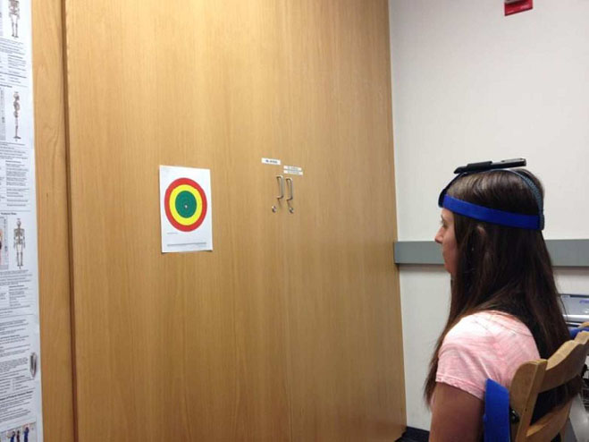
The procedures established by Revel et al. with slight modifications were followed to collect NPE [21]. Each participant was positioned exactly 90 cm from a target placed on the wall and strapped into a straight backed chair to limit accessory movement of the torso see Fig. 1. Following positioning the participants were instructed to position their head in the most comfortable natural resting head position staring straight at the wall. Blacked-out glasses were worn by each participant to occlude their vision to insure that they could not see the target during each trial. The participant was asked to maximally flex their neck and then actively return to the starting position. The point where the laser came to a rest on the target was marked and the distance from the center point mas measured and marked as the error. The same process was repeated with the participant extending the neck, rotating the neck to the left and rotating to the right representing both horizontal and vertical movements. No practice trials were allowed and the mean of 8 trials was used in the analysis as 8 trials had previously been identified to be the maximum number of trials required to obtain optimal reliability [31]. The degrees of error were derived by calculating the arc tangent of the distance between the center point and the marking on the target in centimeters divided by 90 centimeters (the distance the subject was from the target).
Range of motion
Range of motion measurements were taken using cervical range of motion instrument 3 (Performance Attainment Associates, Manitowoc, WI). Range of motion was taken for flexion, extension, left and right lateral flexion and left and right rotation. During the range of motion measurements participants were seated in a straight back chair and strapped in to minimize any accessory movements at the torso. The intra-tester and inter-tester reliability have previously been reported to be reliable in both normal and chronic neck pain groups with high ICC coefficients ranging from 0.75–*0.92 [32].
Isometric strength
Isometric strength was measured using a microFET2 hand held dynamometer (Hoggan Health, Sandy, UT). To minimize examiner error the hand held dynamometer was mounted to a mobile device. This device allowed for the location and orientation of the dynamometer to be adjusted both vertically and horizontally and to be lined up with the proper landmark for measuring isometric strength in each direction. The participants were then seated and strapped into a straight backed chair next to the device. Flexion, extension, left and right lateral flexion and left and right rotation were measured. To measure flexion the dynamometer was aligned with the forehead of the individual and they were instructed to, “over the course of 5 seconds try as hard as you can to bring your chin down to your chest”. This procedure was repeated in extension with the dynamometer being aligned with the occiput. For lateral flexion the dynamometer was aligned just superior to the ear and for right and left rotation the dynamometer was aligned slightly superior to the pterion. Three trials were performed for each motion and the average of the highest was used as the maximum isometric contraction. Pilot data revealed that 3 trials was sufficient to attain reliable measurements with intra-session ICC values ranging from 0.88–0.95 for 5 pilot subjects.
Statistical analysis
There were two groups of subjects, classified as either with neck pain or healthy. The most important response variables included joint reposition error (four variables: flexion, extension, right and left), and tactile acuity at six cervical dermatomes. A standard independent two-sample t-test with equal variance was reported for each cervical dermatome and for each movement with joint reposition error as well as peak isometric strength and cervical R.O.M.
Tactile acuity threshold (mm)
Tactile acuity threshold (mm)
*Significant difference at 0.05 level.
Neck reposition error
*Significant at level of 0.05.
Neck range of motion in degrees
Neck isometric strength (Kg)
The results of the two-point discrimination test are displayed in Table 2. C5 dermatome showed a significant level of difference in sensation between the neck pain group and the healthy group. While none of the other levels achieved a statistical significant difference the absolute mean in two-point discrimination threshold was higher in the pain group in each of the six measured dermatomes. Results for NRE can be found in degrees in Table 3. Neck flexion and extension were found to vary significantly between the healthy and neck pain groups; however, right rotation, left rotation showed no significant differences between the two groups. The values for range of motion and neck isometric strength are shown in Tables 4 and 5 respectively. No significant difference was found between the neck pain group and healthy group range of motion or isometric strength measurement in any movement direction.
Discussion
In spite of the fact that the mean NDI for our moderate chronic neck pain participants was less than 11 points, statistically significant differences between our chronic moderate neck pain group and the heathy controls were observed in NRE for flexion and extension as well as sensation at the C5 dermatome. Similar to previous research in which differences in NRE ranging from 3–6
The dermatome identified to be most affected by the moderate neck pain was C5. This suggests that the effect of neck pain on tactile acuity is not limited to the area directly surrounding the neck, but may also effect the upper arms as the sensory afferents associated with sensation of the upper arm pass through the neck and pain may at the neck may alter neuronal activity. Previous, literature on the effects of neck pain on tactile sensation are limited however preliminary data using monofilaments has shown so indication of impaired sensory function in neck pain suffers [30]. This evidence combined with our findings suggests that while evaluating neck sensation of an individual following a neck injury, in the presence of neck pain, it may be of interest to measure the sensation of the surrounding dermatomes and not limit the examination to dermatomes C3, C4 surrounding the neck.
Isometric neck strength as well as range of motion have also been reported to be affected by neck pain [12, 34, 35, 36]. Our results however, showed no significant differences in cervical range of motion or isometric neck strength in of the movements measured suggesting that perhaps moderate neck pain is not sufficient to alter the descending motor efferent neurons from the motor cortex to the musculature of the cervical spine. This evidence suggests that perhaps a more inclusive examination including NPE, tactile acuity, cervical range of motion and isometric neck strength may give a more comprehensive view of the patients symptoms prior to treatment and may allow for a more detailed picture of a patient’s neck dysfunction.
Clinical test such as NPE and two-point discrimination allow clinicians to identify deficiencies in these afferent pathways and respective sensory receptors. Increased understanding of the effect neck pain has on these afferent pathways and in turn on neck function has led to a recent increase in vestibular and cervicogenic rehabilitation as a treatment of neck pain as well as the treatment of concussion related symptoms [8]. Recent evidence suggests that neck pain sufferers may benefit from training exercises designed to improve neck position sense and sensation as well as restore normal muscle strength. The two-point discrimination threshold test offers an inexpensive and rapid method for measuring the tactile acuity of the associated dermatomes of the neck within a clinical setting [29].
Conclusion
In spite of using a population with mild to moderate chronic neck pain both the Two-Point Discrimination Test and the NRE appear to give clinicians additional inexpensive and quick objective data that can be used in the diagnostic portion of an examination when combined with other measures such as range of motion and isometric neck strength. The results of such diagnostic exams could in turn be used in designing a rehabilitation program and assisting in monitoring progress during the course of treatment of cervical pain. Further research is needed to establish the clinical significance of this information.
Limitations
Limitations to this study include variation in the duration and intensity of pain for each participant in the pain group. The mean NDI value of our chronic pain group was below the reported cut-off value of the NDI to detect pain associate with disability. All participants had experienced neck pain
Conflict of interest
The authors declare no conflicts of interest.