






Abstract
BACKGROUND:
Electrotherapy is part of a physician’s toolbox for treating various musculoskeletal conditions, including radicular pain, but the preferred modality is yet unclear.
OBJECTIVE:
To compare the short-term efficacy of three electrotherapeutic modalities in relieving lumbar disc herniation (LDH)-induced radicular pain.
METHODS:
Fourteen patients with LDH-induced radicular pain attended a single session of electrotherapy, which included four 10-min consecutive treatments: transcutaneous electrical nerve stimulation (TENS), interferential (IF) stimulation, a combined treatment with pulsed ultrasound and IF current (CTPI), and a sham control. Treatments were randomized and the straight leg raise (SLR) degree was measured immediately before and after each treatment.
RESULTS:
Each of the three active modalities significantly improved the SLR score. The most prominent improvement was observed in the CTPI condition, followed by IF and, finally, TENS. The sham stimulation did not affect the SLR scores.
CONCLUSIONS:
A single session with either TENS, IF current or CTPI is sufficient to improve the range of motion and degree of radicular pain associated with LDH. CTPI appears to be the most effective modality of the three, possibly due to greater penetration efficiency of the induced current. The effects of a long-term treatment schedule are yet to be identified.
Keywords
Background
Lumbar disc herniation (LDH) is the localized displacement of disc material, usually in L–S1, beyond the normal margins of the intervertebral disc space [1]. This condition is a major contributor to low-back pain (affecting around 9% of the population worldwide [2]) and is considered the most common cause of nerve-root irritation (NRI), which affects 1–5% of the population annually [3]. NRI typically causes nerve inflammation [4] which, upon movement of or tension on the nerve root, may induce radicular leg pain: a wide (7–9 cm) band of lancinating pain along the lower limb [5].
Patients with LDH-induced NRI are typically offered various conservative (i.e., nonsurgical) first-line treatments before surgery (e.g., discectomy) is considered [6]. One such conservative treatment, which is usually applied as part of a multimodal treatment program, is electrotherapy, namely, placing electrode pads on the skin to electrically stimulate deeper tissues [7]. While numerous studies support the use of electrotherapy for a wide variety of indications [7, 8, 9, 10, 11, 12], its efficacy in relieving NRI-induced pain is not entirely clear. Several (mostly methodological) reasons appear to account for this lack. First, when electrotherapy is examined as part of a multimodal treatment program (e.g., in [13, 14]), its unique contribution to pain relief cannot be isolated. Second, most previous studies employed either transcutaneous electrical nerve stimulation (TENS) [13, 15, 16, 17, 18, 19] or interferential (IF) current stimulation [14, 20], two of the most commonly used electrotherapeutic modalities [7, 21], while only a handful of studies examined other modalities [15, 18]. Third, most previous studies of electrotherapy in the context of LDH-induced radicular pain employed subjective measures of pain, such as a visual analog scale (VAS) or questionnaires [14, 15, 16, 17, 18, 19, 20], while only a few studies [13] used more objective measures of pain. Thus, taken together, still lacking are systematically comparative and controlled studies, which directly compare between the efficacies of various electrotherapeutic modalities by using more objective measures of radicular pain.
To fill these lacunae, the current study aimed to systematically compare the efficacies of widely used electrotherapeutic currents in instantaneously relieving LDH-induced radicular pain. We focused on three electrotherapeutic modalities: TENS, IF current, and a combined therapy with pulsed ultrasound and IF current (CTPI), a common modality in most electrostimulation devices which has shown some clinical utility in the context of fibromyalgia [8, 22, 23, 24]. Notably, CTPI has been demonstrated to penetrate deeper into the tissue than TENS and IF-induced currents [25]; as the NRI associated with LDH and radicular pain is a relatively deep pathology (usually located 3.7–4.4 cm deep within the tissue [26]), and the high penetration efficiency of CTPI may be especially beneficial for relieving LDH-induced radicular pain.
As a relatively reliable measure of the radicular pain and of the typical reduction in the range of motion associated with LDH, we used the straight leg raise (SLR) test, a simple-to-use, reliable and, at least to some degree, clinically relevant measure of the integrity and mobility of neuronal structures [27, 28, 29, 30]. Notably, this test is considered to have high intra- and inter-rater reliability (namely, 0.95–0.98 [31] and 0.67–0.97 [32], respectively) and is used by 97.9% of physiotherapists to assess pain in patients with LDH-induced radicular pain [21]. During the SLR test, the leg of the patient is raised passively by the experimenter, such that traction is exerted onto the sciatic nerve, lumbo-sacral nerve roots, and dura, thus moving the nerve roots through their intervertebral foramina for distances up to several millimeters [33]. The recorded SLR score is the degree of hip flexion required to elicit the recurrence of symptoms (mean detectable change has been reported to range between 1.50
Methods
Participants
This study included 14 adult participants (eight males and six females; mean age 53.5 years, SD
Selected demographic data, stimulation intensities (Stim; in mA) at each active stimulation condition, and the SLR scores (in degrees) before (pre) and after (post) applying each stimulation modality
Selected demographic data, stimulation intensities (Stim; in mA) at each active stimulation condition, and the SLR scores (in degrees) before (pre) and after (post) applying each stimulation modality
Notes:
The participants were recruited by contacting doctors at the Sheba Medical Center Pain Clinic, who performed the initial screening and obtained the initial consent to participate in the study. Each candidate participant was then contacted by phone and briefed about this study, and those who agreed to participate were sent (by fax or e-mail) a written explanation and a copy of the informed consent form. Upon arrival at the Pain Clinic, the participants were briefed, signed the informed consent form, and completed a short demographic questionnaire. Shortly after the experimental session, all participants received a pre-scheduled epidural lumbar injection under x-ray at the Pain Clinic. The study complied with the Declaration of Helsinki and was approved by the Human Ethics Committee of Sheba Medical Center and Tel Aviv University.
The entire experimental session took place in a dedicated closed and quiet room at the Pain Clinic and lasted about 2 h in total. First, the participant was instructed to lie supine and the initial SLR score (SLR
Experimental timeline. Prior to the experimental session (pre-session), the participants were screened by the Pain Clinic doctors for LDH-induced NRI (according to MRI scans and clinical evaluations) with radicular pain and a positive SLR response. On the day of the experiment, all participants were briefed and completed an informed consent form and a demographic questionnaire. Next, prior to applying each stimulation modality (pre-treatment), the SLR score was measured (SLRpre) by an observer blind to the conditions of the experiment and the pain threshold for the selected modality (except sham) was measured by the experimenter. During the subsequent treatment phase, the selected modality (TENS, IF, CTPI, or sham, randomized across participants) was applied for 10 min, after which (post-treatment) the SLR score was again measured (SLRpost) by the observer. Following a 15-min washout phase, the process (pre- to post-treatment, curved arrow) was repeated until all four conditions were tested.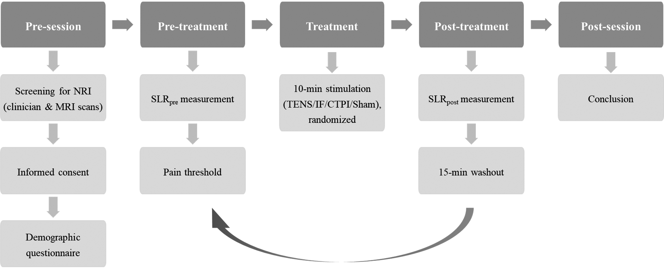
The SLR scores were recorded immediately before and immediately after the application of each of the four stimulation conditions, which yielded the
(A) The inclinometer used in this study to measure the SLR score, attached to the thigh of the participant using a custom-built device made of flexible leather and Velcro straps (insert). (B–C) Electrode placement for TENS (A), IF (B), and CTPI (C). Circles indicate the average LDH level across participants. 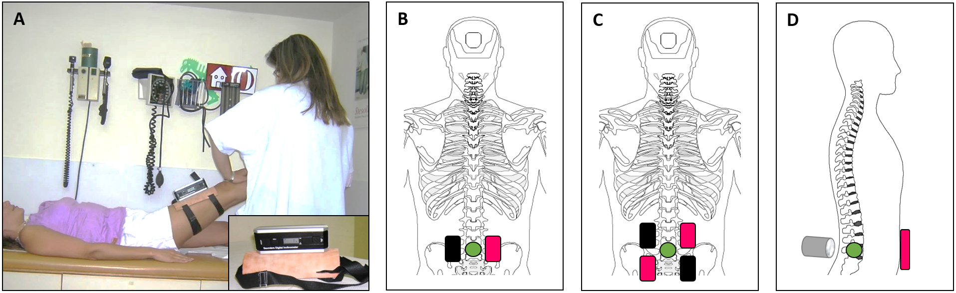
The stimulation electrodes were either 5
The TENS condition included the application of biphasic symmetrical currents through two electrode pads positioned dorsally on the paravertebral muscles, at the height of the herniated disc (Fig. 2B). One electrode pad was placed on each side of the spinous process (1 cm lateral to the spinous process), such that the middle of each pad was placed at the level of the herniated disc. The IF condition included the application of amplitude-modulated sinusoidal currents (4 kHz carrier frequency) through two pairs of electrode pads, positioned in the most commonly used IF configuration [7], in which each two adjacent electrode pads are positioned 2 cm apart, such that the herniated disc is directly between the four stimulating pads (Fig. 2C). The CTPI condition included the application of a pre-modulated IF current (4 kHz carrier frequency) through the ultrasound treatment head (5 cm
Statistical analyses
A power analysis was conducted in Statistica (ver. 12) to indicate the appropriate sample size for this study, based on the results of a pilot experiment. This analysis indicated that, under the assumption that SLR differences between the four test conditions will exceed 3
Due to the relatively low sample size and non-normal distribution of our data, we used the related-samples Friedman’s two-way analysis of variance by rank (a non-parametric test) with the Benjamini-Hochberg correction for multiple testing (FDR
Results
Selected demographic data, the applied stimulation intensities in each condition, and the SLR scores before and after applying each stimulation are shown in Table 1. A Friedman’s non-parametric test indicated that, whereas the Sham condition did not significantly affect the SLR score (
SLR scores before and after applying each stimulation modality. Bars indicate mean 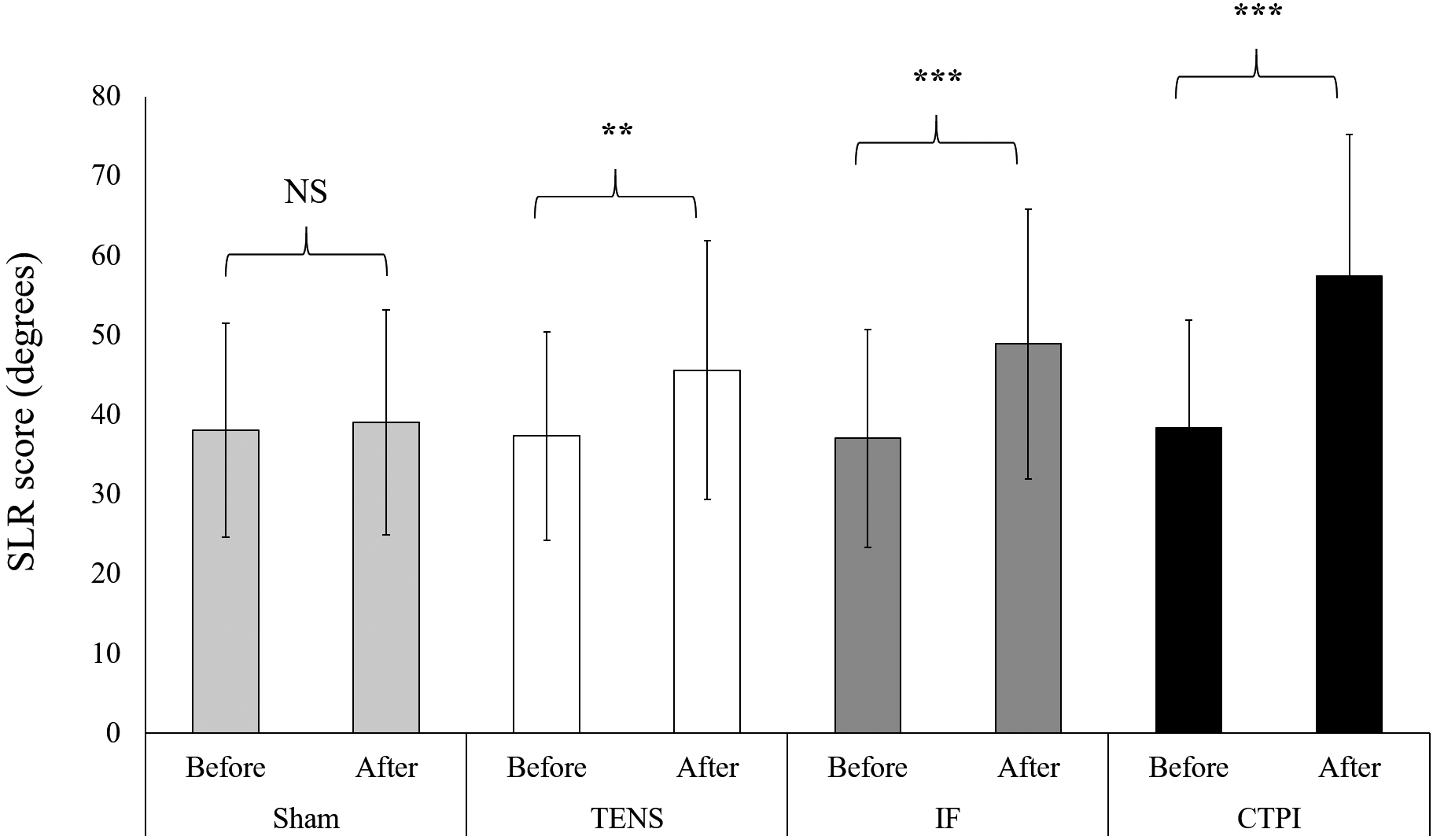
Comparing the
Multiple pairwise comparisons (two-sided Friedman’s non-parametric test with a Benjamini – Hochberg correction) of the
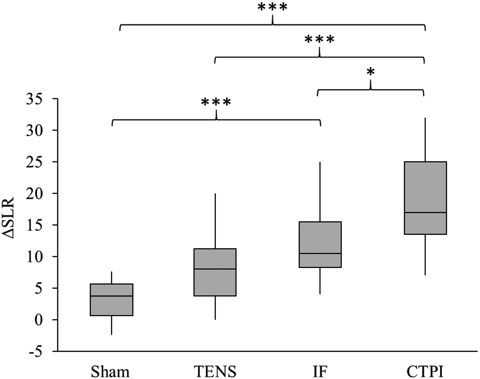
This study aimed to test and compare between the efficacies of three widely available electrotherapeutic modalities in improving LDH-induced radicular pain, as reflected by the SLR scores. We found that an active (but not sham) 10-min electrotherapeutic stimulation is sufficient to significantly improve SLR scores, a finding with both diagnostic and, possibly, clinical relevance [30]. A comparison between the three active modalities revealed that (a) CTPI induced the greatest improvement in SLR, followed by IF and, finally, TENS; and (b) as compared with the Sham stimulation, both CTPI and IF stimulation significantly improved the SLR scores, while the TENS only induced a borderline significant difference. Thus, our findings suggest that, among the three active modalities tested in the current study, CTPI may be the most suitable for potentially relieving LDH-induced radicular pain, while the effects of TENS are more equivocal. Notably, similar results were previously reported in the context of fibromyalgia [8], which is the main context in which CTPI has been examined to date.
To the best of our knowledge, this study is the first to systematically examine the change in SLR scores in response to specific electrotherapeutic modalities. Thus, the degree of improvement in the SLR scores (namely, 8.3
Although deciphering the physiological mechanisms underlying the therapeutic effects of surface-induced electrical currents is beyond the scope of this study, several speculations can be raised. First, LDH is very often accompanied by local inflammation and by inflammation-associated physiological changes (edema, impaired microcirculation, etc. [43]), which induce the radicular pain [44]. Thus, although the effects of electrotherapy on such peripheral physiological processes are controversial [45, 46, 47], it is possible that the stimulation reduced the severity of local inflammation around the herniated disc [45, 48, 49], which decreased the radicular pain. In the case of CTPI, the ultrasound itself may have further reduced inflammation and swelling [7, 47]. Second, various pharmacological and physiological evidence supports a central activation theory for the reduction of pain by electrotherapeutic currents [50, 51, 52, 53], and various types of spinal and/or supra-spinal receptors are known to be involved in electrotherapy-induced antihyperalgesia, including opioid [51, 52], serotoninergic [54], and cholinergic [55] receptors. Notably, the relative degree to which each of the three stimulation modalities improved the SLR scores is concomitant with the degree to which each of these modalities was shown to effectively penetrate biological tissues (namely, the penetration efficiency of CTPI is the highest of the three, while that of TENS is the lowest [25]). As the inflammation in LDH is relatively deep within the tissue, it is plausible that, as compared with more superficial currents, currents that penetrated deeper into the tissue (a) better reduced the LDH-induced inflammation, e.g., by stimulating the paravertebral muscles and reducing edema [56]; and (b) recruited more deep-tissue afferents to further reduce pain [57]. However, more direct mechanistic studies of these and other peripheral and central mechanisms are required to determine whether these speculations are valid.
The unique design of our study, in which all participants received all the types of treatment in a single, randomized 2 h session, increased the participation rate and enabled us to minimize inter- and intra-individual variability. However, while this design enabled us to directly compare between the different modalities, it also necessitated some methodological compromises, which should be considered when interpreting our findings and designing future studies. First, since all treatments were conducted during the same session, we only studied short-term post-stimulation effects; similar long-term effects are also possible and could be mediated, e.g., through a gradual but consistent reduction of inflammation, especially if the treatments were repeated periodically (e.g., 2–3 times a week) as part of a long-term treatment program. These speculations must be tested empirically, and an optimal protocol should be developed. Second, restricting the session to 2 h limited both the number of modalities that could be examined and the stimulation and washout durations. Thus, while we chose to test only three electrotherapeutic modalities and a sham control, future studies should also test other modalities (and, possibly, other types of sham controls; see below) and, especially, the combination of TENS with ultrasound stimulation. In addition, prolonging the stimulation and washout durations would have been methodologically preferable, although it would also increase the session duration and the inter- and intra-subject variability, and lying supine for longer durations during the washout phase might be especially uncomfortable, and even painful, for patients with NRI. Third, due to the need to randomize the treatment modalities, some participants received the sham stimulation after they had already experienced an active stimulation modality. As the physical sensation of active stimulation is quite different from that of a sham stimulation, these patients were probably aware of the fact that they were given a sham treatment, thus compromising our ‘control’ condition. Indeed, while previous studies reported a ‘placebo effect’ when comparing the efficacy of electrotherapy in ‘real’ versus ‘control’ groups, in which case the participants cannot differentiate between the active and sham stimulation [58], such an effect was not observed in our study. Fourth, while the observer in our study was blind to the treatment condition, the experimenter who performed the SLR test was not – which could potentially bias the results. Finally, while we adhered to the sample size indicated by a preliminary power analysis, the sample size is still relatively small and should be increased in future studies.
Conclusions
We tested the short-term efficacy of three widely used electrotherapeutic stimulation modalities (TENS, IF stimulation, and CTPI) in improving SLR scores in patients with LDH-induced radicular pain. We found that a single 10 min stimulation session improved the SLR scores in all three modalities, and that CTPI induced the greatest improvement, followed by IF stimulation and, finally, TENS. Further studies are required to elucidate the mechanisms underlying the improvement in SLR and to examine the ability of other stimulation modalities, treatment protocols, and treatment programs to improve the SLR scores.
Footnotes
Acknowledgments
This work was conducted as part of a PhD thesis by EA, submitted to Tel Aviv University. The authors thank Dr. Ram Gal for his helpful comments on the manuscript.
Conflict of interest
The authors report no conflicts of interest.
Funding
There are no funders to report for this study.