
Abstract
The current feasibility study deals with the elimination of Escherichia coli (gram-negative) and Staphylococcus aureus (gram-positive) bacterial strains isolated from swimming pools using zinc oxide (ZnO) nanoparticles (NPs) doped with copper (Cu2+) ions (CuX%/ZnO NPs) and co-doped with copper (Cu2+) and silver (Ag+) ions (AgX%/CuY%/ZnO NPs) synthesized by sol-gel method. Antibacterial activity was evaluated by Agar well diffusion assay. As-produced NPs were characterized by X-ray diffraction, Field emission-scanning electron microscopy, Energy Dispersive X-Ray and Transmission electron microscopy techniques. The results showed that the size of the co-doped NPs was smaller than that of mono-doped NPs. Meanwhile, co-doped Ag5%/Cu5%/ZnO NPs had the maximum bactericidal activity, and the destructive effect on Gram-positive bacteria was greater than that on Gram-negative bacteria. The lowest effective nanoparticle concentrations were 0.1 and 0.05 g/mL. The main bactericidal mechanism, in addition to the size of co-doped NPs, was due to the formation of reactive oxygen species, so that the destruction of the bacterial cell wall and finally death occurred through the radicals formed.
Keywords
Introduction
Nanoparticles (NPs) are commonly known in the size range of 1 to 100 nm. The properties of NPs change by decreasing their dimensions [1–3]. Advances in nanoscience and the ability to synthesize NPs in different shape and size have led to the production of antibacterial agents. These nanoscale materials have a high surface-to-volume ratio, which enhances antibacterial activity [4–6]. The development of effective agents with high antimicrobial activity and photocatalytic activity is of great importance. NPs can form antimicrobial agents due to their high surface-to-volume ratio and high reactivity [1, 7].
Metal NPs are synthesized through chemical and physical methods. NPs are typically made by chemical reactions with appropriate metal ions [7, 8]. Synthesis of NPs by sol-gel method has advantages, such as high purity and uniformity, low synthesis temperature and obtaining new stable compounds. It is a suitable method in the synthesis of metal NPs due to good uniform products [2, 9].
The main mechanisms of antibacterial effects of NPs have been reported to be protein degradation, DNA damage, cell wall destruction [4], formation of reactive oxygen species (ROS) on oxide surface, and release of Zn2+ ions on the membrane of the microorganism [10, 11]. Inorganic metal oxide NPs (MONPs) such as ZnO, MgO, CuO, TiO2 and SiO2 have antimicrobial activity and are used as therapeutic and diagnostic factors [1, 12–15]. MONPs have been more widely used as antibacterial agents than organic NPs because of their stability. Among these MONPs, zinc oxide (ZnO) NPs are known as a potential antibacterial agent [8, 16]. Pure, doped and co-doped ZnO is known as an antimicrobial compound and is used in various fields such as biological activities [16]. ZnO has attracted attention due to its unique optical, electrical and chemical properties, high chemical and physical stability, high catalytic activity, strong antibacterial activity and effective photocatalytic applications in the destruction of environmental and toxic organic pollutants [1, 10]. It is also one of the NPs used on an industrial scale [4]. Doping various metals on ZnO reduces the particle size, resulting in the emergence of admirable properties such as non-toxicity and cost-effectiveness with a variety of applications like antibacterial activity [17]. ZnO has shown high antibacterial activity against bacteria such as Bacillus subtilis and Escherichia coli [10].
By investigating the effect of ZnO, TiO2 and CuO NPs in removing Gram-positive and Gram-negative bacteria from municipal wastewater, the results showed a direct relationship between the concentration of nanoparticles and the efficiency of eliminating bacteria [18]. By synthesizing ZnO for the photocatalytic removal of E. coli from aqueous solutions, the data revealed that the efficiency of removing bacteria was maximum at pH 7 and the efficiency increased from 40 to 98.5% as the concentration of nanoparticle increased from 0.25 to 5 g/L; also, prolonging the contact time and reducing the initial bacterial density increased the removal efficiency [19]. The ZnO NPs was reported to have a significant antibacterial effect on Pseudomonas aeruginosa, so that the bacterial growth reached zero at a concentration of 2μg/mL and at a contact time of 150 min in the presence of ZnO [20]. By synthesizing ZnO and investigating the antimicrobial effect on E. coli and Klebsiella pneumonia, data reported the minimum inhibitory concentration (MIC) of 800μg/mL, and attributed the cause of bacterial cell death to the loss of cell membrane and wall integrity according to evidence obtained from imaging of killed bacteria [21].
By exploring the combined effect of MgO and Fe2O3 NPs on the growth and morphology of E. coli and Staphylococcus aureus in fruit juice, data clarified that these NPs reduce the growth of both bacteria in the aqueous solutions [22]. The combined effect of NPs causes the destruction of the cell membrane and the leakage of intracellular contents and ultimately the death of bacterial cells. According to evidence, the copper oxide (CuO) NPs exert significant antimicrobial activity on Gram-positive and Gram-negative bacteria [23]. A researcher evaluated the toxicity of ZnO NPs and reported significant growth inhibition in E. coli at a concentration of 3 mM [24]. It was also found that ZnO NPs had more antibacterial properties on B. subtilis than E. coli. In the investigation of the antimicrobial activity of ZnO NPs on E. coli and S. aureus strains, the obtains elucidated more susceptibility of S. aureus towards ZnO NPs [25]. The findings revealed that increasing the concentration of ZnO NPs enhanced the antibacterial activity against S. aureus and E. coli, but showed no effect on Salmonella [26].
The current feasibility study deals with the elimination of two different E. coli (gram-negative) and S. aureus (gram-positive) bacterial strains isolated from swimming pools using ZnO NPs doped with copper (Cu2+) ions (Cu (1–6 wt%)/ZnO NPs) and co-doped with copper (Cu2+) and silver (Ag+) ions (AgX% /CuY% /ZnO NPs) synthesized by sol-gel method at calcination temperature of 400°C. Considering that in the previous work, Ag+ ion increased the antibacterial activity of ZnO by doping in the ZnO network. In this work, Cu2+ and Ag+ ions were used for co-doping.
Experiments
Materials
The materials used in this study were zinc acetate dihydrate, oxalic acid dihydrate, silver nitrate, Copper(II) nitrate trihydrate, ethanol 99.99% from Merck products, Muller Hinton Agar (MHA) medium, Staphylococcus aureus (ATCC 25923), Escherichia coli (ATCC 25922) and double distilled water. The required tools and equipment were: flask, volumetric flask, graduated cylinder, beaker, pipette, burette, thermometer, petri dish, desiccator, porcelain mortar and pestle, rubber suction bulb, digital scale (accuracy: 0.0001 gr, model: CP 124 S Sartorius), electric furnace (model: ALF-18-iran, Atbin Co., Iran), oven (model: E24-Sherwood Co., Germany), Hot plate with magnetic stirrer (model: PT 1000 Medium, Germany), X-Ray Diffractometer (model: X’pert Pro-X-ray diffractometer, Panalytical Co.), Transmission electron microscope (TEM, model: EM10C-100 kV-Zeiss, Germany), Field emission-scanning electron microscope (FE-SEM, model: Sigma VP - ZEISS, Germany), and Energy-dispersive X-ray spectroscopy (EDX).
Synthesis of Metal/ZnO NPs (doping and co-doping)
A certain amount of zinc acetate dihydrate (6.585 g, calculated based on stoichiometry) was dissolved in 100 mL of ethanol at 60°C, and then the alcoholic solution was stirred for 30 min with a temperature control of 60°C on the stirrer. According to stoichiometric calculations based on Table 1 (with a concentration of 1 to 6 wt%), we used copper(II) nitrate trihydrate for doping and also silver nitrate plus copper(II) nitrate for co-doping with variable weight percent in 20 mL of ethanol while stirring to completely dissolve the salt with temperature control at 60°C. The solution of the dissolved salts was transferred to a burette and added drop by drop to the alcoholic solution of zinc acetate.
Calculated stoichiometric values of silver and copper salts in doping and co-doping ZnO
Calculated stoichiometric values of silver and copper salts in doping and co-doping ZnO
After adding the desired salts for doping and co-doping to ZnO, 7.5640 g of oxalic acid dihydrate was dissolved in a separate beaker containing 40 mL of ethanol with a temperature control of 60°C. After complete dissolution, the alcoholic oxalic acid solution was added dropwise with the help of a buret into the zinc acetate solution (with temperature control of 60°C). The resulting solution was stirred for 2 h at 60°C. Then, the sample was transferred to the oven to dry at 90°C for 14 h. The obtained product was calcined at 400°C for 2 h in the electric furnace and then placed in the desiccator to cool the NPs and to prevent environmental pollution and next powdered in the mortar. The powders synthesized by sol-gel method were the ZnO NPs doped and co-doped with silver and copper metal ions (CuX% /ZnO, AgX% /CuY% /ZnO) [27].
Cultivation and passage of bacteria and all bacterial experiments were performed in a completely sterile and disinfected environment. Bacterial culture was done in solid culture medium in test tubes and plates. The media were prepared according to the manufacturer’s instructions and then dispersed in the sterile plates, solidified and kept in a refrigerator for 24 h. For passaging, the studied bacteria were cultured onto the medium in a zigzag manner using a loop under aseptic conditions, and incubation was carried out for 24 h at 37°C. Next, a portion of the grown bacterial colony was harvested by swap and cultivated onto solid medium by lawn culture method.
Evaluation of the antibacterial activity of NPs via agar well diffusion assay
In this study, antibacterial activity was determined by agar well diffusion assay. Thus, under completely sterile conditions under the laminar flow cabinet, after the bacteria were cultured by the lawn culture method, 10 min time was given for the cultured bacteria to stabilize on the culture medium. On the surface of the culture medium in the plate, a number of wells were created with a diameter of 5 mm and a distance of 2 cm from each other using a sterile Pasteur pipette for the number of nanoparticle concentrations. The wells were sealed with 1 mL of liquefied medium to prevent leakage from the bottom of the well to the plate floor. Then, 100μ λ of nanoparticle stocks was poured into the wells and allowed to absorb in the medium for 45 minutes. During this time, the plates containing the medium should not be shaken. After nanoparticle dilutions were absorbed, the plates were incubated at 37°C for 24 h. The results were analyzed by measuring the diameter of the zone of inhibition (ZOI) using a caliper (in mm) and their average was calculated [28].
Characterization of synthesized NPs
X-ray diffraction analysis (XRD) was used to determine the crystal structure, degree of crystallization and identify the phases formed in the synthesized nanomaterials. XRD patterns were measured using Cu/kα radiation at a wavelength of 0.154 nm at 40 kV and 40 mA with a diffraction angle (2θ) between 5 and 80 degrees. The average crystallite size (D, nm) was calculated in accordance with Debye–Scherrer equation (Equation 1) [29].
Where, D stands for the crystallite size (in nm), λ for the wavelength (0.154 nm), θ for the diffraction angle (in degrees) corresponding to the peak (101), β for full width at half maximum or FWHM [2]. The value of corrected β was obtained from Equation (2):
Where, βsample stands for FWHM of the desired peak and βstandard for FWHM of the characteristic peak of the standard sample used in the laboratory (=0.01). The FWHM value is reported in degrees. This value needs to be converted to radians to be included in the Debye–Scherrer equation using Equation (3). Also, the microstress of NPs was calculated using Equation (4).
The FE-SEM images of the samples were prepared by Day Petronic Company (Tehran, Iran) using an apparatus from (ZEISS, SIGMA VP-500, Germany). EDX method was used to determine the elemental composition of NPs. To determine the size and shape of NPs, TEM images were provided by Day Petronic Company (Tehran, Iran) using an apparatus from (Zeiss-EM10C-100 kV, Germany).
Determination of crystallite size of NPs using XRD
Cu5% /ZnO NPs synthesized by sol-gel method
Figure 1 shows the XRD pattern of synthesized Cu5% /ZnO NPs. These NPs were calcined at 400°C for 2 h to form a crystallite structure. The 2θ values for the (101) diffraction peak (the main peak of ZnO) in the XRD pattern of Cu5% /ZnO NPs was equal to 36.386°. The ionic radius of Cu2+ (0.69 Å) was smaller than that of Zn2+ (0.74 Å), and there was a shift in the 2θ value and broadening. The mean crystallite size of Cu5% /ZnO NPs was calculated by the X-ray line broadening the (101) diffraction peak using the Debye- Scherrer equation, the results of which are given in Table 2, and it was equal to 12.58 nm. The microstress of NPs was calculated using the Equation (4), see Table 3.
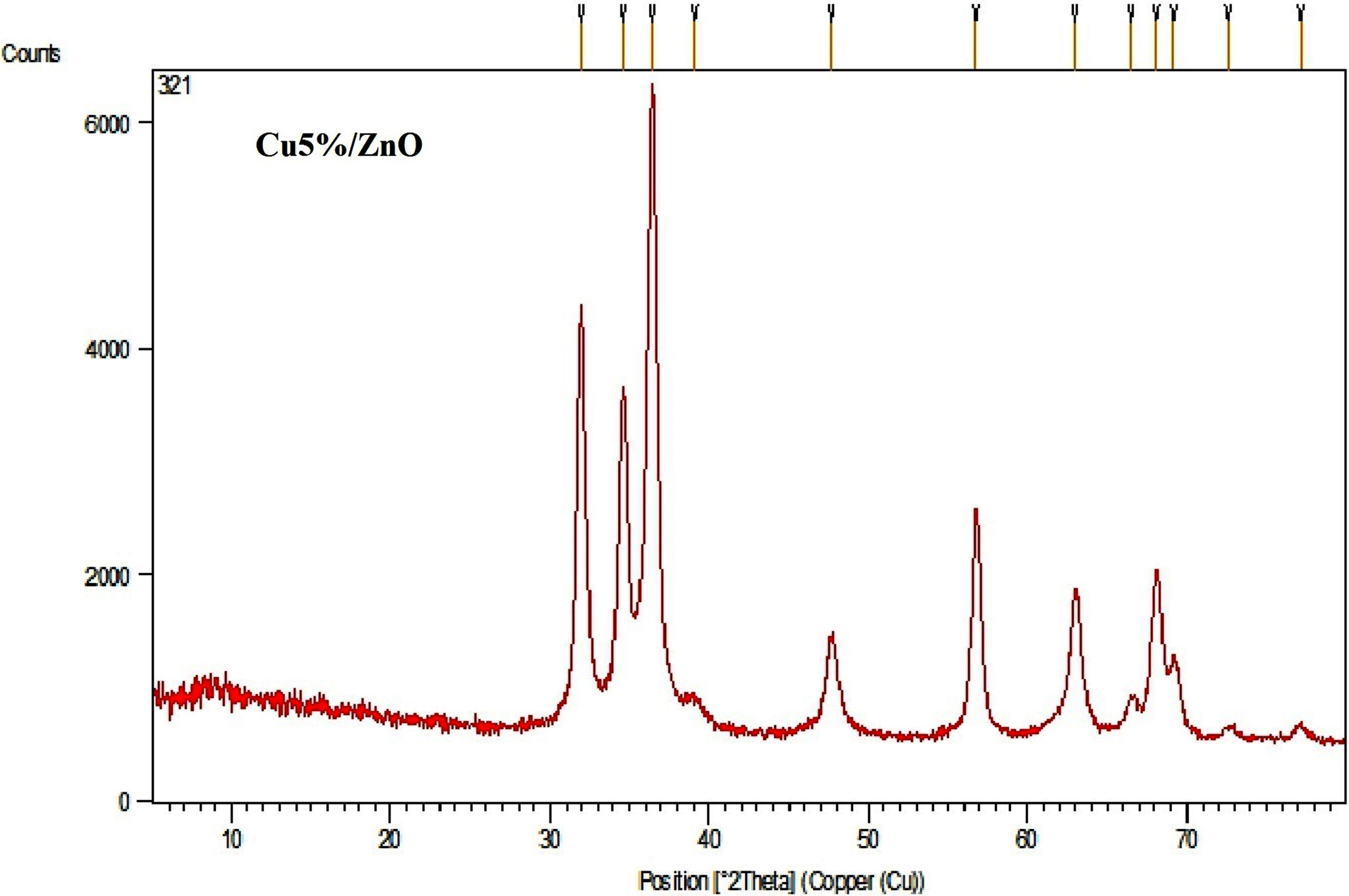
XRD spectrum of Cu5% /ZnO nanoparticles synthesized using sol-gel process.
Calculation of the mean crystallite size of synthesized nanoparticles using Debye–Scherrer equation
Microstress level of nanoparticles synthesized using sol-gel process
The XRD pattern of synthesized Ag5% /Cu5% /ZnO NPs is given in Fig. 2. These NPs were calcined at 400°C for 2 h to form a crystal structure. The 2θ value for the (101) diffraction peak (the main peak of ZnO) in the XRD pattern for Ag5% /Cu5% /ZnO NPs was 36.334°. The ionic radius of Cu2+ (0.69 Å) was smaller and the ionic radius of Ag+ (1.26 Å) was larger than the ionic radius of Zn2+ (0.74 Å). There was a shift in the 2θ value and the broadening of diffraction peaks. The mean crystallite size of Ag5% /Cu5% /ZnO NPs was calculated by the X-ray line broadening the (101) diffraction peak using the Debye-Scherrer equation, the results of which are given in Table 4, and it was equal to 12.03 nm and a decrease was observed in NPs due to co-doping.
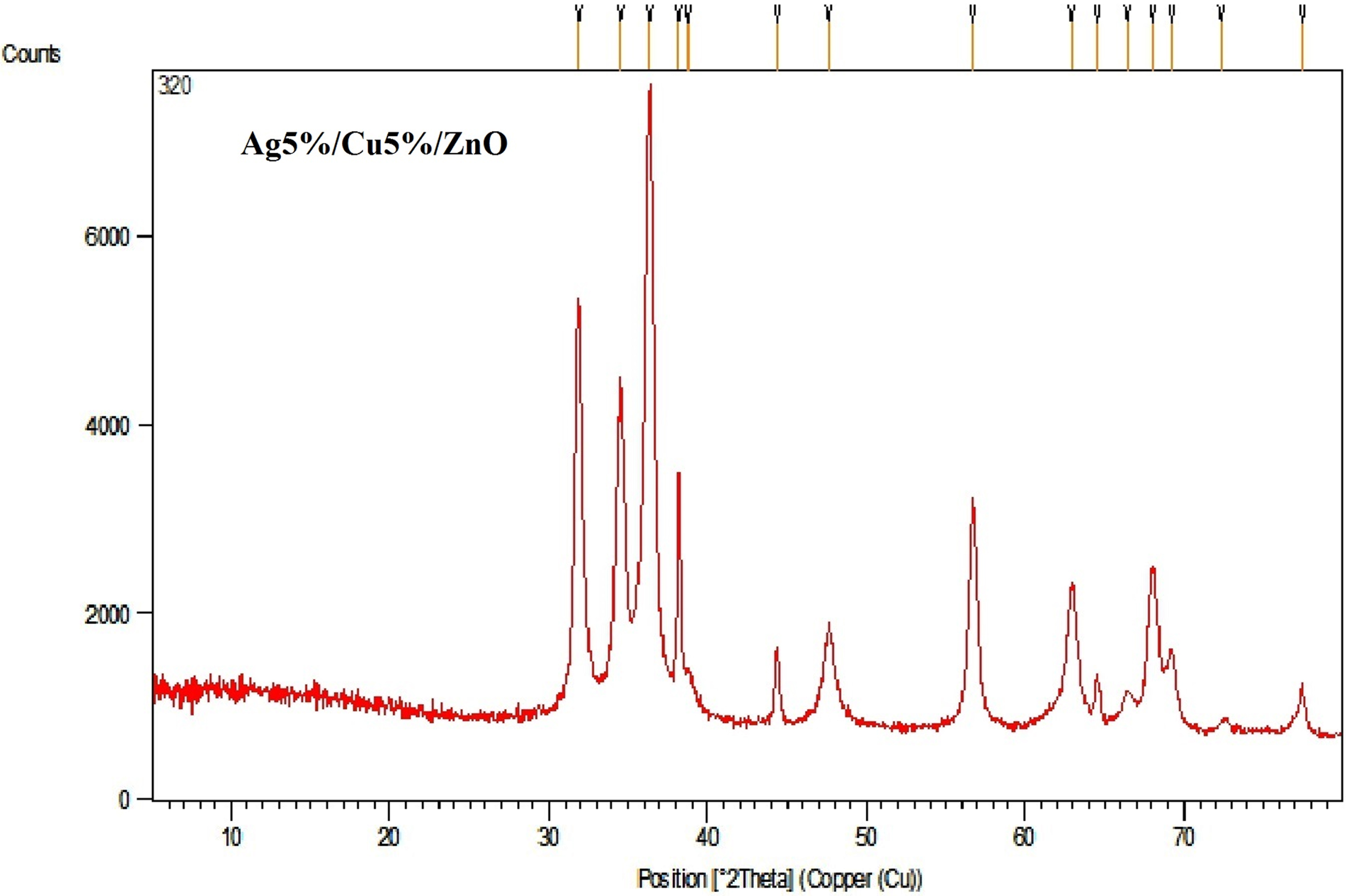
XRD spectrum of Ag5% /Cu5% /ZnO nanoparticles synthesized using sol-gel process.
Calculation of the mean crystallite size of synthesized nanoparticles using Debye–Scherrer equation
The microstress of NPs was calculated using the Equation (4), see Table 5.
Microstress level of nanoparticles synthesized using sol-gel process
Mono-doped NPs (Cu5% /ZnO) synthesized by sol-gel method
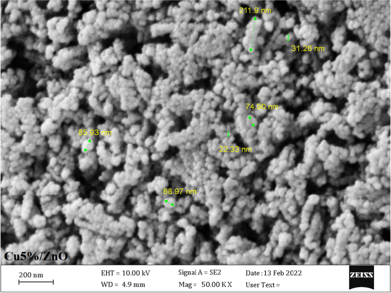
The FE-SEM image for NPs synthesized by sol-gel method is presented in Fig. 3. The size of Cu5% /ZnO NPs ranged from 22.3 nm to 211.9 nm with an average size of 82.54 nm. In the mono-doped sample, a decrease was observed in the nanoparticle size.
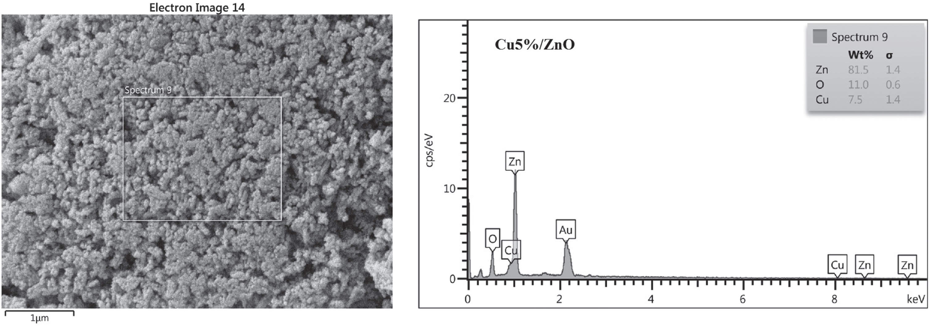
FE-SEM images of Cu5% /ZnO nanoparticles synthesized using sol-gel process.
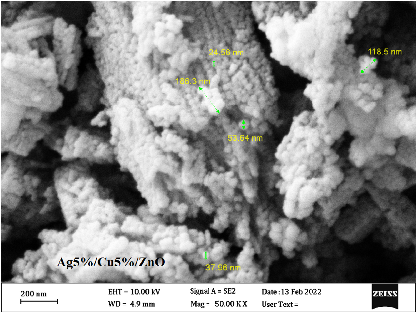
The FE-SEM image for NPs synthesized by sol-gel method is presented in Fig. 5. The size of Ag5% /Cu5% /ZnO NPs ranged from 24.56 nm to 186.3 nm with an average size of 84.19 nm. In the co-doped sample, an insignificant decrease was observed in the nanoparticle size. EDX analysis was used to investigate the chemical composition of NPs. Figure 6 shows the EDX spectrum of NPs synthesized by the sol-gel method.
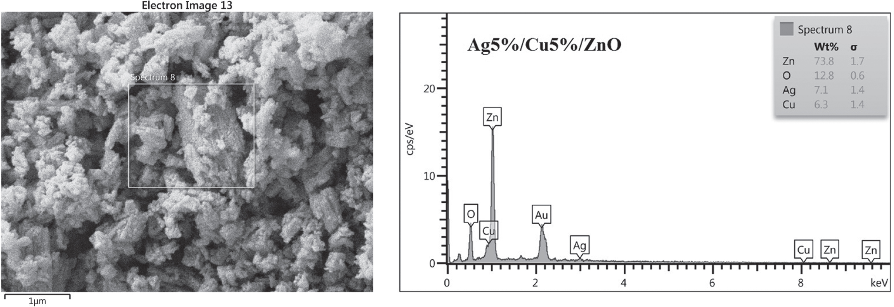
FE-SEM images of Ag5% /Cu5% /ZnO nanoparticles synthesized using sol-gel process.
EDX spectrum of Ag5% /Cu5% /ZnO nanoparticles synthesized using sol-gel process.
Mono-doped NPs (Cu5% /ZnO) synthesized by sol-gel method
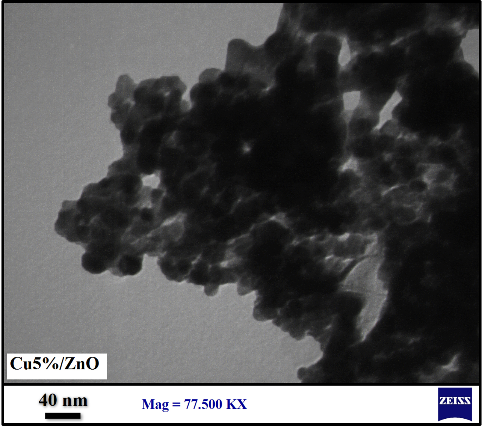
Figure 7 shows the particle shape, size and dispersion of the synthesized NPs determined by TEM images. The size range of synthesized Cu5% /ZnO NPs was 11.67 to 33.33 nm with an average size of 21.67 nm.
TEM image of Cu5% /ZnO nanoparticles synthesized using sol-gel process.
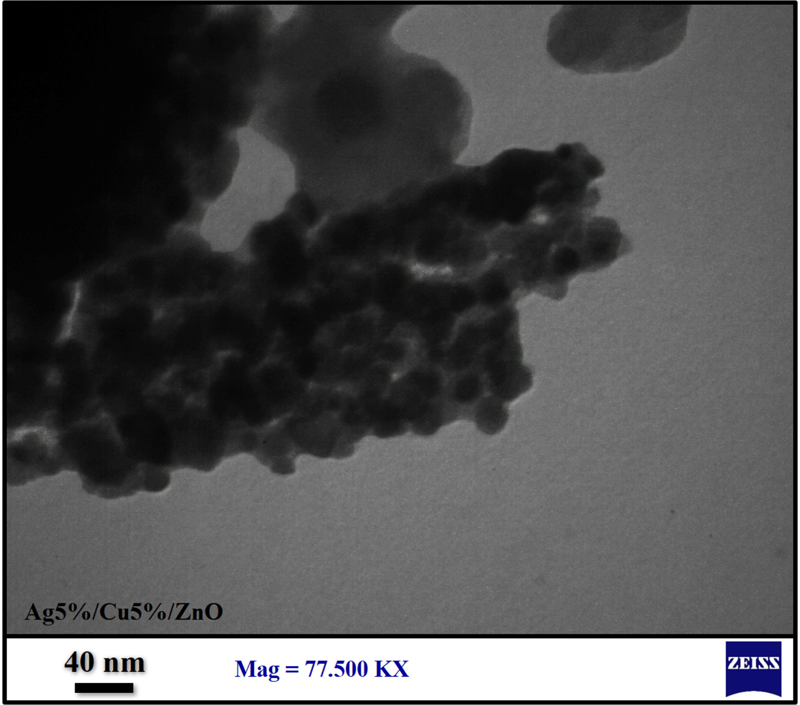
Figure 8 shows the particle shape, size and dispersion of the synthesized NPs determined by TEM images. The size range of synthesized Ag5% /Cu5% /ZnO NPs was 14.55 to 25.45 nm with an average size of 20.0 nm.
TEM image of Ag5% /Cu5% /ZnO nanoparticles synthesized using sol-gel process.
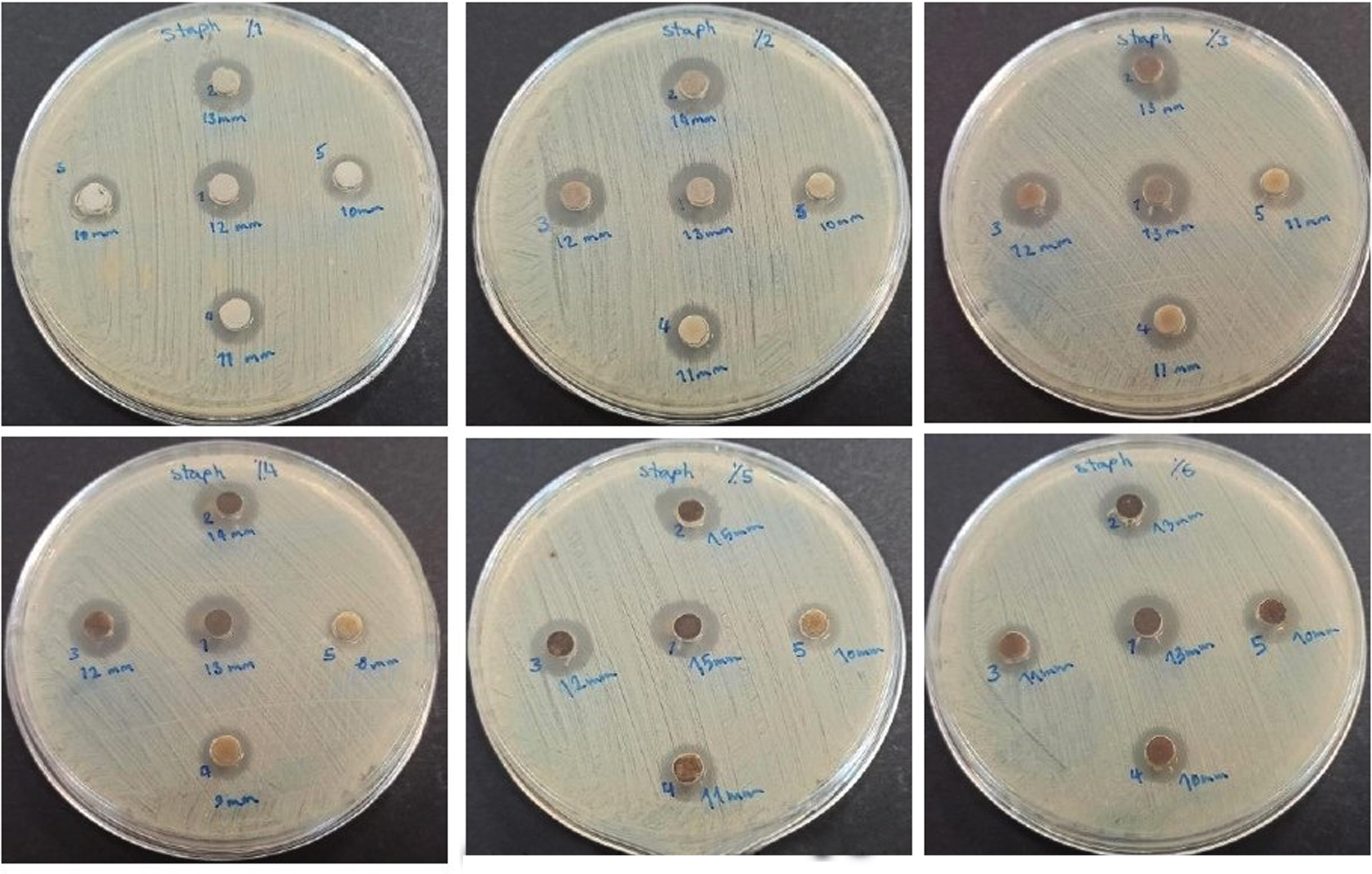
Antibacterial activity of CuX% /ZnO (X = 1–6 wt%) nanoparticles against Staphylococcus aureus within 24 h
The antibacterial activity of synthesized CuX% /ZnO (X = 1–6 wt%) NPs was determined against S. aureus (Fig. 9) within 24 h. The mean diameters of ZOI generated by the agar well diffusion assay at different concentrations of NPs were calculated as shown in Table 6.
Antibacterial susceptibility test results and zone of inhibition generated by CuX% /ZnO (X = 1–6 wt%) NPs in the presence of Staphylococcus aureus within 24 h.
Mean diameter of zone of inhibition (mm) against Staphylococcus aureus at different concentrations of synthesized nanoparticles using agar well diffusion method within 24 h
The antibacterial activity of synthesized CuX% /ZnO (X = 1–6 wt%) NPs was determined against S. aureus (Fig. 10) within 48 h. The mean diameters of ZOI generated by the agar well diffusion assay at different concentrations of NPs were calculated as shown in Table 7.
Antibacterial susceptibility test results and zone of inhibition generated by CuX% /ZnO (X = 1–6 wt%) NPs in the presence of Staphylococcus aureus within 48 h.
Mean diameter of zone of inhibition (mm) against Staphylococcus aureus at different concentrations of synthesized nanoparticles using agar well diffusion method within 48 h
The antibacterial activity of synthesized CuX% /ZnO (X = 1–6 wt%) NPs was determined against E. coli (Fig. 11) within 24 h. The mean diameters of ZOI generated by the agar well diffusion assay at different concentrations of NPs were calculated as shown in Table 8.
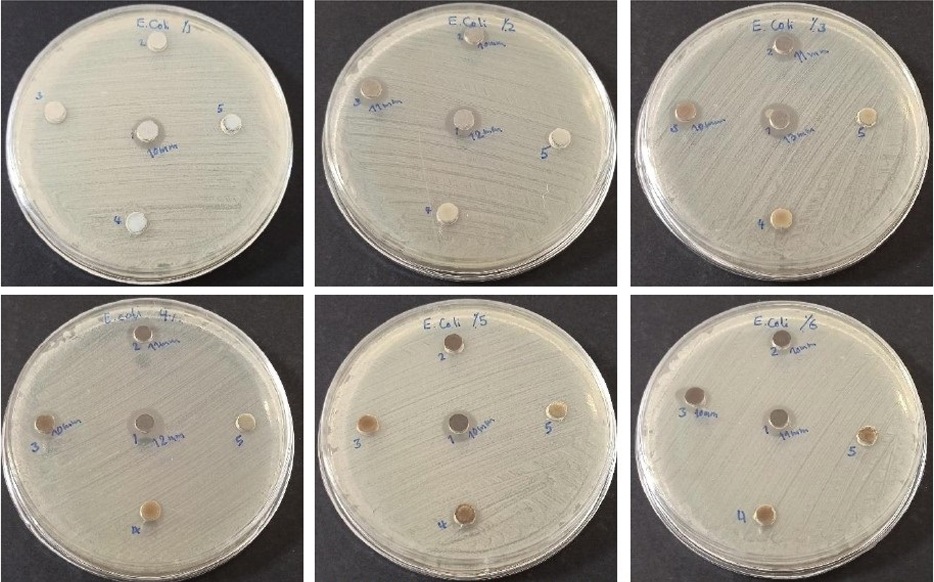
Antibacterial susceptibility test results and zone of inhibition generated by CuX% /ZnO (X = 1–6 wt%) NPs in the presence of Escherichia coli within 24 h.
Mean diameter of zone of inhibition (mm) against Escherichia coli at different concentrations of synthesized nanoparticles using agar well diffusion method within 24 h
The antibacterial activity of synthesized CuX% /ZnO (X = 1–6 wt%) NPs was determined against E. coli (Fig. 12) within 48 h. The mean diameters of ZOI generated by the agar well diffusion assay at different concentrations of NPs were calculated as shown in Table 9.
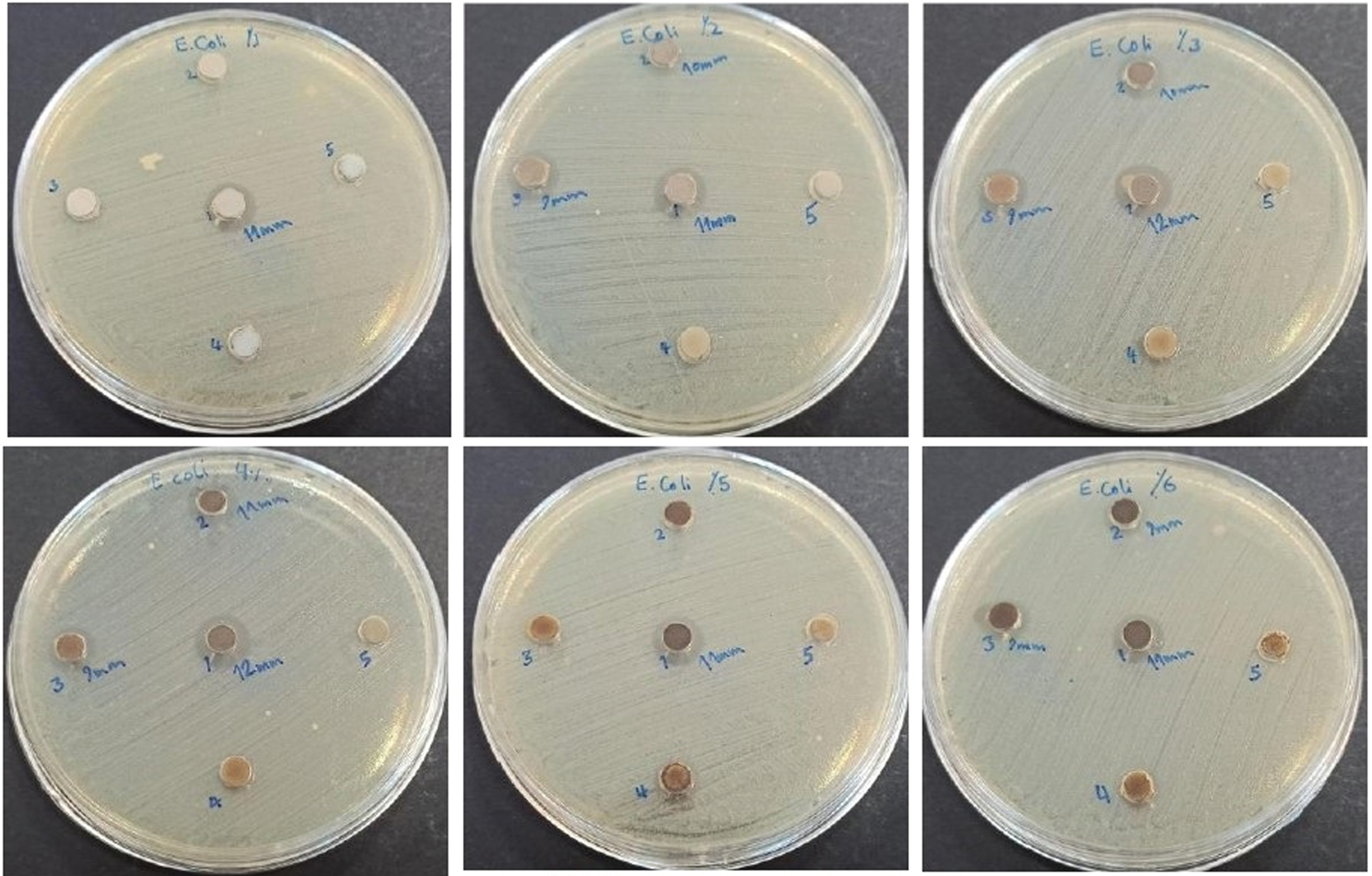
Antibacterial susceptibility test results and zone of inhibition generated by CuX% /ZnO (X = 1–6 wt%) NPs in the presence of Escherichia coli within 48 h.
Mean diameter of zone of inhibition (mm) against Escherichia coli at different concentrations of synthesized nanoparticles using agar well diffusion method within 48 h
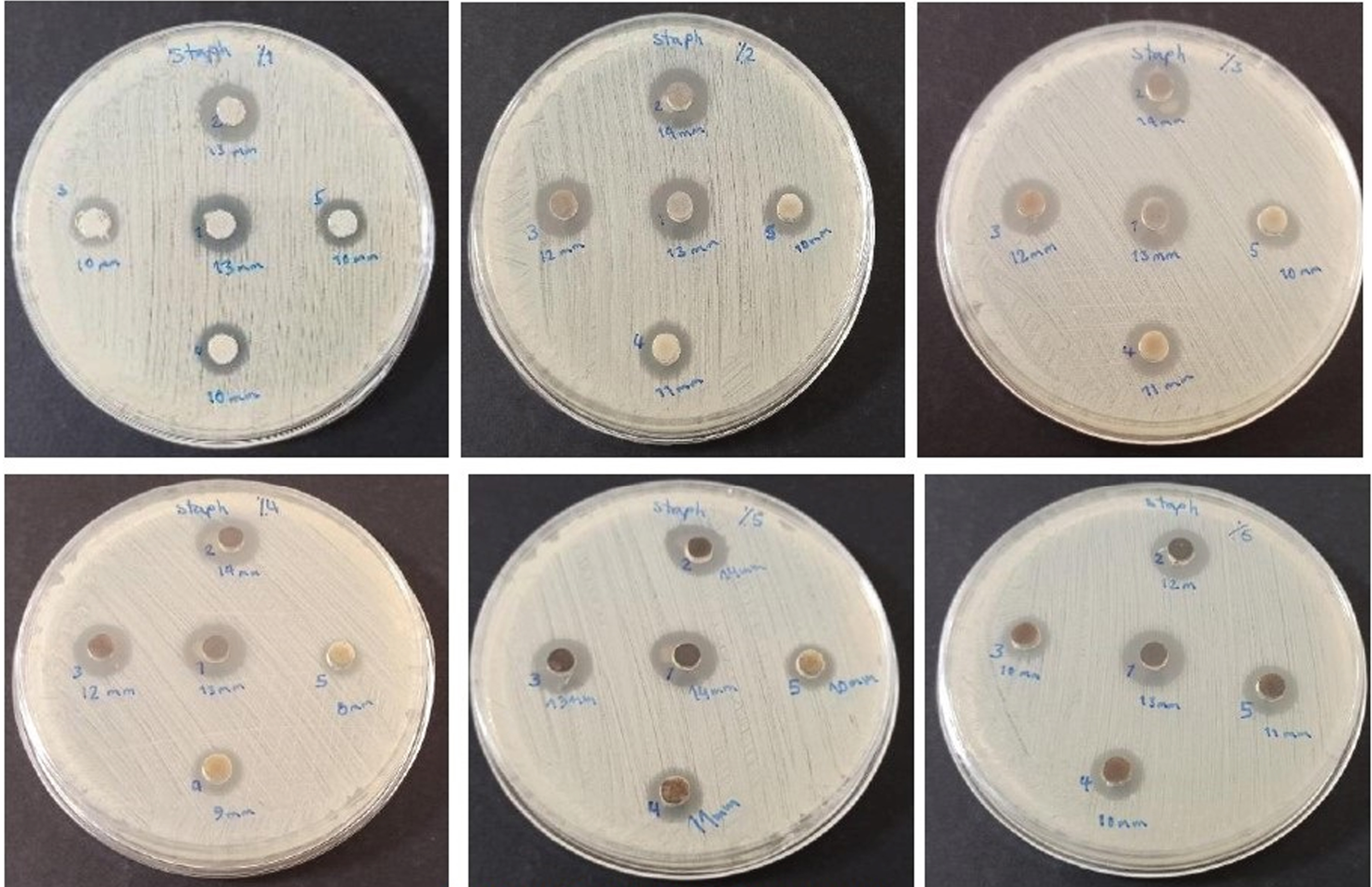
Antibacterial activity of co-doped nanoparticles against Staphylococcus aureus within 24 h
The antibacterial activity of synthesized co-doped NPs was determined against S. aureus (Fig. 13) within 24 h. The mean diameters of ZOI generated by the agar well diffusion assay at different concentrations of NPs were calculated as shown in Table 10.
Antibacterial susceptibility test results and zone of inhibition generated by co-doped NPs in the presence of Staphylococcus aureus within 24 h; (a) Ag5% /Cu3% /ZnO, (b) Ag5% /Cu4% /ZnO and (c) Ag5% /Cu5% /ZnO
Mean diameter of zone of inhibition (mm) against Staphylococcus aureus at different concentrations of synthesized co-doped nanoparticles using agar well diffusion method within 24 h
The antibacterial activity of synthesized co-doped NPs was determined against S. aureus within 48 h by the agar well diffusion assay and no changes were observed in the mean diameters of ZOI.
Antibacterial activity of co-doped nanoparticles against Escherichia coli within 24 h
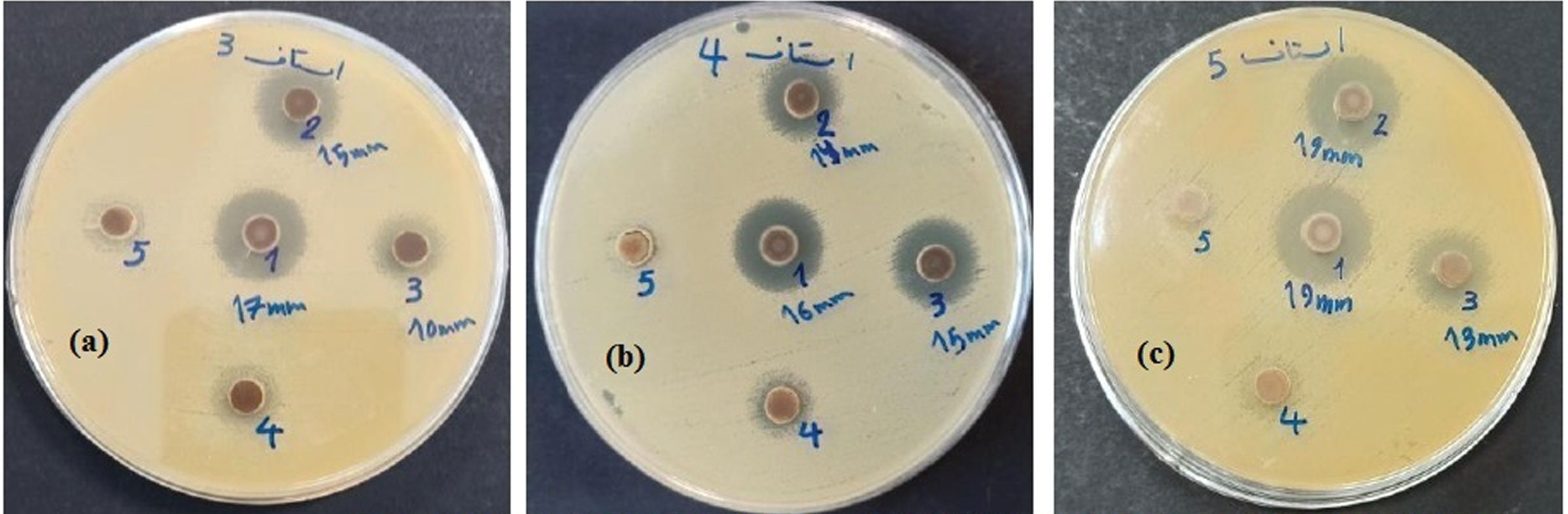
The antibacterial activity of synthesized co-doped NPs was determined against E. coli (Fig. 14) within 24 h. The mean diameters of ZOI generated by the agar well diffusion assay at different concentrations of NPs were calculated as shown in Table 11.
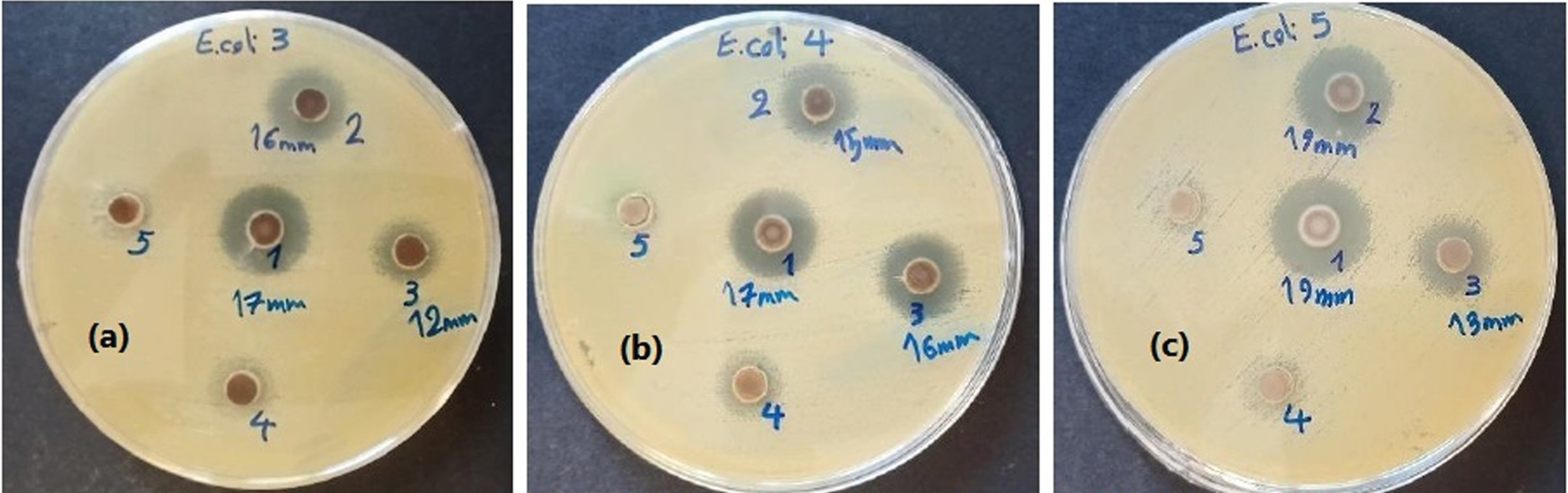
Antibacterial susceptibility test results and zone of inhibition generated by co-doped NPs in the presence of Escherichia coli within 24 h; (a) Ag5% /Cu3% /ZnO, (b) Ag5% /Cu4% /ZnO and (c) Ag5% /Cu5% /ZnO.
Mean diameter of zone of inhibition (mm) against Escherichia coli at different concentrations of synthesized co-doped nanoparticles using agar well diffusion method within 24 h
The antibacterial activity of synthesized co-doped NPs was determined against E. coli within 48 h by the agar well diffusion assay and no changes were observed in the mean diameters of ZOI.
Evaluating the XRD findings
XRD spectrum of Cu5% /ZnO NPs synthesized by sol-gel method
The XRD spectrum for Cu5% /ZnO NPs (Fig. 3) with wurtzite hexagonal crystal structure follows well the standard ZnO pattern with the ID JCPDS36-1451 [30]. Based on the XRD pattern for ZnO NPs and Cu5% /ZnO NPs, no impurity peaks related to Cu or CuO were observed in the spectrum. The absence of Cu2+- specific diffraction in the XRD pattern can be due to the small amount of Cu2+-doped element in the ZnO network or proper diffusion in the ZnO network. The 2θ values for the (101) diffraction peak (the main peak of ZnO) in the XRD pattern of ZnO and Cu5% /ZnO NPs were 24.027 and 36.386 degrees, respectively, which confirms the shift in the 2θ value and the broadening of the diffraction peaks in Cu5% /ZnO NPs, and means that the doped ion has diffused well into the ZnO network. The ionic radius of Cu2+ (0.69 Å) was smaller than that of Zn2+ (0.74 Å), and the small ionic radius was a proof of the proper diffusion of dopant ions in the ZnO network. In these results, a decrease in ZnO crystallite size with doping was observed (shifting crystallite size from 32.56 nm to 12.58 nm in Cu5% /ZnO NPs). According to the calculations of the microstress level (Table 3), this value has reached 2.73 in Cu5% /ZnO from 1.1 in ZnO. The increase in microstress in the doped NPs caused a change in the broadening of the diffraction peak, which led to a decrease in the particle size and a small shift in the XRD peak.
XRD spectrum of Ag5% Cu5% /ZnO NPs synthesized by sol-gel method
The XRD spectrum for Ag5% Cu5% /ZnO NPs (Fig. 2) with wurtzite hexagonal crystal structure follows well the standard ZnO pattern with the ID JCPDS36-1451 and shows successful synthesis of NPs. Based on the XRD pattern for ZnO NPs and Ag5% Cu5% /ZnO NPs, no impurity peaks related to Cu, Ag, Ag2O or CuO were observed in the spectrum. The absence of impurity peak in the XRD pattern can be due to the small amount of co-doped elements in the ZnO network or the proper diffusion in the ZnO network. The 2θ values for the (101) diffraction peak (the main peak of ZnO) in the XRD pattern of ZnO and Ag5% Cu5% /ZnO were 24.027 and 36.334 degrees, respectively, which confirms the shift in the 2θ value and the broadening of the diffraction peaks in Ag5% Cu5% /ZnO NPs, and means that the co-doped ion has diffused well into the ZnO network. The ionic radius of Cu2+ (0.69 Å) was smaller and the ionic radius of Ag+ (1.26 Å) was larger than that of Zn2+ (0.74 Å), which means that the two silver and copper ions, with respectively larger and smaller radii than Zn2+ ion, because of proper diffusion in the ZnO network, caused a reduction in the crystallite size. The results of these calculations clearly show a decrease in crystallite size of ZnO due to co-doping (shifting crystallite size from 32.56 nm to 12.03 nm in co-doped NPs). According to the calculations of the microstress level (Table 5), this value has reached 2.85 in Ag5% Cu5% /ZnO from 1.1 in ZnO. The increase in microstress in co-doped NPs caused a change in the broadening of the diffraction peak, which led to a decrease in the particle size and a small shift in the XRD peak.
FE-SEM and EDX findings of NPs synthesized by sol-gel method
Evaluating FE-SEM and EDX findings of Cu5% /ZnO NPs synthesized by sol-gel method
Figure 3 shows FE-SEM images for Cu5% /ZnO NPs synthesized by sol-gel method. The NPs had a unified spherical morphology, and the images showed that Cu2+ ion doping had no effect on nanoparticle morphology, and there was a significant decrease in the particle size, with a size of 22.3 nm in NPs. The size of Cu5% /ZnO NPs ranged from 22.3 nm to 211.9 nm with an average size of 82.54 nm. Figure 4 shows the EDX spectrum of NPs synthesized by sol-gel method, so that NPs contain only three elements of Zn, Cu and O, confirming the purity of Cu5% /ZnO NPs.
EDX spectrum of Cu5% /ZnO nanoparticles synthesized using sol-gel process.
Figure 5 shows FE-SEM images for Ag5% /Cu5% /ZnO NPs synthesized by sol-gel method. The co-doped NPs had a unified spherical morphology, and the images showed that co-doping of Cu2+ and Ag+ ions had no effect on nanoparticle morphology, and there was a significant decrease in the particle size, with a size of 24.56 nm in NPs. The size of Ag5% /Cu5% /ZnO NPs ranged from 24.56 nm to 186.3 nm with an average size of 84.19 nm. Figure 6 shows the EDX spectrum of NPs synthesized by sol-gel method, so that NPs contain only four elements of Zn, Cu, Ag and O, confirming the purity of Ag5% /Cu5% /ZnO NPs.
TEM images of NPs synthesized by sol-gel method
Evaluating TEM images of Cu5% /ZnO NPs synthesized by sol-gel method
The shape and size of synthesized NPs provided by TEM images are shown in Fig. 7. The image of NPs showed that the synthesized NPs had a regular dispersion and spherical morphology, and the size of synthesized Cu5% /ZnO NPs was in the range of 11.67 to 33.33 nm with an average size of 21.67 nm. The results indicated that the nanoparticle size was reduced as a result of Cu2+ doping to the ZnO network.
Evaluating TEM images of Cu5% /ZnO NPs synthesized by sol-gel method
The shape and size of synthesized Ag5% /Cu5% /ZnO NPs provided by TEM images are shown in Fig. 8. The image of NPs showed that the synthesized NPs had a regular dispersion and spherical morphology, and the size of synthesized Ag5% /Cu5% /ZnO NPs was in the range of 14.55 to 25.45 nm with an average size of 20.00 nm. The results indicated that the nanoparticle size was reduced as a result of Cu2+ and Ag+ co-doping to the ZnO network.
Evaluating the antibacterial activity of NPs synthesized by sol-gel method on tested bacterial strains
The antibacterial activity of CuX% /ZnO (X = 1–6wt%) NPs against S. aureus within 24 h and 48 h (Figs. 9 10, Tables 6 7) based on ZOI diameter showed that the largest ZOI diameter (14 mm) related to Cu5% /ZnO NPs in 24 h of incubation at a nanoparticle concentration of 0.1 g/mL. By reducing the concentration of NPs used to 0.05, 0.025 and 0.0125 g/mL, the largest ZOI diameter corresponding to Cu5% /ZnO NPs was 14, 13 and 11 mm, respectively. It should be noted that the ZOI diameter of pure and doped ZnO NPs at the concentration of 0.0625 g/mL was the same. The results showed that Cu5% /ZnO NPs had the highest bacterial inhibitory activity among CuX% /ZnO (X = 1–6wt%) NPs against S. aureus during 24 h of incubation. During 48 h of incubation, the largest ZOI diameter (15 mm) was related to Cu5% /ZnO NPs at a concentration of 0.1 g/mL. By reducing the concentration of NPs to 0.05 g/mL, the largest ZOI diameter (15 mm) was related to Cu5% /ZnO NPs, and the ZOI diameter of NPs doped with 2–5wt% was 12 mm at a concentration of 0.025 g/mL. At concentrations of 0.0125 and 0.00625 g/mL, ZnO NPs had the highest antibacterial activity among NPs against S. aureus.
In investigating the antibacterial activity of CuX% /ZnO (X = 1–6wt%) NPs against E. coli (Figs. 11 12; Tables 8 9), the data on the average diameter of ZOI showed that the largest ZOI diameter (13 mm) belonged to Cu3% /ZnO NPs at a concentration of 0.1 g/mL within 24 h of incubation. By reducing the concentration of NPs to 0.05, 0.025, 0.0125 and 0.00625 g/mL, the maximum ZOI diameter corresponding to ZnO NPs was 13, 12, 12 and 11 mm, respectively and the rest of the NPs were less effective and the doped NPs at low concentrations of 0.0125 and 0.00625 g/mL had no effect on inhibiting bacterial growth. During 48 h of incubation at a concentration of 0.1 g/mL of NPs, the largest ZOI diameter (13 mm) corresponding to ZnO NPs and NPs doped with 2–5wt% was 12 mm. By reducing the concentration of NPs to 0.05 g/mL, the largest ZOI diameter (13 mm) was related to ZnO NPs and the rest of the doped NPs had no effect on inhibiting bacterial growth. At the ZnO NP concentrations of 0.025, 0.0125, and 0.00625 g/mL, the ZOI diameter was 12, 12, and 11 mm, respectively and the rest of the doped NPs had no effect such that E. coli bacteria were resistant.
The larger size of the ZOI diameter for S. aureus compared to E. coli was due to the resistance of E. coli to these NPs; at lower concentrations, E. coli was completely resistant to these NPs, especially the doped ones; by Cu doping into ZnO, E. coli was practically and completely resistant and the doped NPs had no effect on the bacteria. For S. aureus, increasing the incubation time had a positive effect on increasing the ZOI diameter, especially with Cu5% /ZnO NPs, and the ZOI diameter increased from 14 to 15 mm at a concentration of 0.1 g/mL. By decreasing the nanoparticle concentration, an increase in the diameter of the ZOI was observed for S. aureus and prolonged incubation showed an appreciable effect. For E. coli, 24 h and 48 h of incubation periods had no effect and bacterial resistance to NPs was observed with nanoparticle doping. These results showed the resistance of E. coli to NPs compared to S. aureus, especially at lower concentrations and also to doped NPs. The antibacterial activity of co-doped NPs (including: Ag5% /Cu5% /ZnO, Ag5% /Cu4% /ZnO and Ag5% /Cu3% /ZnO) against S. aureus (Fig. 13) was evaluated at 24 and 48 h incubation periods. According to Table 10, the data on the average diameter of the ZOI for S. aureus showed that the largest ZOI diameter (19 mm) during 24 h of incubation at the nanoparticle concentration of 0.1 g/mL was related to Ag5% /Cu5% /ZnO NPs compared to ZnO with the ZOI diameter of 13 mm. By reducing the nanoparticle concentration to 0.05 and 0.025 g/mL, the maximum diameter of ZOI corresponding to Ag5% /Cu5% /ZnO and Ag5% /Cu4% /ZnO NPs was 19 and 15 mm, and this value for ZnO was 13 and 12 mm, respectively. At the nanoparticle concentration of 0.0125 and 0.0625 g/mL, bacteria were resistant to co-doped NPs and the ZOI diameter for ZnO was 12 and 11 mm, respectively. The results showed that Ag5% /Cu5% /ZnO NPs had the highest bacterial inhibitory activity among co-doped NPs against S. aureus at the concentrations of 0.1 and 0.05 g/mL. During 48 h of incubation, there was no change in the ZOI of bacteria, and increasing the incubation time had no effect on the removal of bacteria.
The antibacterial activity of synthesized co-doped NPs (including: Ag5% /Cu5% /ZnO, Ag5% /Cu4% /ZnO and Ag5% /Cu3% /ZnO) was evaluated against E. coli (Fig. 14) during the incubation period of 24 and 48 h. Based on Table 11, the data on the mean diameter of the ZOI for E. coli showed that the largest ZOI diameter (19 mm) during 24 h of incubation at the nanoparticle concentration of 0.1 g/mL was related to Ag5% /Cu5% /ZnO NPs versus ZnO with the ZOI diameter of 13 mm. By reducing the nanoparticle concentration to 0.05 g/mL, the largest ZOI diameter (19 mm) was related to Ag5% /Cu5% /ZnO NPs, and this value was 13 mm for ZnO. At the concentration of 0.025 g/mL, the largest ZOI diameter (16 mm) was related to Ag5% /Cu4% /ZnO NPs, and this value was 12 mm for ZnO. At the concentrations of 0.0125 and 0.00625 g/mL, bacteria were resistant to co-doped NPs, and the ZOI diameter for ZnO was 12 mm and 11 mm, respectively. The results showed that Ag5% /Cu5% /ZnO NPs had the highest antibacterial activity among co-doped NPs against E. coli at the concentrations of 0.1 and 0.05 g/mL. During 48 h of incubation, there was no change in the ZOI of bacteria.
The NPs synthesized and studied based on existing mechanisms can release Zn2+, Cu2+ and Ag+ ions, which can interact with the bacterial cell wall and deactivate the compounds in the bacterial membrane, which leads to membrane deactivation and prevents the penetration of nutrients into the cell and this eventually leads to the death of the bacterial cell [31]. The studied bacteria, E. coli and S. aureus, were destroyed due to the contact of the NPs suspension and binding to the surface of the bacteria and applying mechanisms such as inhibiting DNA and RNA, inhibiting protein synthesis and inhibiting other cell enzymes. The findings showed that Ag+ ions are released from NPs and destroy bacteria by interacting with the membrane and affecting its proteins and DNA damage [16]. These ions can destroy bacteria through other mechanisms, such as collision with the negatively charged cell wall and changes in its permeability and changes in cell enzymes. The activity of NPs in general, regardless of their type and structure, based on previous studies [32, 33], is attacking and creating holes in the cell wall. In general, the existing mechanisms can be named as follows: (a) Oxidative stress induction through the production of reactive oxygen species (ROS), leading to the formation of various radicals; (b) Release of Zn2+, Ag+ and Cu2+ ions from NPs and binding to the bacterial membrane, leading to the accumulation of NPs in the bacterial membrane and the loss of cell integrity by destroying the bacterial membrane; and (c) Inhibition of enzyme activity and DNA synthesis, leading to cell destruction.
One of the early mechanisms of photocatalyst is the production of H2O2. A suspension of pure, doped and co-doped ZnO with metal ions produces an electron-hole pair system due to the collision of visible light, which converts water molecules into OH– and H+. The dissolved oxygen molecule can collide with electrons and H+ to form H2O2 [10]. Cu/ZnO and Ag/Cu/ZnO NPs can result in the following reactions:
Visible radiation on the surface of NPs leads to the formation of electron-hole pairs (Cu/ZnO, Ag/Cu/ZnO + hv = Cu/Ag/ZnO (e– + h+)).
The high oxidation potential of the created hole can form H+ and OH° through water degradation (h+ + H2O = OH° + H+).
Dissolved oxygen by electron absorption of NPs causes the generation of superoxide anion radical (e– + O2 = O2°–).
With the reaction of HO2°, H+ and e– leads to the production of hydrogen peroxide (O2°– + H+ = HO2°) and (HO2° + H+ + e– = H2O2).
The produced radicals can attack the organic compounds on the surface of the bacterial wall and kill the bacteria [10, 14].
Doping on the surface of metal oxides increases the photocatalytic activity, and preventing the rapid recombination of electrons and holes leads to the production of better antimicrobial properties [13].
Conclusion
The current research investigated the antibacterial effects of ZnO NPs doped with Cu2+ ions and co-doped with Ag+ and Cu2+ ions in different weight percentages. NPs were synthesized by sol-gel method and calcined at 400°C. NPs were characterized by XRD, FE-SEM, EDX and TEM techniques. The crystal structure determined by XRD showed that the synthesized samples had wurtzite hexagonal crystal structure. Particle size was calculated by Debye-Scherrer equation and a decrease was observed in the size of doped and co-doped particles. The increase in the microstress level due to the broadening of the diffraction peak highlighted the decrease in the nanoparticle size and the slight shift in the XRD peak. FE-SEM and TEM images confirmed the size reduction of co-doped NPs. The shape of synthesized NPs was spherical with regular dispersion. The EDX spectrum showed that the NPs were composed only of their constituent atoms and were pure. Evaluating the antibacterial effects of synthesized NPs on Escherichia coli (gram-negative) and Staphylococcus aureus (gram-positive) bacterial strains showed that co-doped Ag5% /Cu5% /ZnO NPs had the most destructive effect on bacteria, but with a greater effect on S. aureus. The lowest effective nanoparticle concentrations were 0.1 and 0.05 g/ml. The main mechanism of elimination can be attributed to the size reduction of the co-doped NPs. The resistance of E. coli to NPs, vs. S. aureus, was due to the fact that ions released from NPs, such as Zn2+, must accumulate on the surface of the bacteria. But the results showed the opposite of this action and the reason for the removal of the bacteria was due to the production of ROS, so that the radicals formed were responsible for the destruction and removal of the bacterial cell wall.
Footnotes
Acknowledgments
This research has been extracted from the PhD dissertation in Environmental Engineering written by Mrs. Shadi Ashraf Nohegar at the Islamic Azad University of Tabriz. Therefore, the authors appreciate the president, educational and research deputies of Tabriz Islamic Azad University for their cooperation in facilitating the implementation of this project.
Ethical issues
None.
Competing interests
Authors have no conflict of interests.
Authors’ contributions
All authors contributed equally to the writing, review and final approval of the article.