






Abstract
BACKGROUND:
Fetal pleural effusions are a rare fetal anomaly that may result from congenital chylothorax. Severe cases lead to chest compression with resulting pulmonary hypoplasia and possible neonatal demise. Fetal thoracoamiontic shunt (TAS) placement may decrease the amount of pleural effusion and improve lung expansion.
CASE:
A 30-year-old primigravida at 29 2/7 weeks’ gestation presented with fetal bilateral pleural effusions with no identifiable genetic or structural abnormalities. TAS placement accomplished decompression of the left fetal chest. The neonate was delivered at 33 3/7 weeks and required minimal respiratory support with no apparent long term complications at discharge.
CONCLUSION:
This case demonstrated that fetal intervention with TAS placement can improve neonatal outcomes. Referral to an MFM specialist capable of TAS should be considered for isolated fetal bilateral pleural effusion.
Introduction
Fetal pleural effusion, which may be unilateral or bilateral, results from a non-specific accumulation of fluid in the pleural space of the fetal chest [1]. Isolated pleural effusions are a rare fetal anomaly occurring in approximately 1 in 10,000 pregnancies [2]. Multiple pathologic processes may result in fetal pleural effusion including chromosomal abnormalities, fetal structural defects, immune or nonimmune hydrops, and chylous leak [3].
Congenital chylothorax results from leakage of lymphatic fluid into the pleural space, either by direct leakage from the thoracic duct, overproduction, or impaired drainage of lymph [4]. The condition has a variable outcome ranging from spontaneous resolution to severe chest compression with progression to hydrops. In severe cases, pulmonary hypoplasia may lead to neonatal mortality [4–8].
Thoracoamniotic shunting, in the setting of congenital chylothorax, allows for continuous decompression of the fetal chest and resulting lung expansion, thereby decreasing the risk for pulmonary hypoplasia and neonatal demise [3]. The following case presents a diagnosis of isolated bilateral pleural effusions in the third trimester fetus that was successfully treated in utero with thoracoamniotic shunt (TAS) placement.
Case
The patient was a 30-year-old G1 P0 at 29 2/7 weeks’ gestation who presented for evaluation and possible TAS placement for bilateral fetal pleural effusions that were initially identified by outside ultrasound at time of growth scan for gestational diabetes. Detailed anatomy ultrasound performed by maternal fetal medicine at 29 3/7 weeks revealed large bilateral pleural effusions with mediastinal shift, polyhydramnios, normal fetal growth, otherwise normal fetal anatomy, and 6/8 biophysical profile with absent fetal breathing movements (Figs. 1 and 2).
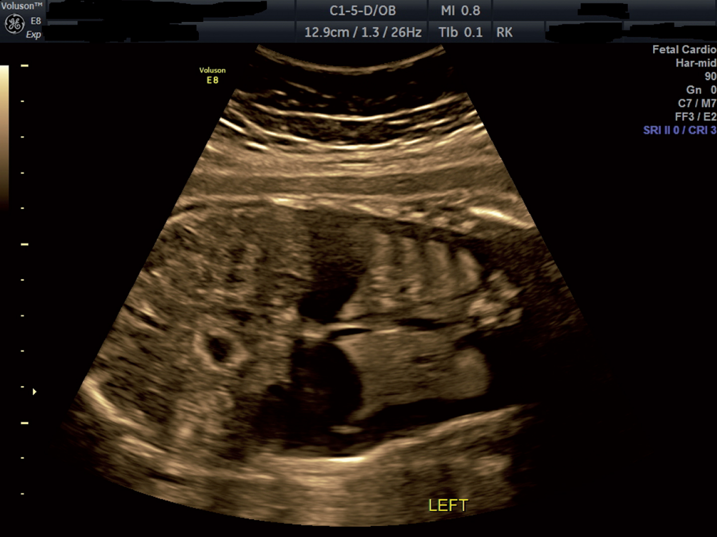
Coronal image of the fetal chest demonstrating bilateral pleural effusions.
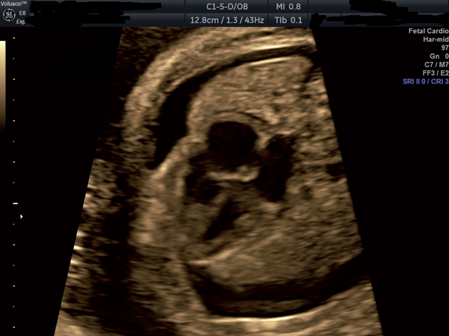
Axial image of the fetal chest demonstrating bilateral pleural effusions.
The pregnancy had otherwise been complicated by fetal echogenic bowel, diet-controlled gestational diabetes and a short cervix for which the patient was using vaginal progesterone nightly. Prenatal labs revealed maternal Rh negative status with negative antibody screen. The patient was also found to be a cystic fibrosis carrier but the father of the baby had negative cystic fibrosis screening. Previous evaluation for fetal echogenic bowel included genetic screening with non-invasive prenatal testing that revealed a female fetus with low risk for trisomies 21, 18, and 13. Maternal serum CMV IgM/IgG and toxoplasmosis IgM/IgG titers were also negative.
After extensive counseling regarding risks, benefits, and alternatives, the patient underwent TAS placement in the right fetal chest at 29 5/7 weeks’ gestation. Of note, the patient received a course of antenatal steroids for fetal benefit at 29 3/7 and 29 4/7 weeks and twelve hours of magnesium sulfate for fetal neuroprotection prior to the procedure.
For the TAS placement, the patient was placed under general anesthesia after confirmation of fetal position with ultrasound. The procedure was performed in a sterile fashion under ultrasound guidance. A Harrison fetal catheter trocar was inserted into the maternal abdomen. The trocar was then advanced through the uterus, into the amniotic cavity, and then into the right fetal chest. The Harrison fetal bladder stent was inserted through the catheter trocar and deployed in the right fetal chest without difficulty. The trocar sheath was then removed under direct visualization. Normal fetal heart rate was confirmed throughout the entirety of the procedure. Amniocentesis was then performed to complete genetic evaluation and obtain fetal infectious studies. After completion of the procedure, ultrasound confirmed inflation of the right fetal lung with minimal effusion remaining. Complete details regarding the Harrison fetal bladder stent and its instructions for assembly and use can be found within a Cook Medical Harrison Fetal Bladder Stent Set.
The patient was seen for follow up ultrasound at 31 3/7 weeks, at which time the previous shunt was found to be dislodged from the fetal chest and floating in the amniotic sac. Bilateral pleural effusions were once again visualized with polyhydramnios and fetal skin edema present. Due to the worsening fetal hydrops on ultrasound, the decision was made to perform a repeat TAS procedure. At 31 4/7 weeks, TAS was placed in the left fetal chest without difficulty and the left fetal lung was noted to fill the fetal chest after completion of the procedure (Figs. 3 and 4).
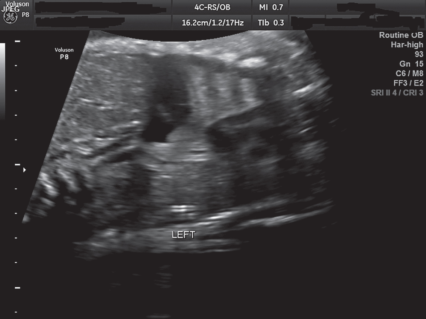
Coronal image of the fetal chest showing left thoracoamniotic shunt in place with improvement of the left pleural effusion.
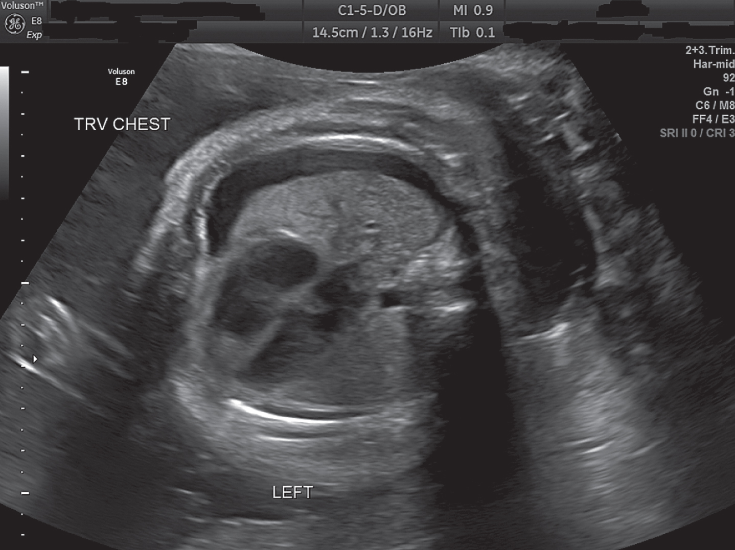
Axial image of the fetal chest status post thoracoamniotic shunt placement.
The patient was monitored closely for the remaining pregnancy with twice weekly antenatal testing and TAS evaluation. The left TAS remained in place for the remainder of the pregnancy. The left pleural effusion remained minimal. Fetal skin edema resolved and polyhydramnios improved but remained present. Amniocentesis revealed normal 46XX karyotype and negative infectious studies including HSV 1 and 2, CMV, Parvovirus, and Toxoplasmosis. Amniocentesis microarray results were not available until after delivery but were significant for 9q34.3 microdeletion with unknown clinicalsignificance.
The decision was made to move forward with delivery via primary cesarean section at 33 3/7 weeks due to persistence of the right fetal pleural effusion and polyhydramnios. A second course of antenatal steroids was administered for fetal benefit prior to delivery at 33 1/7 and 33 2/7 weeks’ gestation. Delivery was accomplished at a tertiary care facility and cesarean section was performed by a maternal fetal medicine specialist. The patient gave birth to a 2480g female infant with Apgars of 8 and 9 at one and five minutes, respectively.
Initial neonatal assessment noted the left TAS which had been placed in utero. This was removed shortly after birth. Chest x-ray on day of life 1 was significant for right pleural effusion and left pneumothorax. The neonate was admitted to the NICU, received curosurf and remained stable on CPAP. On day of life 2, repeat chest x-ray revealed almost complete re-expansion of the left lung, no large pleural effusions noted, and minimal residual respiratory distress syndrome without any interventions necessary. During the initial days after birth, the infant weaned to room air but then had to resume nasal cannula support. On day of life 12, a pleurocentesis was performed due to moderate left pleural effusion and pleural fluid evaluation revealed increased triglycerides and lymphocytes, which was indicative of congenital chylothorax. The neonate was started on Enfaport for feeds with plan for effusion evacuation if neonatal oxygen requirements were greater than 40% FiO2 or greater than 2 L/min on nasal cannula. The neonate never required evacuation of pleural effusion. By day of life 30, pleural effusions had improved and the infant was discharged home on nasal cannula at 0.5 L/min baseline with up to 2 L/min if needed during feeds with an FiO2 of 21%. The infant was to follow up with pediatric pulmonology on an outpatient basis.
Multiple recent studies have evaluated TAS placement for isolated primary hydrothorax. Witlox et al. identified 48 fetuses with pleural effusions who underwent at least one TAS procedure and the overall perinatal survival rate was 63% with 10 neonates receiving a diagnosis of congenital chylothorax [4]. Jeong et al. reviewed 68 singleton pregnancies with massive fetal pleural effusion that underwent TAS and found a 75.4% perinatal survival rate [5]. Mon et al. identified 15 out of 175 fetal pleural effusion cases that represented primary hydrothorax, 10 of which underwent TAS placement with a 76% survival rate [6]. Both Carr et al. and Derderian et al. found that fetuses with bilateral pleural effusions and resulting hydrops have better survival rates when they undergo TAS placement [7, 8].
Most studies reported an average gestational age of approximately 27-28 weeks’ gestation at the time of initial TAS placement and found that multiple TAS procedures were necessary in many patients due to shunt migration. Furthermore, procedure-related complications such as preterm labor (PTL) and prelabor premature rupture of membranes (PPROM) were commonly encountered and should be considered when performing TAS. The above studies also agree that neonatal survival is much improved when delivery occurs after 32–34 weeks’ gestation [4–6].
Our case highlighted the importance of early diagnosis of fetal pleural effusions, in order to successfully treat congenital chylothorax with TAS placement. It also demonstrated that the TAS procedure can have complications including shunt dislodgement and may require multiple shunt procedures throughout the pregnancy. We did not encounter PTL or PPROM but delivery was accomplished after 32 weeks’ gestation due to persistent unilateral pleural effusion on the fetal side without TAS. Fetal intervention in a hydropic fetus due to bilateral pleural effusions resulted in a live neonate with minimal respiratory complications.
In conclusion, congenital chylothorax is associated with severe neonatal morbidity and mortality with reported mortality rates as high as 53% [3–7]. However, early in utero diagnosis of bilateral pleural effusions and fetal intervention with TAS can improve neonatal outcomes. Referral to a maternal fetal medicine subspecialist capable of TAS placement should be considered in all cases of isolated fetal bilateral pleural effusion.
Disclosure statements
There are no financial disclosures or conflicts of interest. This research did not receive any specific grant from funding agencies in the public, commercial, or not-for-profit sectors.
Footnotes
Acknowledgments
Patient consent was obtained for the publication of this case.