






Abstract
OBJECTIVE:
We sought to determine the clinical and histopathological factors associated with intestinal hemorrhage and its correlation with clinical outcomes in neonates with surgical necrotizing enterocolitis (NEC).
METHODS:
A retrospective study compared clinical and histopathology information in neonates following surgical NEC with severe hemorrhage and those with mild/moderate hemorrhagic lesions seen on resected intestine pathology.
RESULTS:
The infants with severe hemorrhage (Grade 3-4, 81/148, 54.7%) had significantly lower exposure to antenatal steroids (52.5 % vs 76.9 %; p = 0.004), had higher gestational age (28.5 weeks [7.14] vs. 26.58 [2.90]; p = 0.034), lost more bowel length (p = 0.045), had higher CRP levels at 2 weeks (p = 0.035), and had less intestinal failure ([30.3 % vs 52.5 %]; p = 0.014) than mild/moderate (Grade 0–2, 67/148, 45.2%) hemorrhage group. Those with severe hemorrhage had significantly higher mean inflammation score (2.67 [0.94] vs. 1.63 [0.92]; p = <0.001), higher necrosis scores (1.95 [1.28] vs. 1.49 [1.35]; p = 0.037), higher neovascularization (p = 0.01), higher fibroblasts (p = 0.023) and higher lymphocyte percentages up to 48 hours (p < 0.05) following NEC than mild/ moderate hemorrhage group.
On multivariable regression, less exposure to antenatal steroids (OR 0.18 [95% CI 0.05–0.58]; p = 0.005), higher inflammation (OR 3.7 [95% CI 2.09–7.32]; p = 0.001), and lymphocyte count on the day of onset/24 hours following NEC (OR 1.06 [95% CI 1.02–1.11]; p = 0.005) were independently associated with a higher odd of severe intestinal hemorrhage.
CONCLUSION:
The surgical NEC infants with intestinal hemorrhage were less likely to have antenatal steroid exposure but had higher inflammation grade and lymphocyte counts following NEC onset on multivariable regression modeling.
Introduction
Necrotizing enterocolitis (NEC) is inflammatory bowel necrosis seen in 5–10% of very low birth weight infants [1]. The disease remains a leading cause of morbidity and mortality, surgical intervention, and prolonged medical needs in the surviving infants [2–8]. Infants with advanced NEC usually undergo bowel resection after an exploratory laparotomy, and the remaining intestine is managed with primary anastomoses and/or intestinal stoma.
The resected bowel typically shows coagulation necrosis, inflammation, interstitial hemorrhages, and reparative changes on the pathology examination. The severity of these pathological abnormalities may correlate with the disease’s severity and the pace of progression and may help predict clinical outcomes [9, 10]. For example, our previous study reported worse clinical outcomes for infants with incomplete resected margins in neonates with surgical necrotizing enterocolitis [11]. In addition, we have recently reported an association between bowel hemorrhage (OR 7.79 [95% CI: 2.19–27.72]; p = 0.002) and Grade 3-4 white matter brain injury in preterm infants with surgical NEC [12].
We have previously reported that neonates with fulminant NEC (death within 48 hours of NEC onset) frequently developed thrombocytopenia, lymphopenia, neutropenia, and leukopenia. Neonates who had also received platelet transfusions before and red blood cell transfusions after the onset of NEC were associated with a higher likelihood of developing fulminant disease [13]. This suggests active bleeding at the intestinal tissue level contributing to subsequent intra-vascular volume depletion, causing hypotension and shock. In clinical practice, occult or frank blood in stools as a presenting symptom is seen in less than 10% of infants with NEC. In the current literature, there is no study analyzing the clinical impact and factors linked with intestinal hemorrhage at the histopathological level in preterm infants with surgical necrotizing enterocolitis. This study aims to determine the clinical and histopathological predictors of intestinal hemorrhage after injury secondary to NEC and its correlation with clinical outcomes in preterm infants.
Methods
After Institutional Review Board approval, this retrospective study was conducted at the University of Mississippi Medical Center (UMMC) in Jackson, Mississippi. Our center is a regional referral center for infants with surgical NEC. A detailed review of the medical records identified 193 patients who underwent exploratory laparotomy for advanced NEC (NEC Bell stage III) [14] between January 2000 and December 2018. After excluding 15 infants due to incomplete clinical data and 30 with confounding disorders such as congenital heart disease, intestinal atresia, and spontaneous intestinal perforation (SIP), we identified 148 infants as eligible for this study.
Clinical information
Demographic data such as birth weight, gestational age, gender, ethnicity (African American, Caucasian, or Latino), and mode of delivery were noted. Other important clinical information included Apgar scores≤6 at 5 minutes, age at initiation of feedings, culture-proven sepsis at the time of NEC onset, central line use, patent ductus arteriosus, indomethacin therapy, and the age of onset and the clinical presentation of NEC (abdominal distension, bloody stools, and feed intolerance). In addition, we collected radiological data such as pneumatosis, pneumoperitoneum, or portal venous gas from the abdominal X-ray and time to surgery after NEC onset. In addition, we recorded data on the Penrose drain, length, and region of bowel resected. At our center, preterm infants with pneumoperitoneum who weigh less than 1 kg at NEC diagnosis and are hemodynamically unstable are treated first with a Penrose drain at the bedside but may later receive laparotomy.
We also recorded data on the length of hospital stay, postoperative morbidities, and mortality in neonates with and without intestinal hemorrhage. Postoperative morbidities included the duration of postoperative ileus, parenteral nutrition days, time to full feeds, surgical complications, and short bowel syndrome. The time to full feeds was defined as the time from surgery until 120 mL/kg/day of enteral feeding. Surgical complications included the presence of adhesions, dehiscence, fistula, and wound infection in the postoperative time. Finally, intestinal failure was defined as the need for parenteral nutrition for more than 90 days or at the date of discharge.
Pathology
Sample assessment
Hematoxylin & eosin (H&E)-stained sections of surgically resected intestine were evaluated by a team of a board-certified gastrointestinal pathologist and a senior pathology trainee. The trained grossing personnel determined the tissue sections were taken from the representative most affected area of the intestine. See Fig. 1.
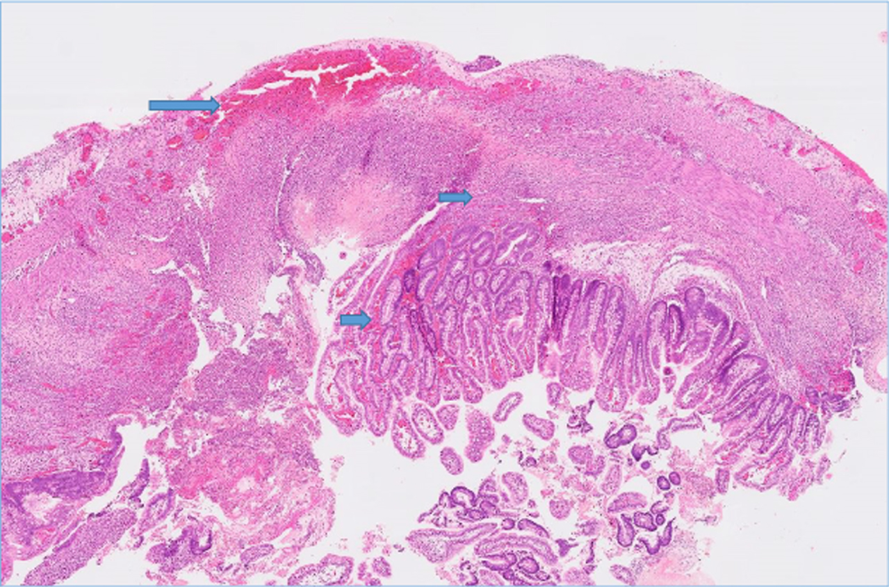
The H&E stain (20X) of resected intestinal tissue showing grade 4 hemorrhage. In infants with grades 1-2, the hemorrhage was limited to mucosa and submucosa. The arrow indicates areas of hemorrhage.
We evaluated the resected bowel for histopathological evidence of NEC, including coagulative necrosis, inflammation, hemorrhages, and reparative changes. Coagulative necrosis in intestinal cells was defined by the loss of hematoxylin staining of the nuclei and diminished cytoplasmic staining with eosin but with relatively preserved, ‘ghost-like’ crypt-villus histoarchitecture. Inflammation was noted as infiltration by inflammatory cells such as monocytes and neutrophils. Hemorrhages, marked by the extravasation of blood cells, were recorded in various layers of the intestine. Reparative changes included microscopic evidence of neovascularization, increased fibroblasts or myofibroblasts, and epithelial regeneration. The severity of NEC was assessed by the depth to which these histopathological changes (coagulative necrosis, inflammation, hemorrhages, and reparative changes) were seen: Grade 1 was limited to the mucosa, Grade 2 changes extended to the submucosa, Grade 3 to the muscularis, and Grade 4 change was transmural. Histopathological changes were also evaluated based on percentage of tissue involved (low = <30%, medium = 30–60%, and high = >60%).
Statistical methods
Continuous variables are presented as mean (standard deviation) if normally distributed and median (interquartile range) if normality does not hold. If normality criteria were satisfied, differences in continuous measures’ statistical associations were tested using a pooled t-test for equal variances or a Welch’s t-test for unequal variances. When the normality assumption was not satisfied, continuous measures’ statistical association was tested with the Wilcoxon rank sums test for equal variances or the Kolmogorov-Smirnov test for unequal variances. Categorical variables are presented as counts (column percentage).
Differences in categorical measures’ associations with and without severe intestinal hemorrhage were tested using the χ2 test when cell counts were adequate; otherwise, Fisher’s exact test was used with low expected cell counts. The unadjusted association between histopathological, clinical risk factors, and intestinal hemorrhage status (severe vs. mild/moderate) on pathology was investigated through univariate logistic regression. In addition, multivariable logistic regression models were performed to assess the adjusted relationship between the hemorrhagic status (severe vs. mild/moderate, mild/moderate as reference group) and histopathological findings. The univariate and multivariable logistic regression results are presented as odds ratios (ORs). All tests were two-sided, with an alpha value set to be 0.05. The statistical analyses were performed in the R foundation statistical software.
Results
One hundred and forty-eight infants were included in the analysis. Eighty-one infants (81/148, 54.7%) had severe-grade (Grade 3-4) hemorrhage, and sixty-seven infants (67/148, 45.2%) had mild to moderate intestinal hemorrhage.
Comparison of severe intestinal hemorrhage (Grade 3-4) vs mild to moderate (Grade 0–2) hemorrhage
The infants with severe hemorrhage (Grade 3-4) had significantly lower exposure to antenatal steroids (42 [52.5%] vs 50 [76.9%]; p = 0.004), had higher gestational age (28.57 [SD 7.14] vs. 26.58 [SD 2.90]; p = 0.034), lost more bowel length (28.8 cm [SD 28.04] vs. 20.2 cm [SD 22.13]; p = 0.045), exhibited higher mean CRP levels 2 weeks following the onset of N.E.C. (7.06 [SD 6.87] vs. 4.20 [SD 3.72]; p = 0.035), and had lower frequency of intestinal failure (TPN > 90 days, 23 [30.3% ] vs. 32 [52.5%]; p = 0.014) compared to preterm infants with mild to moderate (Grade 0–2) hemorrhage. The clinical data has been summarized in Table 1.
Clinical and demographic information in infants with intestinal hemorrhage
Clinical and demographic information in infants with intestinal hemorrhage
NEC = necrotizing enterocolitis; CRP = C reactive protein.
Histopathological, the infants with severe hemorrhage (Grade 3-4) had significantly higher mean inflammation score (2.67 [SD 0.94] vs. 1.63 [SD 0.92]; p = <0.001) and higher necrosis scores (1.95 [SD1.28] vs. 1.49 [SD 1.35]; p = 0.037), as anticipated higher neovascularization (1.57 [1.07] vs 0.95 [1.03]; p = 0.01), higher fibroblasts (1.47 [SD 1.04] vs. 0.92 [SD1.07]; p = 0.023) compared to preterm infants with mild to moderate (Grade 0–2) hemorrhage. Infants with severe hemorrhage (Grade 3-4) had significantly lower WBC count, lower absolute neutrophil counts (ANC), and higher lymphocyte percentages up to 48 hours following the onset of NEC compared to the other group. Those with severe hemorrhage had significantly lower platelet counts 24–48 hours after NEC onset. The histopathological data has been summarized in Table 2, and additional laboratory data in Table 3.
Histopathological and clinical outcomes in infants with intestinal hemorrhage
Bold p value = <0.05.
Lab values in preterm infants with intestinal hemorrhage
WBC = white blood cells, ANC = absolute neutrophil count, ALC = absolute lymphocyte counts, NEC = necrotizing enterocolitis, Bold P value = <0.05.
On multivariable logistic regression, less exposure to antenatal steroids (OR 0.18 [95% CI 0.05–0.58]; p = 0.005) and higher inflammation (OR3.7 [95% CI 2.09–7.32]; p = 0.001), lymphocyte count on the date of onset and 24 hours following NEC (OR 1.06 [95% CI 1.02–1.11]; p = 0.005) were significantly associated with a higher risk of severe intestinal hemorrhage compared to those with mild to moderate intestinal hemorrhage. The data is summarized in Table 4.
Predictors of intestinal hemorrhage by multivariable logistic regression
WBC = white blood cells; ANC = Absolute neutrophil count; CI = confidence interval. Bold p value = <0.05.
Our study reports clinical correlates of intestinal hemorrhage in preterm infants with surgical NEC. The infants with severe hemorrhage (Grade 3-4) had significantly lower exposure to antenatal steroids, had higher gestational age, lost more bowel length, had higher mean CRP levels two weeks following the onset of NEC, and had a lower frequency of intestinal failure in the bivariate analysis compared to preterm infants with mild to moderate hemorrhage. However, factors such as exposure to antenatal steroids, lymphocyte count on the onset and 24 hours following NEC diagnosis, and tissue inflammation remained significant on multivariable logistic regression modeling.
In this cohort, infants with severe intestinal hemorrhage received less antenatal steroids, most likely increasing the risk of intestinal hemorrhage in preterm infants. Randomized control trials and meta-analysis have shown that maternal administration of a single course of antenatal corticosteroids is commonly associated with an overall reduction in neonatal death and morbidity, including respiratory distress syndrome, intraventricular hemorrhage, NEC, and neonatal intensive care unit admissions [15]. The effects of glucocorticoids on vascular health and dysfunction may be mediated in part by the activation of endothelial nitric oxide synthase (eNOS) [16].
Infants with severe intestinal bleeding had a median gestation age of 28.5 weeks vs. 26.5 weeks, pointing towards gestational age-specific changes in the vascular structure and responses to various stimuli and functions. The endothelial glycocalyx (EG) has been recognized as an essential determinant of vascular integrity and health. This delicate structure of proteoglycans, glycoproteins, glycosaminoglycans, and associated plasma proteins covers the endothelial cells of the whole vasculature. It is involved in important vascular functions such as the regulation of vessel tone, permeability, cell-endothelial interaction, and hemostasis, as reviewed by Cosgun et al. [17]. EG maturity is gestational and postnatal age-dependent [18].
In this cohort, infants with intestinal hemorrhage had significantly higher coagulative necrosis than other groups. The intestinal vascular resistance is determined by the balance between vasodilatation by nitric oxide and endothelin (ET-1) mediated vasoconstrictor inputs [19]. Furthermore, studies have shown that vascular endothelial growth factor (VEGF) and the VEGF receptor-2 signaling pathway play a vital role in intestinal mucosal microvasculature [20]. In addition, endothelial TLR4 activation is required for NEC development, which impairs intestinal perfusion [21] via inhibition of the vasodilator molecule endothelial nitric oxide synthase (eNOS).
Animals with NEC (rat pups) demonstrated significantly smaller inflow and premucosal arterioles than control animals. The rat pups with NEC also had an altered intestinal arteriolar flow with a distinct “stop-and-go” pattern, suggesting severe vascular dysfunction compared to control animals [22]. The experimental animals also demonstrated an increased expression of inflammatory mediators. In our cohort, we noticed a higher severity of inflammation in infants with intestinal hemorrhage, suggesting a positive correlation between the degree of inflammation and vascular dysfunction leading to intestinal bleed. These studies underline that ischemia and intestinal microcirculation play an important role in NEC pathogenesis; however, it is still unclear whether vascular dysfunction is a primary initiator or a secondary response to intestinal injury, which needs further study.
The infants with intestinal hemorrhage had moderate to high neovascularization and fibroblasts in the intestinal submucosa, suggesting reparative changes in the intestine following the coagulative intestinal necrosis secondary to intestinal ischemia, vascular dysfunction, and inflammation. The inflammatory response following tissue injury plays a vital role in tissue repair [23]. Studies have shown that two-thirds of the leukocyte infiltrate in NEC consists of macrophages and play a crucial role in the pathogenesis of necrosis, inflammation, hemorrhage, and repair in necrotizing enterocolitis [10, 24]. The data has shown that macrophages in neonates with NEC have suppressed TGF-β signaling and augmented nuclear factor-kappa B (NF-κB) activation and cytokine production in areas of tissue damage and high bacterial load [25]. In addition, the macrophages promote healing by removing inflammatory cells, such as neutrophils, by actively ingesting them [24]. In our cohort, infants with severe hemorrhage (Grade 3-4) had significantly lower WBC count, absolute neutrophil counts (ANC), higher lymphocyte percentages up to 48 hours, and elevated CRP up to 2 weeks following the onset of NEC.
In this cohort, infants were thrombocytopenic following NEC onset. Platelets play an important role in controlling hemorrhage within the body. Several previous studies [26–29] have reported thrombocytopenia in surgical NEC. In our previous report, we recorded significant thrombocytopenia in fulminant and non-fulminant NEC (73.0 [41.8; 166] and 68.0 [33.8; 101] surgical non-fulminant) lasting 48 and 96 hours, respectively, and noticed recovery generally after four days [13]. The exact mechanism causing thrombocytopenia in these cases of NEC is still uncertain, but consumptive disorders are likely and include the formation of microthrombi in the diseased intestine [30] and platelet activation from bacterial products leading to aggregation in the microvasculature [31]. Coagulative parameters such as prothrombin time (PT), PT international normalized ratio, activated partial thromboplastin time (APTT), fibrinogen, and platelet count at the time of NEC diagnosis were independently associated with surgical NEC [32]. Our study lacks data on the coagulation parameters.
Our study also has several limitations. First, a single-center, retrospective experience may reduce the study’s generalizability. The retrospective study design affects the completeness of each patient’s available data. Secondly, the small sample size coupled with multiple factors and outcomes yields a high probability of Type I errors from multiple comparisons. The study is observational and descriptive; causality cannot be established with or without formal adjustment for multiple comparisons. Thirdly, our study lacks data on coagulation parameters (PT/INR). We acknowledge the proper determination of each histology category by biomarkers would be ideal, but we did not perform those determinations, which is an entirely different study that would require appreciable funding. In this study, we focused on H &E evaluation of intestinal pathology and its correlation with clinical outcomes in infants with surgical NEC. Finally, the infant cohort was predominantly African American, reflecting the racial distribution in Mississippi.
The study’s strengths are the data describing the clinical correlates of severe intestinal hemorrhage and the association of histopathological and hematological factors from a consecutive single-center cohort in a comprehensive manner.
In conclusion, 54.7% of infants had a severe intestinal hemorrhage in preterm infants with surgical NEC. Those with severe intestinal hemorrhage likely had lower exposure to antenatal steroids, a higher grade of inflammation, and higher lymphocyte counts following NEC onset on multi regression modeling. There is a need for further clinical, animal, and translational studies to evaluate the effect of gestational age, intestinal inflammation, and hematological factors on an intestinal hemorrhage. There is also a need for investigating and discovering biomarkers of vascular injury in preterm infants with surgical NEC. In the future, larger, prospective multi-center studies that include additional clinical detail (e.g., mesenteric perfusion using NIRS) and laboratory predictors such as hematological and stool biomarkers may inform earlier recognition of intestinal bleed in preterm infants with surgical NEC.
Author Contribution
P.M.G designed the study, P.M.G., MD, RT, MO, CM, HS; NV, WBH collected and analyzed the data. All the authors contributed to and approved the manuscript.
Footnotes
Acknowledgment
The Mississippi Clinical and translational research center for supporting the N.E.C. research. We would like to Thank Dr. Isabelle De Plaen from Northwestern University, IL, and Dr. Misty Good from UNC, Chapel Hill, NC, for reviewing and all the feedback provided for the project. We thank Emma L. Draughn, grant editor for MCCTR, for editing and proofreading the article.
Conflicts of interest
The authors disclose no conflicts.
Funding
Dr. Parvesh Garg and William Hillegass are partially supported by the National Institute of General Medical Sciences of the National Institutes of Health under Award Number 5U54GM115428. The content is solely the responsibility of the authors and does not necessarily represent the official views of the National Institutes of Health.
Category of study
Clinical science.
Consent
Patient consent is not required as per I.R.B.