






Abstract
OBJECTIVE:
We sought to investigate the clinical determinants of intestinal failure and death in preterm infants with surgical NEC.
METHODS:
Retrospective comparison of clinical information between Group A = intestinal failure (Parenteral nutrition (PN) >90 days) and death and Group B = survivors and with PN dependence < 90 days in preterm infants with surgical NEC.
RESULTS:
Group A (n = 99/143) had a lower mean gestational age (26.4 weeks [SD3.5] vs. 29.4 [SD 3.5]; p = 0.013), lower birth weight (873 gm [SD 427g] vs. 1425 gm [894g]; p = <0.001), later age of NEC onset (22 days [SD20] vs. 16 days [SD 17]; p = 0.128), received surgery later (276 hours [SD 544] vs. 117 hours [SD 267]; p = 0.032), had cholestasis, received dopamine (80.6% vs. 58.5%; p = 0.010) more frequently and had longer postoperative ileus time (19.8 days [SD 15.4] vs. 11.8 days [SD 6.5]; p = <0.001) and reached full feeds later (93 days [SD 45] vs. 44 [SD 22]; p = <0.001) than Group B.
On multivariate logistic regression, higher birth weight was associated with lower risk (OR 0.35, 95% CI 0.15–0.82; p = 0.016) of TPN > 90 days or death. Longer length of bowel resected (OR 1.76, 95% CI 1.02–3.02; p = 0.039) and longer postoperative ileus (OR 2.87, 95% CI 1.26–6.53; p = 0.011) were also independently associated with TPN >90days or death adjusted for gestational age and antenatal steroid treatment.
CONCLUSION:
In preterm infants with surgical NEC, clinical factors such as lower birth weight, longer bowel loss, and postoperative ileus days were significantly and independently associated with TPN >90 days or death.
Introduction
Necrotizing enterocolitis (NEC) affects 6–10% of preterm infants with a BW of less than 1500 grams [1, 2]. NEC remains a leading cause of death and intestinal failure (Parenteral nutrition more than 90 days following NEC) among preterm neonates, leading to increased hospital care and economic burden [3].
Infants with surgical NEC have a mortality risk of almost 50% [4]. Intestinal failure in newborn infants can be secondary to necrotizing enterocolitis or congenital gastrointestinal anomalies requiring surgery, such as gastroschisis, omphalocele, or bowel atresia requiring extensive bowel resection [5]. The prevalence of intestinal failure ranges from 13% to 45% following surgical necrotizing enterocolitis [6, 7]. Infants with intestinal failure are associated with longer hospital stay, growth failure, cholestasis, and liver injury [5]. Studies have reported higher prevalence and mortality in African American infants with surgical necrotizing enterocolitis [8]. Few studies have reported predictors of intestinal failure [9–12]. The clinical predictors of combined death and intestinal failure outcomes in preterm infants with surgical necrotizing enterocolitis (NEC) are poorly studied in a predominantly African American population.
Our previous retrospective observational cohort studies have reported the demographics, clinical outcomes, and systemic morbidities in preterm infants with NEC [13–17]. In this report, we sought to investigate the clinical determinants of intestinal failure (IF) and death in preterm infants with surgical NEC. The published reports have many definitions of IF or short bowel syndrome (SBS), including dependence on parenteral nutrition for 42 days or 90 days following NEC surgery [11, 12]. Our study defined intestinal failure as any patient requiring parenteral nutrition duration equal to or more than 90 days following NEC surgery.
In this study, we made a retrospective comparison of clinical information between Group A = intestinal failure (Parenteral nutrition (PN) >90 days) and death and Group B = survivors with PN dependence <90 days in preterm infants with surgical NEC. Univariate and multivariable logistic regression analyses assessed the associations between clinical factors and outcomes.
Methods
Population and study design
The study was conducted at the University of Mississippi Medical Center (UMMC) Neonatal Intensive Care Unit, a Level IV unit with 900–1000 admissions yearly and referrals from the entire state. The UMMC Institutional Review Board approved this retrospective study with a waiver of informed parental consent. All infants admitted between January 2013 and December 31, 2018, with an NEC (Bell stage III)/SIP diagnosis were included in the study [18]. In our research, we had 143 cases of surgical NEC included in the study. Neonates diagnosed with medical NEC, isolated ileal perforation, kidney anomalies, congenital heart disease, gastroschisis, omphalocele, and intestinal atresia were excluded from the analysis. The cohort derivation is summarized in Fig. 1.
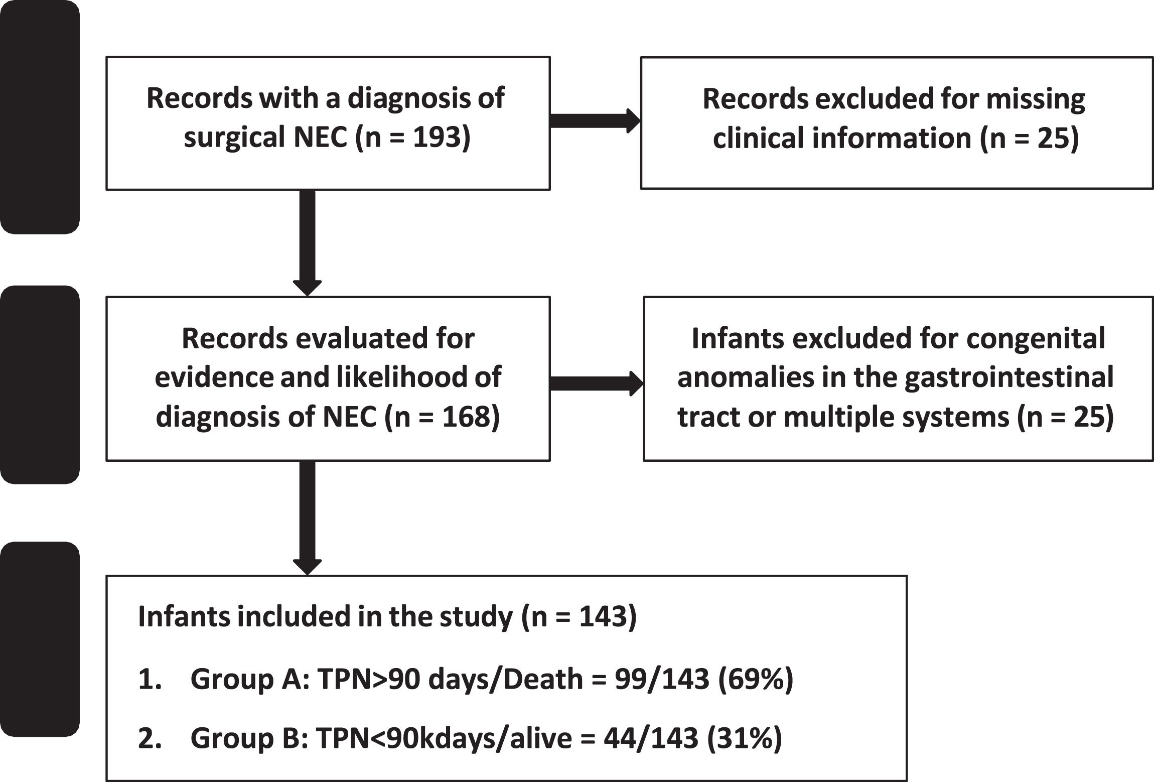
The patient included in the study.
Demographic data collected included birth weight (BW), gestational age (GA), appropriate for GA status (AGA), race and sex, mode of delivery, outborn status, and Apgar score≤6 at 5 min. Maternal information collected included clinical chorioamnionitis, antenatal steroids, and pregnancy-induced hypertension (PIH). Clinical data included patent ductus arteriosus (PDA), respiratory support data, inotrope (dopamine) use 24 hours after NEC onset, hematological information, ibuprofen /indomethacin treatment (before NEC), and frequency of cholestasis (direct bilirubin > 2 mg/dl) at any time after NEC diagnosis. Sepsis-related variables included blood culture-proven sepsis at NEC onset and duration/type of antibiotics.
NEC information
NEC was defined using Bell’s criteria [18], and a diagnosis of NEC was made on abdominal X-ray findings, including portal venous gas, pneumatosis, and pneumoperitoneum. Bell stage III/surgical NEC frequency was gathered [18]. In addition, we recorded information on the age at NEC diagnosis [19].
At our center, preterm infants with pneumoperitoneum who weigh less than 1 kg at NEC diagnosis and are hemodynamically unstable are treated first with a PD at the bedside but may later receive laparotomy. The timing of laparotomy after placement of Penrose was based on clinical deterioration. We also recorded information on intestinal failure (parenteral nutrition > 90 days) and surgical morbidity. Postoperative information such as postoperative ileus days (defined as infants being NPO after bowel surgery), time to reach full feeds (≥120 ml/kg/day), total parenteral nutrition days, length of stay, and surgical morbidities. The surgical morbidity was classified as strictures, fistulas, wound dehiscence, surgical site infections (including abscesses), adhesions, and perforations.
Histopathological evaluation
Gross and microscopic examination was performed on all resected intestinal segments. The tissue sections were taken from the representative most affected intestine area, which was determined by the trained grossing personnel. Hematoxylin & eosin-stained surgical resected intestinal tissue sections were evaluated by a team of a board-certified gastrointestinal pathologist and a senior pathology trainee for histologic evidence of necrosis, inflammation, hemorrhage, and reparative changes. The severity of NEC was assessed by the depth to which these histopathological changes (coagulative necrosis, inflammation, hemorrhages, and reparative changes) were seen; grade 1 was limited to the mucosa, grade 2 changes extended to the submucosa, grade 3 to the muscularis, and grade 4 change was transmural. The resected intestine was evaluated by the depth up to which these changes were seen. A scoring system was devised for assessment. A score of 0 was assigned when the exam appeared normal, 1 for 1–25% necrosis/inflammation, 2 when 25–50% area involved, 3 when 50–75% area was affected, and 4 when > 75% changes were seen [20]. The reparative changes included the presence of neovascularization, fibroblasts, and epithelial regeneration.
Outcome data
We collected data on mortality and intestinal failure. We defined mortality as death due to any reason before hospital discharge. We defined intestinal failure as requiring parenteral nutrition for 90 days or more following NEC surgery. We sought to assess the determinants of severe morbidity and mortality in preterm infants with surgical NEC.
Neonatal MRI data
Brain MRI without contrasts were routinely obtained at the corrected age of 36 weeks or before discharge, whenever clinically indicated, in all infants with BW less than 1500 grams per routine hospital clinical practice. The most common reason for not obtaining an MRI was death or transfer to a higher center for bowel transplantation. All term equivalent age (TEA) MRI scans were scored independently by two pediatric neuro-radiologists aware of the infants’ clinical diagnosis but unaware of the initial MRI read. We used a scoring system of eight scales for white and gray matter injury developed by Woodward et al. [21].
Statistical methods
Demographic and clinical information was compared between Group A (those preterm infants with intestinal failure (Parenteral nutrition (PN) >90 days) and/or death and Group B (surviving preterm infants with surgical NEC and those with PN dependence < 90 days). Continuous data were summarized as median (1st quartile, 3rd quartile) with Mann-Whitney U or Kruskal-Wallis tests for differences. The Categorical data were summarized as counts and percentages. Group differences were tested with Chi-squared or Fisher’s exact tests. The association between groups and other factors was evaluated with multivariable logistic regression. Variables with significant association in bivariate analysis were candidates for the multivariable models. Some variables were not included due to collinearity. All continuous clinical candidate factors such as gestational age, birth weight, length of bowel resected, and the postoperative ileus days were centered and standardized prior to multivariable logistic regression modeling. This uniform scaling permits more objective comparisons of the relative magnitudes of the effect of the associations of each factor with the intestinal failure or death endpoint. The multivariable-adjusted odds ratios can thus be interpreted per standard deviation of each clinical factor adjusted for the other factors in the model.
A p-value < 0.05 was considered statistically significant for all the analyses. All analyses were performed in SAS version 9.4.
Results
A total of 143 infants were included in the analysis. The demographic data, the clinical features, and the NEC features have been summarized in Tables 1 and 2.
Clinical and demographic information
Clinical and demographic information
NEC features
Group A (n = 99/143) had lower mean gestational age (26.4 weeks [SD3.5] vs. 29.4 [SD 3.5]; p = 0.013), lower mean birth weight (873gm [SD 427g] vs. 1425 gm [894g]; p = <0.001), later age of NEC onset (22 days [SD20] vs. 16 days [SD 17]; p = 0.128), received surgery later (276 hours [SD 544] vs. 117 hours [SD 267]; p = 0.032), had cholestasis (77% vs. 44%; p = 0.003), received invasive ventilation (95% vs 78%; p = 0.004) and dopamine (80.6% vs 58.5%; p = 0.010)more frequently than Group B infants. Group A had significantly higher mean WBC count, lower mean lymphocyte percentages, and higher absolute monocyte counts than Group B. There was no significant difference in the two groups: necrosis, inflammation, hemorrhage, and reparative changes in intestinal pathology, white matter injury, and surgical complications.
Group A infants had significantly longer postoperative ileus time (19.8 days [SD 15.4] vs. 11.8 days [SD 6.5]; p = <0.001) and took longer time to reach full feeds (93 days [SD 45] vs. 44 days [SD 22]; p = <0.001) than Group B [Table 3].
Clinical outcomes
On multivariate logistic regression, higher birth weight was associated with lower risk (OR 0.35, 95% CI 0.15–0.82; p = 0.016) of TPN > 90 days or death. Longer length of bowel resected (OR 1.76, 95% CI 1.02-3.02; p = 0.039) and longer postoperative ileus days (OR 2.87, 95% CI 1.26–6.53; p = 0.011) were independently associated with TPN > 90days or death adjusted for gestational age and antenatal steroid treatment. See Table 4 and Fig. 2.
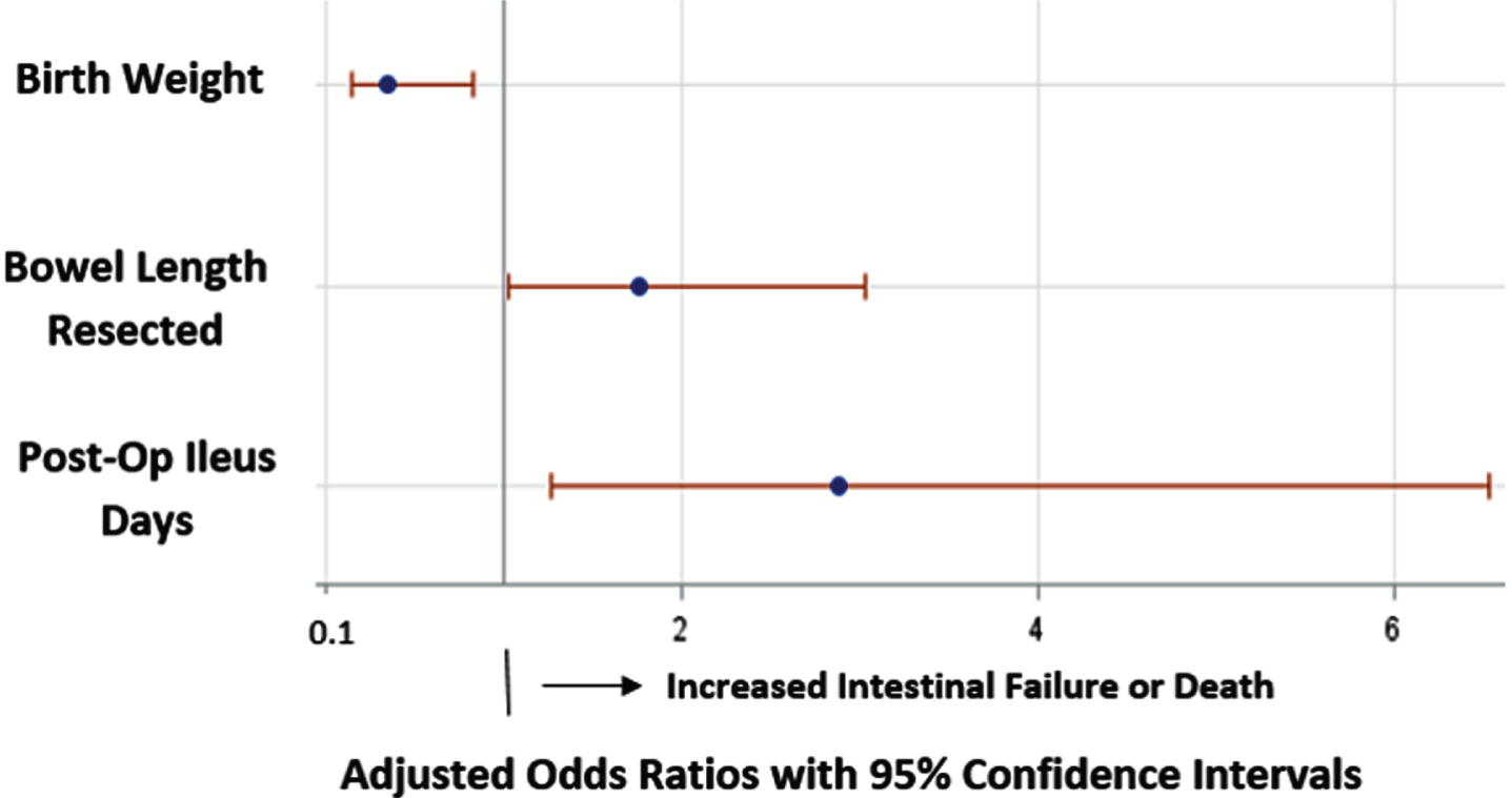
Adjusted Odds ratio of factors independently associated with intestinal failure and death.
Regression analysis relating to the risk of TPN > 90 days or death
Continuous variables gestational age, birth weight, bowel length resected, postoperative ileus days were centered and standardized so the adjusted odds ratios could be uniformly interpreted per one standard deviation of the covariate. The reference group for the binary factor of antenatal steroids is no antenatal steroid exposure.
In our consecutive cohort of surgical NEC infants, approximately 70% experienced the adverse composite outcome of parenteral nutrition for > 90 days or death. Lower birth weight, longer length of bowel resection, and longer duration of postoperative ileus days were significantly and independently associated with worse outcomes adjusted for gestational age and antenatal steroid treatment.
In a multicenter prospective cohort study of 129 infants, Duro et al. reported intestinal failure (PN > 90 days) in 54/129 (42%) of cases. In their cohort, characteristics of severe NEC (low birth weight, antibiotic use, ventilator use, and greater extent of bowel resection) were associated with the development of intestinal failure (IF) [22]. In our cohort, we also observed similar trends. In this cohort, infants in Group A were operated on later. They were more acutely sick than the other Group, most likely due to their lower birth weight, and received Penrose drain therapy more likely than Group B infants. The initial Penrose drain treatment also explains the longer time to laparotomy following NEC onset in Group A infants. In a report by Wang et al., lower birth weight, greater extent of bowel resection, and larger percentage of small bowel resection were also associated with the incidence of IF [11].
The Elfvin study [12] compared IF in preterm infants with medical and surgical NEC and reported low birth weight, low gestational age, need for surgical intervention, and gram-negative bacteremia were predictors of intestinal failure [12]. Similarly, in our cohort, the infants at risk of death and PN dependence more than 90 days were significantly younger and smaller; however, the sepsis rate was not significant in the two groups.
A study of 80 pediatric patients with short bowel syndrome by Spencer et al. [7] found that cholestasis and age-adjusted small bowel length are the major predictors of mortality in pediatric short bowel syndrome. Other clinical factors, such as age-adjusted small bowel length and ileocecal valve, are the major predictors of weaning from PN [7]. However, in their cohort, only 45% of infants had necrotizing enterocolitis. Our study similarly showed that infants at risk for death or intestinal failure had significantly more cholestasis on bivariate comparison and had longer bowel length resected on the multivariable regression analysis. A prospective study by Duro et al. reported cholestasis in 56% to 85% of cases in preterm infants with surgical necrotizing enterocolitis [23]. Their multivariable logistic regression analysis identified small-bowel resection or creation of jejunostomy (odds ratio [OR] 4.96, 95% confidence interval [CI] 1.97–12.51, P = 0.0007) and duration of PN in weeks (OR 2.37, 95% CI 1.56–3.60, P < 0.0001) as independent risk factors for parenteral nutrition-associated liver disease [23]. In a retrospective study of 225 infants with surgical NEC in the Finnish population from 1986–2014 by Karlia et al., the Group noticed intestinal failure associated with cholestasis in 42% of cases, and risk factors such as septicemias and the reoperations were associated with cholestasis. Interestingly, cholestasis did not increase the overall mortality [24].
Kelleher et al. study [10] of 240 infants with surgical NEC reported no difference between peritoneal drainage and laparotomy in infants with surgical necrotizing enterocolitis when comparing the combined mortality outcome or survival with intestinal failure. However, they noticed increased intestinal failure in surviving infants treated with peritoneal drain with subsequent salvage or secondary laparotomy compared to peritoneal drainage alone [10]. In our study, the infants at risk of death and intestinal failure were more likely to receive the Penrose drain treatment, although the difference did not reach statistical significance.
Our study did not see a significant difference in intestinal histopathology in infants with and without intestinal failure and death. However, in our previous study [25], we noted that Infants with necrosis in the intestinal margins were most likely associated with significantly higher mortality and a longer length of stay.
Our study’s strengths include a detailed evaluation of the clinical risk factors associated with intestinal failure and death in preterm infants with surgical NEC. Identifying these risk factors can help bedside providers better predict outcomes in infants with surgical necrotizing enterocolitis.
Our study has important limitations. First, this was a single-center experience with most neonates with NEC as African American, which may reduce the study’s generalizability to other demographic groups. The predominantly African-American cohort is partly due to race distribution in Mississippi. However, this may also be related to adverse social determinants of maternal/infant health or potentially genetic risk factors for NEC. Second, sample size limits our power to detect associations between clinical factors and the outcomes. Thirdly, the small sample size coupled with multiple factors, outcomes, and comparisons may result in type I errors. Finally, we did not include infants with congenital intestinal anomalies such as atresia, gastroschisis, or omphalocele in our analysis as we had designed our study to assess the predictors of intestinal failure and death secondary to surgical NEC. The exclusion of infants with congenital conditions may have influenced our results compared to other reports.
In conclusion, in preterm infants with surgical NEC, clinical factors such as lower birth weight, longer segment of bowel resections, and a longer postoperative ileus period were significantly and independently associated with TPN > 90days or death adjusted for gestational age and antenatal steroid treatment. Infants with large bowel loss and longer postoperative ileus course should perhaps receive more intensive bowel rehabilitation therapy with further evaluation of whether this improves outcomes.
Identifying these risk factors would help design these prospective studies and, ideally, randomized clinical trials evaluating management strategies and clinical prognostication of infants with surgical necrotizing enterocolitis.
Conflicts of interest
The authors disclose no conflicts.
Footnotes
Acknowledgments
The Mississippi Center for clinical and Translational research for supporting the NEC research.
Funding
Dr. Parvesh Garg and Dr. William Hillegass are partially supported by the National Institute of General Medical Sciences of the National Institutes of Health under Award Number 5U54GM115428. The content is solely the responsibility of the authors and does not necessarily represent the official views of the National Institutes of Health.
Consent
Patient consent was not required for de-identified retrospective data.