






Abstract
BACKGROUND:
Few quantitative analyses have been performed on muscle activation and gait function according to cane dependence.
OBJECTIVE:
The purpose of this study was to measure changes in the lower limb muscle activation and gait function according to reduced cane dependence using a weight-bearing feedback cane (WBFC) that had been designed to quantitatively measure cane dependence in stroke patients during walking.
METHODS:
Twenty-four subjects were recruited from a local rehabilitation hospital. The WBFC measured the average weight support (AWS, kg) loaded on the cane during walking through a Bluetooth connection to laptop software. All subjects walked 20 m using a WBFC set in the three levels of weight support (WSR, 100%, 60%, and 20%) based on the measured AWS. Paretic lower limb muscle activation and gait function (velocity, cadence, paretic side stride length, and symmetry index) were measured using wireless surface EMG and a 3-axis accelerometer during walking.
RESULTS:
The paretic side lower limb muscle activation of the 20% WSR on the cane was significantly higher than that of the 100% WSR on the cane (
CONCLUSION:
These findings suggest that indiscreet weight support on the cane during walking can interfere with lower limb muscle activation and gait function. Therefore, in a clinical setting, reducing cane dependence during stroke gait training should be carefully considered.
Introduction
Stroke patients often present with muscle weakness, spasticity, sensory impairment, reduced postural control and asymmetrical gait patterns [1]. Specifically, 61–80% of body weight is loaded on the non-paretic side lower limb because of muscle weakness in the lower limb on the paretic side [2], resulting in asymmetrical gait patterns, including decreased duration of the stance phase on the paretic side and decreased stride length, as well as reduced walking speed [3]. Persistent asymmetric gait patterns cause lower limb joint damage and pain, reduced gait efficiency, and increased incidence of falls [4]. For these reasons, recovery of muscle strength and walking function of the paretic-side lower limb are among the main goals of stroke rehabilitation.
Canes are commonly used in clinical practice for gait training in stroke patients. Canes provide external support to enhance gait safety and independence for stroke patients [5], and also provide a wider base of support to compensate for the muscle strength and balance ability of the affected lower limb [6]. The use of a cane during gait rehabilitation in stroke patients encouraged an increase in the paretic side step and stride length, cadence, and step width [7]. In contrast, previous studies reported that cane-assisted walking in stroke patients caused decreases in weight-bearing on the paretic lower limb: Chen et al. reported that the use of a cane during gait transferred 12.7% of the weight-bearing from paretic-side lower limb to the cane [8]. Such asymmetric weight-bearing was reported to be more prominent in hemiparetic stroke patients [9]; Buurke et al. reported that the use of a cane during initial gait training in stroke patients caused incremental increases in base of support, and decreased muscle activation of paretic-side lower limb, thereby impairing gait independence in the long term [10].
According to motor control theories with respect to recovery of balancing mechanisms in stroke patients, the need for a cane in gait training is lower after enhancement of postural stability and gait stability through initial gait training with the cane [11, 12]. Therefore, in clinical practice, the indiscriminate use of canes that does not consider the progressively reduced weight support during cane-assisted gait training for stroke patients is thought to interfere with independent gait in stroke patients [13].
The most important factor of cane gait training is provision of normal patterns of lower limb muscle activation on the paretic side via progressive reduction of weight support on the cane, to restore balancing mechanisms, and to improve gait independence by increasing weight support on the paretic side lower limb [12, 14]. Nevertheless, in a clinical setting, it is difficult to quantitatively measure cane dependence during gait training for stroke patients. Furthermore, there are few studies on the changes of paretic-side lower limb muscle activation and gait function according to reduced cane dependence during walking.
Therefore, this study aimed to measure changes in paretic-side lower limb muscle activation and gait function according to reduced cane dependence using a weight-bearing feedback cane (WBFC) that had been designed to quantitatively measure cane dependence in stroke patients during walking.
Methods
Subjects
Twenty-four stroke patients were recruited from a local rehabilitation hospital. After attaining a full understanding of the purpose and methods of the research, they signed an informed consent form. The sample size was determined after a calculation based on results of the G-power 3.1.9.3 software. The power and alpha were set at 0.95 and 0.05, respectively, and the effect size was set at 0.74 in accordance with prior analysis, requiring at least 24 subjects.
The inclusion criteria were: (1) hemiparesis from a single stroke that occurred at least six months prior to the time of recruitment, (2) adequate cognition levels to follow simple instructions and understand the content and purpose of the study (Korean version of the Mini-Mental State Examination score
General characteristic of the subjects
General characteristic of the subjects
Values are expressed as mean
This study applied a cross-sectional design to investigate changes in lower limb muscle activation and gait function according to cane dependence in chronic stroke patients. We explained the objective and experimental procedure of the study to all subjects, and they voluntarily signed informed consent forms. Ethical approval was granted by the Korea National University of Transportation (KNUT IRB 2018-18).
Weight-bearing feedback cane (WBFC). The WBFC measures the degree of weight support (kg) loaded on the cane during walking. Measurement of the degree of weight support occurs through a load cell located inside the bottom of the cane handle, and the measured degree of weight support is displayed in real time on the display at the top of the cane handle.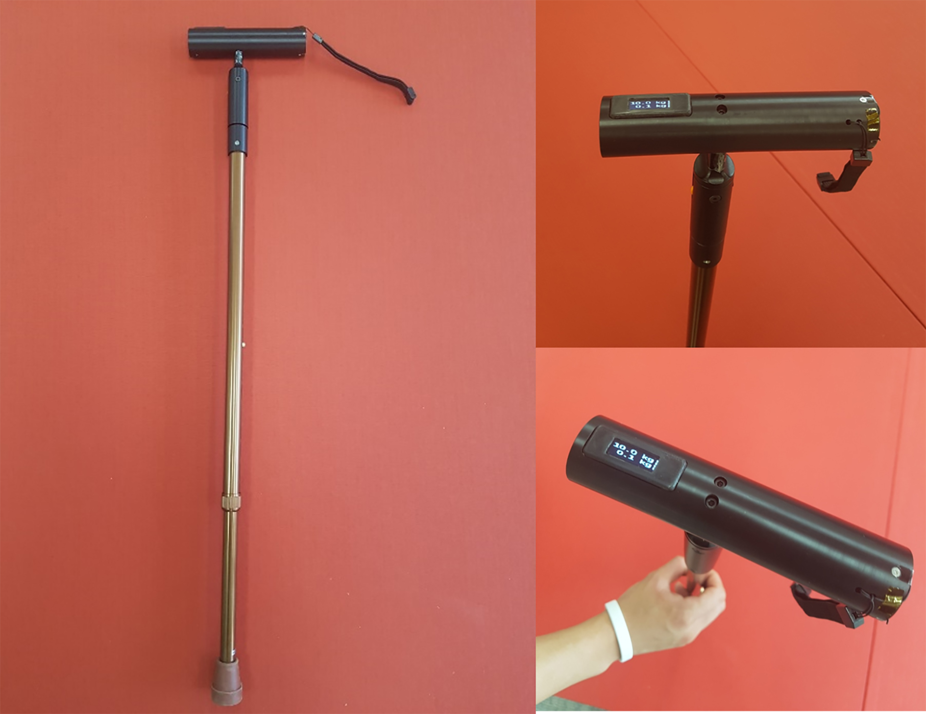
A weight-bearing feedback cane (WBFC) was used to quantitatively measure cane dependence during walking (Fig. 1). The WBFC measures cane dependence (degree of weight support, DWS (kg)) loaded on the cane during walking. While a subject walks 20 m with the WBFC, the average cane dependence (average weight support, AWS (kg)) was measured using laptop software. The formula used for average weight support (AWS) calculation is:
Measurement of the degree of weight support occurs through a load cell located inside the bottom of the cane handle. The degree of weight support is displayed in real time on the display at the top of the cane handle. The WBFC measures the average weight support (kg) loaded on the cane during walking via a Bluetooth connection to laptop software. Based on the measured average weight support (kg), the user can set the desired weight support rate (1–100%) using laptop software (Fig. 2). If a weight above the preset weight support rate (%) was loaded on the cane during walking, auditory feedback (beeping sounds) was generated from a buzzer installed in the cane handle.
System configuration for cane dependence measurement. The WBFC measures the average weight support (kg) loaded on the cane during walking through connected to the laptop software via Bluetooth. Based on the measured the average weight support (kg), the user can set the desired weight support rate (1–100%) through the laptop software.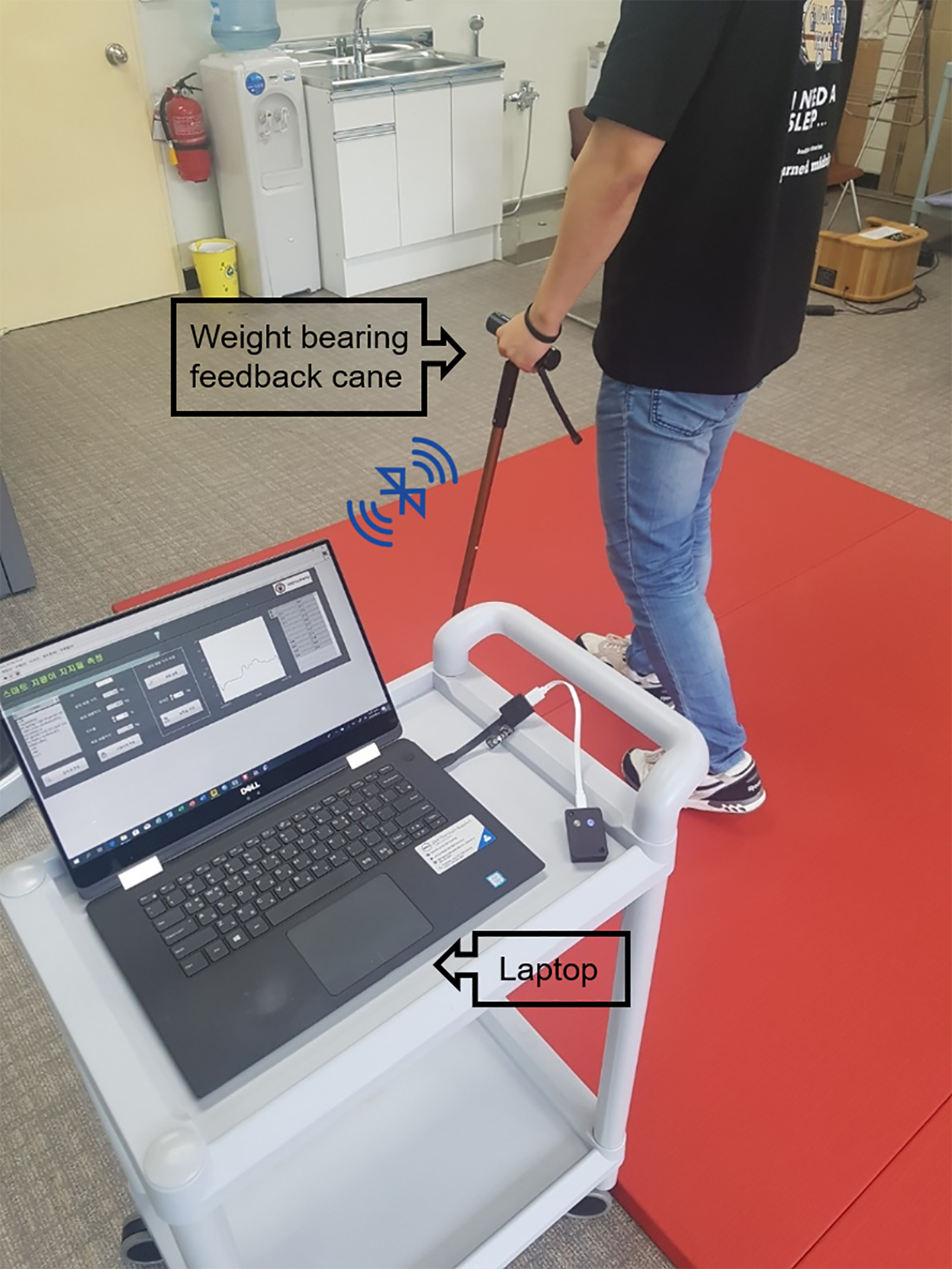
Prior to the experiment, all subjects naturally walked 20 m using a WBFC, while the average weight support (kg) for 20 m walking was measured simultaneously. Only subjects whose average weight support loaded on the WBFC was greater than 7% of their body weight were included in the study [8]. Subsequently, three levels of weight support (100%, 60%, and 20%) were set based on the measured average weight support of each subject. All subjects walked 20 m three times using a WBFC set in the three levels of weight support (100%, 60%, and 20%) based the measured average weight support. During walking, paretic-side lower limb muscle activation (rectus femoris, biceps femoris, medial gastrocnemius, and gluteus medius) and gait function (velocity, cadence, paretic side stride length, and symmetry index) were measured. With the weight support rate set at 100%, the subjects were asked to walk below measured average weight support. With the weight support rate set at 60% and 20%, subjects were asked to walk below 60% and 20% of measured average weight support.
All subjects were instructed to hold the WBFC at the height of the greater trochanter using the non-paretic side hand. During the experiment, one assistant stood beside the subject for safety reasons.
We used a wireless surface EMG (sEMG) (FreeEMG1000, BTS Bioengineering, Milano, Italy) and 3-axis accelerometer (G-Walk, BTS Bioengineering, Italy) to measure lower limb muscle activation and gait function during WBFC gait with three levels of weight support.
The wireless sEMG was used to measure paretic-side lower limb muscle activation during the stance phase of WBFC gait. Eight wireless sEMG electrodes were attached to the following four major muscle groups of the paretic side lower limb: rectus femoris, biceps femoris, medial gastrocnemius, and gluteus medius. To minimize skin resistance, we removed skin hair at the site of attachment, cleaned the site with alcohol, and attached the electrodes according to the direction of the muscle fibers. Muscle activation data were obtained using EMG Analyzer v2.9.37.0 (BTS Bioengineering, Milano, Italy). The collected sEMG raw data were band-pass filtered at 20–500 Hz to remove artifact and high-frequency noise. The root mean square (RMS) values were computed over a time constant of 50 ms. Muscle activation data were measured three times and then averaged. To normalize the sEMG signal, all values were set to reference voluntary contraction (RVC) and expressed as %RVC.
The 3-axis accelerometer was used to measure gait function during WBFC gait. The 3-axis accelerometer was attached at the S1 level of the subjects using a special Velcro strap. The sampling frequency was set to 100 Hz. The gait functions (velocity, cadence, paretic side stride length, and symmetry index) were obtained using G-Studio (BTS Bioengineering, Milano, Italy).
Statistical analysis
Data analysis was performed using SPSS (version 21.0; IBM Corp., Armonk, NY, USA). Descriptive statistics were used to describe the characteristics of the subjects. The Shapiro-Wilk test was used to confirm that all outcome variables were normally distributed. For dependent variable measures, one-way repeated measure analysis of variance (ANOVA) (post hoc analysis used Bonferroni correction) was used to compare changes in lower extremity muscle activation and gait function according to the dependence of the cane. A significance level of 0.05 was used for all tests.
Results
A summary of the general characteristics of the 24 subjects is shown in Table 1. Tables 2 and 3 show the changes in lower limb muscle activation during stance phase and gait function during WBFC gait with the three levels of weight support (100%, 60%, and 20%).
Changes of paretic side lower limb muscle activation during stance phase according to cane dependence (
24)
Changes of paretic side lower limb muscle activation during stance phase according to cane dependence (
Values are expressed as mean
Changes of gait function according to cane dependence (
Values are expressed as mean
Paretic-side lower limb muscle activation during the stance phase showed significant differences in the rectus femoris, biceps femoris, and gluteus medius muscles according to the weight support level on the WBFC. In the post hoc analyses, the muscle activation of the 20% weight support level on the cane was significantly higher than that of the 100% weight support level.
With respect to gait function, significant differences were observed in velocity, cadence, paretic side stride length, and symmetry index according to the weight support level on the WBFC. In the post hoc analyses, the velocity, cadence, paretic side stride length, and symmetry index of the 20% weight support level on the cane were significantly lower than those of the 100% weight support level.
Higher dependence on the cane has been reported to negatively affect paretic-side lower limb muscle activation [10]. Nevertheless, there is a lack of quantitative analysis in lower limb muscle activation according to cane dependence. Therefore, we investigated changes in paretic-side lower limb muscle activation according to three levels of cane dependence (100%, 60%, and 20%) during gait with a cane in stroke patients. We found that paretic-side lower limb muscle activation during the stance phase was significantly different in the rectus femoris, biceps femoris, and gluteus medius muscles according to the three levels of cane dependence. In particular, the paretic-side lower limb muscle activation of the 20% weight support rate on the cane was significantly higher than that of the 100% weight support level on the cane.
According to the stroke rehabilitation practice guidelines in Canada [16] and the guidelines for adult stroke rehabilitation in the USA [17], it is strongly recommended that stroke patients use walking aids such as canes if this is necessary for standing and walking; based on this recommendation, many clinicians recommend canes during gait training in stroke rehabilitation [18]. The cane was used to improve the balance during walking by increasing the base of support and supporting the stabilizing of the pelvis [17]. However, stroke patients who walk with a cane impose 7–25% of body weight on the cane [8, 19]. Therefore, there is a significant decrease in antigravity muscle activation when walking with a cane, compared to when walking without a cane. In particular, it was reported that there was a significant decrease in the muscle activation of the erector spinae and the tibialis anterior muscle when walking with a four-point cane, compared to when walking with a single-point cane [10]. The results of previous studies and the present study confirmed that the uncontrolled weight support loaded on the cane during walking may interfere with paretic-side lower limb muscle activation during the stance phase. These findings suggest that incremental reduction in cane dependence during stroke gait training should be considered in future clinical settings. In particular, we believe that cane dependence equivalent to 20% of average weight support during walking may be effective for paretic-side lower limb muscle activation.
We measured changes in gait function (velocity, cadence, paretic side stride length, and symmetry index) according to cane dependence. In terms of gait function, significant differences were observed according to the weight support rate loaded on the WBFC. In particular, velocity, cadence, paretic side stride length, and symmetry index of the 20% weight support rate on the cane were significantly lower than that of the 100% weight support rate on the cane. Previous studies reported that the use of a cane during walking encouraged improvements in velocity, cadence, and step length, increased confidence, stability and comfortability, and enhanced gait symmetry [20, 21]. In particular, previous studies reported that the use of a cane during walking promoted the free movement of the paretic-side lower limb during the stance phase, and enhanced motor control, thereby improving gait symmetry [22, 23]. The evidence of these improvements can be confirmed through the improvement of walking function in the partial body weight support treadmill training [23].
Similar to the results of previous studies [20, 21, 22, 23], we found that restriction of cane dependence during walking caused decreased gait velocity, cadence, stride length and gait symmetry. A major consideration in the prescription of walking assistive device is safety and dynamic balance during walking [24]. Because reduction in gait parameters, particularly gait symmetry, may negatively impact dynamic balance during walking, reducing cane dependence during gait training in the stroke rehabilitation should be considered carefully.
This study has some limitations. First, the WBFC is designed to generate auditory feedback for all weight support that exceeds the preset weight support rate. Thus, the changes of muscle activation and gait function shown in this study may not be precisely based on 100%, 60%, and 20% of average weight support. Second, it is difficult to generalize the results because this study only involved chronic stroke patients who could walk with a cane. Third, because gait ability in stroke patients is an important predictor of social participation [25], further studies are needed to measure changes in community participation according to cane dependence in stroke patients. Finally, we investigated changes in muscle activation and gait function during WBFC walking with only three stages of weight support rate (100%, 60%, and 20%). Therefore, future studies that investigate changes in muscle activation and gait function according to the subdivided weight support rate (more than three stages) are needed.
Conclusions
This study investigated changes in paretic side lower limb muscle activation and gait function according to reduced cane dependence using a weight-bearing feedback cane (WBFC) that had been designed to quantitatively measure cane dependence in stroke patients during walking. We found that the indiscreet weight support loaded on the cane during walking can interfere with the paretic side lower limb muscle activation and gait function during walking. Therefore, in a clinical setting, reducing cane dependence during stroke gait training should be carefully considered.
Footnotes
Acknowledgments
This work was supported by the National Research Foundation of Korea (NRF) grant funded by the Korean government (Ministry of Science and ICT; No. 2018R1C1B5084411).
Conflict of interest
The authors declare no conflict of interest.