






Abstract
Purpose:
To evaluate and correlate anthropometric, biometric, and refractive error changes in thalassemia major (TM).
Methods:
One hundred children with TM and another hundred healthy controls were recruited. Height, weight, body mass index (BMI), and occipitofrontal circumference (OFC) were the anthropometric parameters recorded. Full ophthalmologic examination was performed, including best-corrected visual acuity, cycloplegic refraction, slit-lamp examination, Goldmann applanation tonometry, indirect ophthalmoscopy, keratometry (K readings), and ocular biometry.
Results:
Compared to controls, children with TM were shorter and lighter, with a smaller BMI (p<0.001); however, no significant difference existed in OFC. Regarding ocular biometric data, patients with thalassemia had steeper mean K readings (p = 0.03), shorter axial length (AXL) (p = 0.005), shorter vitreous chamber depth (p<0.001), and thicker crystalline lens (p<0.001) than controls. Patients with thalassemia had a significant myopic shift (p = 0.003). Multiple regression analyses only showed a significant correlation between corneal astigmatism and both weight and height (β = -0.05 and p = 0.03 and β = 0.06 and p = 0.04, respectively). Spherical equivalent was significantly correlated to K readings, lens thickness, and anterior chamber depth (p<0.0001 for all parameters).
Conclusions:
Compared to controls, children with TM have significant retardation in general and ocular growth (smaller BMI and shorter AXL). Ocular growth changes probably resulted in compensatory biometric changes (steeper corneas and thicker lenses) to reach emmetropization, with an exaggerated response and subsequent myopic shift. However, growth retardation is not directly related to ocular growth changes, myopic shift, or variations in biometric parameters.
Introduction
Thalassemia major (TM) is a hereditary chronic hemolytic anemia that especially affects those living in the Mediterranean region, having a high frequency in consanguineous marriages. The disease results in erythrocyte destruction and requires lifelong follow-up, blood transfusions, and iron chelators use, creating social and economic problems (1).
Eye complications of TM are related to various factors, including iron accumulation in tissues due to erythrocyte destruction, the chelating agents used with regular blood transfusions, and effects of orbital bone marrow expansion (2).
Patients with TM can develop characteristic skeletal changes due to bone marrow expansion, including typical craniofacial changes and deformities of the long bones. Craniofacial changes include bossing of the skull, depressed nasal bridge, and prominent malar eminence, and can increase up to a mongoloid slant of the eye and hypertrophy of the maxillae (3). Ocular growth is intimately related to the growth of the adjacent bony orbit. In principle, craniofacial changes in patients with TM might lead to an abnormal bony orbit and this may result in distinctive ocular biometry or refraction in patients with thalassemia (4).
Impaired weight and height have been widely reported in patients with thalassemia. Previous studies suggest a significant incidence of growth deficits, depending on severity of the disease (5-7). Growth failure is multifactorial in thalassemia, related to chronic hypoxia of chronic anemia, chelation toxicity, low serum zinc level, hepatic iron overload with hepatic dysfunction, and iron-associated endocrinopathies such as hypogonadism, hypothyroidism, and growth hormone deficiency (5-7). Moreover, patients with growth hormone deficiency have been reported by some studies to have shorter AXL, thicker lenses, steeper corneas, and more refractive errors in comparison to healthy controls (8-10).
The aim of our study was to evaluate the possible anthropometric, biometric, and refractive error changes in children with thalassemia and to correlate them together, assessing whether general growth retardation is directly related to ocular growth changes, or if possible ocular growth retardation could adversely affect patients’ refraction or biometric measurements.
Methods
The study was conducted in accordance with the ethical standards stated in the Faculty of Medicine, Ain Shams University, with informed consent obtained from the patients’ guardians. The study was carried out in accordance with the Declaration of Helsinki.
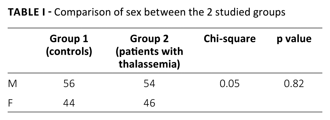
This cross-sectional observational case-control study was performed between May 2014 and January 2015. The study included 100 children (54 M/46 F) with TM and another 100 age- and sex-matched (56 M/44 F) healthy volunteers who served as controls (Tab. I). All participants were examined in the Ophthalmology Department at Ain Shams University Hospitals.
Comparison of sex between the 2 studied groups
All the patients had been diagnosed with TM of 10 to 15 years duration and are regularly followed up at the Paediatric Haematology Clinic of Ain Shams University Hospitals. We excluded patients with previous ocular trauma, history of corneal disease, contact lens wear, or previous ocular surgery. Furthermore, all recruited patients were medically evaluated and determined to be free of systemic diseases other than TM.
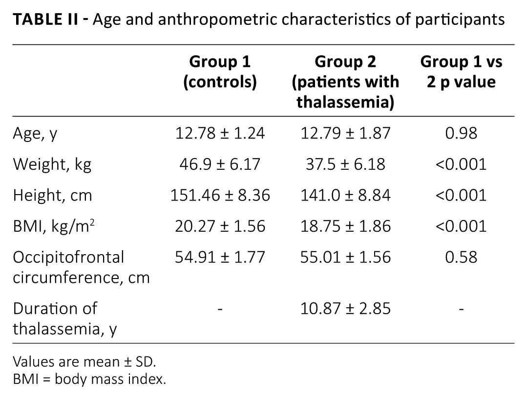
Height, weight, body mass index (BMI), and occipitofrontal circumference (OFC) were recorded for all participants (Tab. II).
Age and anthropometric characteristics of participants
Values are mean ± SD.
BMI = body mass index.
Full ophthalmologic examination was performed for both eyes of all participants, including best-corrected visual acuity (BCVA), cycloplegic refraction, slit-lamp examination, Goldmann applanation tonometry, indirect ophthalmoscopy, keratometry, and ocular biometry.
Refraction
Cycloplegic refraction was performed using an autorefractometer (Topcon RM-A2000; Topcon Medical Systems, Tokyo, Japan). Full cycloplegia was achieved using cyclopentolate 1% eyedrops. The drops were instilled 3 times, 10 to 15 minutes apart, then refraction measurements were captured. The spherical equivalent (SE) was determined by adding half the minus cylinder to the sphere dioptric value.
Keratometry
Keratometry was performed using an automated keratometer. We recorded the mean keratometry (K readings) and the corneal astigmatism (which is the difference between the dioptric values of flat and steep axes). Axis of corneal astigmatism was also recorded and analyzed.
Conventional A-scan ultrasonography
Contact ultrasound biometry (nonimmersion) was performed using an A-scan device (Echo Scan US-800; Nidek, Aichi, Japan). Topical anesthesia (1 drop of benoxinate 0.5%) was instilled in each eye before the measurements were taken. Analyzed biometric data included axial length (AXL), anterior chamber depth (ACD), vitreous chamber depth (VCD), and lens thickness. We took 5 measurements per eye, provided that their standard deviation (SD) was not more than 0.1, and averaged their values.
Statistical analysis
We analyzed data using Statistica software, version 8. Quantitative variables were expressed as mean ± SD. Student t test was used to evaluate the statistical significance of differences between 2 groups. Descriptive statistics were prepared using chi-square analysis. Correlation analyses were performed by calculating Pearson correlation coefficient (r). Regression analyses were done to evaluate the relation between anthropometric measurements and ocular biometric data. p Values were considered statistically significant if <0.05.
Results
One hundred children with TM with a mean age of 12.79 ± 1.87 years and 100 age- and sex-matched healthy controls were recruited.
Compared to controls, children with thalassemia were shorter and lighter, with a smaller BMI (p<0.001). However, no significant difference in OFC was found between the groups (Tab. II).
Slit-lamp examination and indirect ophthalmoscopy showed no abnormalities in the 2 groups. Moreover, no significant differences between controls and patients were found as regards BCVA (20/20 for both groups) and intraocular pressure (13.06 ± 1.08 mm Hg and 13.12 ± 1.15 mm Hg, respectively).
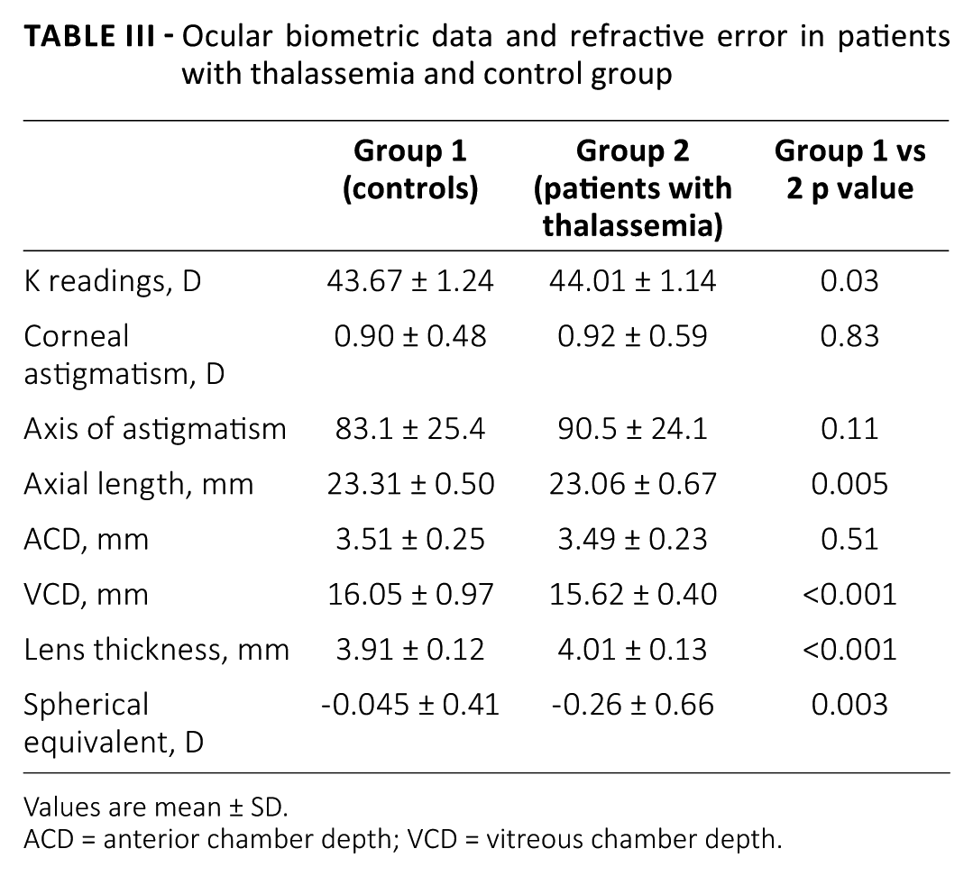
Regarding ocular biometric data, patients with thalassemia had steeper K readings (p = 0.03), shorter AXL (p = 0.005), thicker crystalline lens (p<0.001), and shorter VCD (p<0.001) in comparison to controls. However, no significant difference was found regarding ACD (p = 0.51). Furthermore, patients with thalassemia had greater myopic refractive error (p = 0.003). Both patients and controls had a strong trend of against-the-rule astigmatism (ARA), with an insignificant p value (p = 0.11) (Tab. III).
Ocular biometric data and refractive error in patients with thalassemia and control group
Values are mean ± SD.
ACD = anterior chamber depth; VCD = vitreous chamber depth.
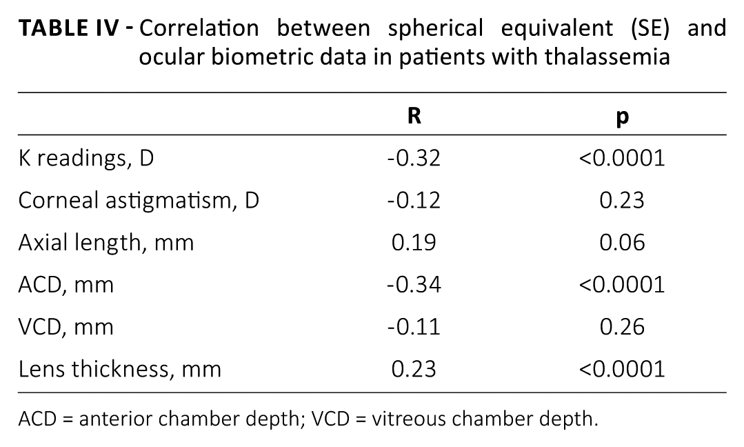
On performing correlations between spherical equivalent and ocular biometric parameters, spherical equivalent was significantly correlated to K readings, lens thickness, and ACD (Tab. IV).
Correlation between spherical equivalent (SE) and ocular biometric data in patients with thalassemia
ACD = anterior chamber depth; VCD = vitreous chamber depth.
As regards the correlations performed between anthropometric and biometric parameters, multiple regression analyses of children with thalassemia only showed a significant correlation between corneal astigmatism and weight and height (β = -0.05 and p = 0.03; β = 0.06 and p = 0.04, respectively) (Tabs. V and VI). The OFC did not have any significant correlations with other variables in our studied groups.
Multiple regression analyses of different factors affecting weight in patients with thalassemia
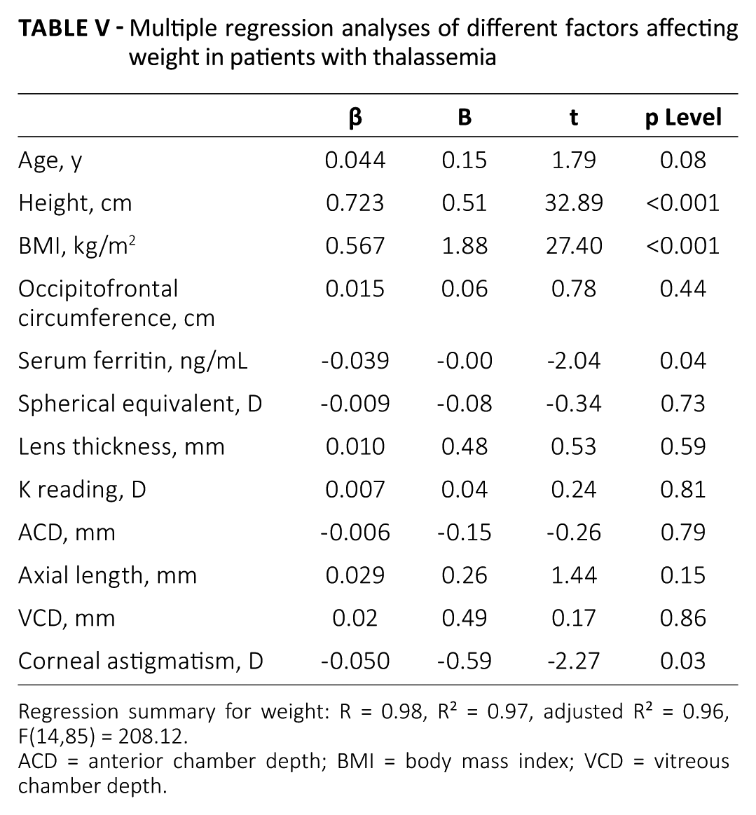
Regression summary for weight: R = 0.98, R² = 0.97, adjusted R² = 0.96, F(14,85) = 208.12.
ACD = anterior chamber depth; BMI = body mass index; VCD = vitreous chamber depth.
Multiple regression analyses of different factors affecting height in patients with thalassemia
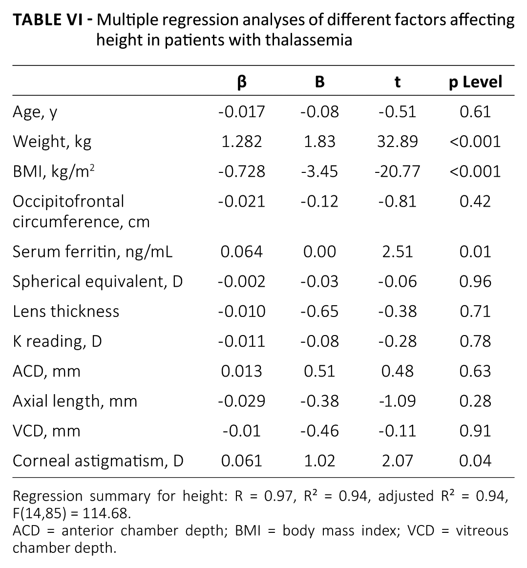
Regression summary for height: R = 0.97, R² = 0.94, adjusted R² = 0.94, F(14,85) = 114.68.
ACD = anterior chamber depth; BMI = body mass index; VCD = vitreous chamber depth.
Weight and height also correlated significantly with serum ferritin (p = 0.04 and 0.01, respectively) (Tabs. V and VI), while BMI showed a significant positive correlation with duration of disease (p = 0.04).
Discussion
β-Thalassemia is a major socioeconomic problem in countries where it is prevalent, as it requires lifelong follow-up and regular blood transfusions. The combination of transfusion and chelation therapy has prolonged the life expectancy of patients with thalassemia, who can now survive longer than before (11-13).
Some studies have evaluated anthropometric changes in patients with thalassemia, as a part of assessing other general manifestations and complications of the disease. Shamshirsaz and colleagues (6) assessed weight, height, and BMI in patients with thalassemia. They reported stunted growth and affected anthropometric measurements. Moreover, Fahim and coworkers (14) assessed the growth parameters in children with TM receiving packed red cells transfusion with chelation therapy. They concluded that children with TM have a significant delay in growth and metabolic abnormalities. The results of both studies are comparable to ours, as we found that weight, height, and BMI were significantly lower in children with thalassemia compared to controls (p>0.001).
Our study revealed significant ocular biometric and refractive error changes in children with TM, including steeper K readings (p = 0.03), shorter AXL (p = 0.005), shorter VCD (p<0.001), thicker crystalline lens (p<0.001), and greater myopic refractive error (p = 0.003).
The study by Nowroozzadeh and colleagues (4) assessed the different biometric and anthropometric changes in patients with thalassemia. The age of their sample was higher, with a wider age range (16.66 ± 6.20 years) compared to our study, which only included children (12.78 ± 1.24 years). Regarding their results for anthropometric data (weight, height, BMI, and OFC), they were comparable to ours, with a significant stunted growth in patients with thalassemia compared to controls, and no significant difference in OFC between the 2 studied groups.
Our results for ocular biometric parameters are almost comparable to those of Nowroozzadeh et al (4), who found steeper corneal K readings, shorter AXL, and thicker lenses in patients with thalassemia compared to controls.
However, Nowroozzadeh et al (4) found no statistically significant difference between patients with thalassemia and controls regarding the spherical equivalent, in contradiction to the significant myopic shift detected among our studied patients compared to controls (p>0.003). We can explain this myopic shift by the ocular growth retardation, manifested by shorter AXLs, resulting in a compensatory emmetropization process of attaining steeper corneas and thicker lenses to counteract the hyperopic effect of shorter eyes. This possibly occurred in an exaggerated form resulting in the established myopic shift. This assumption was enforced on plotting correlations between spherical equivalent and all studied biometric parameters, where we found significant correlations between spherical equivalent and both K readings and lens thickness.
The significant correlation between the spherical equivalent and ACD can be explained by the consequent change in ACD due to changes in corneal curvature and lens thickness.
As regards to the pattern of corneal astigmatism, Nowroozzadeh and coworkers (4) found more trend of ATA among patients with thalassemia compared to controls. On the contrary, we found an insignificant difference between the patients with thalassemia and controls, with both having ATA pattern.
Aksoy and colleagues (2), who performed a full ophthalmologic examination, including only AXL as a biometric parameter, found no significant difference in AXL between patients with thalassemia and controls, contradicting our results.
Our study investigated the relationship among physical anthropometry, ocular biometry, and refractive error in patients with TM, especially how the factors involved in growth retardation could adversely affect ocular growth as well (8-10), besides the possible ocular changes related to craniofacial abnormalities or bony orbit variations resulting from bone marrow expansion (4).
No significant correlations were observed between evaluated anthropometric variables and biometric changes, except for a significant correlation between corneal astigmatism and both weight and height. We do not consider this correlation of clinical relevance; hence, physical size changes are not directly related to ocular growth retardation.
Evaluation of bony orbital dimensions for detecting any changes and correlating them to refractive error and biometric parameters would have been of benefit to the study, especially as craniofacial changes in TM may result in an abnormal bony orbit and subsequent variations in ocular biometry and/or refraction (4). However, ethical limitations of exposing children with thalassemia to a potentially hazardous dose of irradiation for performing a computed tomography (CT) scan precluded us from surveying this aspect. We recommend future studies to address this point, perhaps through recruiting patients already undergoing CT scan for other systemic aspects of the disease. Moreover, exploring the orbit through exophthalmometry, which was previously estimated in the normal pediatric population (15-17), would be another relevant evaluation for further studies.
We also noted that weight and height correlated significantly with serum ferritin (p = 0.04 and 0.01, respectively). This is expected, as high ferritin indicates poor disease control, which can affect the rate of patients’ growth. Moreover, BMI was found significantly correlated to TM duration, reflecting progressive growth retardation along the course of the disease.
Conclusion
Compared to controls, children with thalassemia have significant retardation in general and ocular growth (smaller BMI and shorter AXL). Ocular growth changes probably resulted in compensatory biometric changes (steeper corneas and thicker lenses) to reach emmetropization, with an exaggerated response and subsequent myopic shift. However, growth retardation is not directly related to ocular growth changes, myopic shift, or variations in biometric parameters.
Footnotes
Disclosures
Financial support: No financial support was received for this submission.
Conflict of interest: None of the authors has conflict of interest with this submission.