






Abstract
Cementation of a polyethylene liner into the well-fixed shell is a convenient option for revision total hip arthroplasty. We retrospectively reviewed 45 patients who had liner cementation to investigate the risk factors which gave rise to major complications and reoperation. Patients were observed for a minimum of 7 years (range 7.8-14 years). Relevant risk factors (age, BMI, surgical approach, previous cup size and position, types of coated surface) were assessed with Cox regression analysis. The mean Harris Hip Score was improved from 62.5 (range 57-68) preoperatively to 87.1 (range 70-97). A total of 7 hips (15.5%) had acetabular component loosening that was treated with reoperation. Prevalence of acetabular component loosening was statistically significantly higher in hydroxyapatite-coated group (5 of 13) than in the Ti-coated group (2 of 32, p = 0.015). All recurrent dislocations occurred in patients treated with a posterior approach. Diameter of the previous metal shell of below 54 mm showed a lower 10-year survival rate than those greater than 54 mm in diameter. PE liner cementation in stable metal cup is a useful alternative option for carefully selected patients. Pre-existing HA-coated cups as well as small sized cups were indicative of poor outcomes.
Keywords
Introduction
The use of conventional polyethylene (PE) liners and metal heads has shown that liner wear and osteolysis have been implicated as common complications of total hip arthroplasty (THA) (1). Isolated acetabular polyethylene wear and periacetabular osteolysis in a well-fixed acetabular component is considered as an indication for revision surgery (2).
In contrast to cemented acetabular components, which commonly require revision because of a linear pattern of bone resorption and subsequent loosening (3), osteolysis associated with a cementless component is usually localised and expansile, leaving the prosthesis well fixed (4).
Some have recommended revision of the entire acetabular component when a liner fails (5). However, removal of osteointegrated cementless shell is accompanied by potential complicationsevere bone loss. Simple exchange or cementation of a liner into the well-fixed shell is a convenient and attractive option, and the associated osteolytic lesion can be treated by local grafting through the holes of the metal shell that ensures better bone preservation with reduced morbidity as compared with complete acetabular component revision (1, 6). Moreover, the exchanged or cemented acetabular liner allows patients to immediately weight bear. Therefore, the cementation of PE liner into a well-fixed component is an effective method when a locking mechanism is not intact due to damage of metal shell or a liner.
A few series have demonstrated the outcome of this technique, however most investigations to date are limited by small patient numbers and short follow-up (5). The purpose of the current study was to determine the risk factors (age, BMI, surgical approach, previous cup size and position, types of coated surface) which gave rise to major complications and reoperation.
Patients and Methods
Between 1998 and 2005, we performed cementation of a PE liner into a well-fixed shell in 50 hips (48 patients) undergoing revision hip arthroplasty. This study was approved by our institutional review board and all patients gave informed consent. All procedures were performed by a single surgeon (PMS) at a single institution.
Inclusion criteria for enrolment in this study were a cementless acetabular component without radiological and intraoperative loosening. The revised liner had to be a PE liner and the mean follow-up period was more than 7 years. The exclusion criteria applied were an index age of more than 70 years old or less than 30 years, a follow-up period of less than 5 years follow-up after primary total hip arthroplasty, isolated PE liner exchange as well as ceramic and/or metal hard sandwich liners.
In total, 5 patients were excluded: 2 patients due to hard-on-hard bearing liners and 3 due to being more than 70 years old with isolated PE liner exchange. As a result, 45 hips (43 patients) consecutively operated cases constituted the study cohort. No patients were lost to clinical or radiographic follow-up. We investigated the coated types and sizes of the primary implants and of the metal shells by reviewing previous operations and hospital records. All patients were examined clinically and radiographically at each follow-up. Failure was defined as revision of the cup for any reason.
This study cohort included 31 male patients (33 hips) and 12 female patients (12 hips). The mean age was 57.3 years (range 31-69 years) at the time of revision surgery. The mean patient body weight was 63.7 ± 21.1 Kg (range 46-85 kg) and the mean body mass index (BMI) was 25.6 (range 21.7-32.1). The most common underlying diagnosis was osteonecrosis of femoral head (82.2%) followed by primary osteoarthritis (4.4%), traumatic osteoarthritis (4.4%), rheumatoid arthritis (4.4%), fracture of femoral neck (2.2%) and a dysplastic hip (2.2%). The mean time from primary total hip arthroplasty to liner change was 10.9 years (range 7.2-18.9 years), and the mean diameter of the existing acetabular component was 53.7 mm (range 48-68 mm). The minimum follow-up was 7.8 years (mean 10.7 years; range 7.8-14 years) (Tab. I).
Demographic data

BMI = Body mass index; Ti = Titanium; HA = Hydroxiapatite.
The outer surfaces of the metal shells were porous titanium (Ti) coating (32 hips; 26 Zimmer HG II, 2 DePuy Duraloc, 1 Allofit-S, 1 Osteonics Omni fit, and 2 Bicontact plasmapore cup). Furthermore, there were 13 hips with hydroxyapatite (HA) coated cup (11 SEM, Paris, France and 2 ABG I, Howmedica, Europe). All of the titanium cups had 0 to 6 dome screw holes. The HA-coated cups had 6 to 9 holes.
Surgery
Liner cementation procedure was performed using a posterior approach for 27 hips while an anterolateral approach was used for 18 hips. In each case, we assessed metal shell stability by pull/push testing with a vice grip. We also removed the dome screw and curetted screw holes to improve cement fixation. To manage pelvic osteolysis we inserted morselised cancellous allograft or mixed hydroxyapatite (HA) granules (Wright Medical Technology, Arlington, TN) through the empty holes of the metal shell (Figs. 1 and 2). If no holes were available as was the case for 1 Duraloc cup (DePuy, Warsaw, IN), they were drilled using a 6.5 mm drill bit on the upper margins of the shell. We routinely changed the old PE with new conventional PE liner (2 mm downsized) and 28 mm chrome-cobalt alloy for the femoral head (skirted neck used in 1 hip). The backs of the new PE liners were prepared by working them with a high-speed burr to produce cruciate and circumferential grooves in a spider web-like pattern or cruciate with a dimpled (golf ball-like) pattern, while taking care to avoid making deep grooves that might jeopardise liner integrity (7, 8). A new PE liner was cemented into the metal shell in correct orientation.
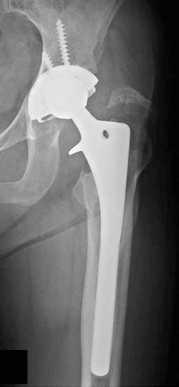
Preoperative radiograph of a 53-year-old woman shows PE liner wear and osteolysis around the screw in a 54 mm metal shell (HG II, Zimmer Warsaw IN).
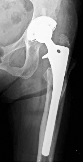
Radiograph taken 8 years after liner cementation with morselised allograft, showing no expansion of pelvic osteolysis.
Postoperatively, patients were mobilised as soon as medically possible. Partial weight bearing was allowed 2 weeks after surgery. In general, partial toe-touch weight-bearing (⅙ of body weight) using crutches or a walker was maintained for 10 to 12 weeks. Progression to full weight bearing was individualised. All patients were clinically evaluated after 6 weeks, 3 and 6 months, and then annually after revision surgery.
Clinical results were evaluated using Harris Hip Scores (HHS) (9) and WOMAC scores (10). Clinically, scores from 90 to 100 points were graded as excellent, from 80 to 89 points as good, from 70 to 79 points as fair, and hips with a score below 70 points as poor.
Four orthopaedic surgeons with over 5 years of orthopaedic experience held consensus building session for radiographic measurements, and 2 of these measured radiographic indices for interobserver reliability determinations. Interobserber reliability was evaluated using intraclass correlation coefficients (11) and mean absolute differences (12). Preoperative and final follow-up radiographs were evaluated to determine the extent of osteolysis in pelvis anteroposterior (AP), cross-table lateral, and oblique view. The size and extent of osteolysis in the pelvis radiograph were measured at their longest extensions. The location of the osteolytic lesion was recorded along the DeLee and Charnley zones (13).
The position of the acetabular cup component was measured using inclination and anteversion. These were measured radiographically as follows: a line joining the inferior margins of the teardrops on AP pelvic radiographs was drawn. The intersection of that line with a line marking the plane of the opening of the acetabular component was defined as the angle of inclination. Acetabular cup anteversion was determined on plain radiographs using Widmer's method (14). Bone ongrowth into the acetabular component was considered present when direct contact of the trabecular striation had occurred between the pelvis and the outer metal shell. All radiographs were qualitatively evaluated using a digitalising tablet powered by PACS (Picture Archiving and Communication System; Maroview, Marotech, Seoul).
During the postoperative follow-up series we checked whether a new osteolytic lesion had occurred or whether an old one had expanded. Preoperative and postoperative radiographs were compared by viewing them on the same screen. Migration of the acetabular component was defined as a change of >5 mm either vertically or horizontally in the position of a cup or a change in cup angle of >5° using the method described by Massin et al (15). To evaluate the position of the acetabular component, vertical migration was determined with respect to the inferior aspect of the teardrop, and the Köhler line which was used to measure horizontal migration. Leg length discrepancy was measured pre- and postoperatively by comparing the interischial line to the top of the lesser trochanter. Clinical outcomes were determined preoperatively and recorded by a research fellow at each clinic visit. Serial radiographs were taken preoperatively and before each follow-up examination.
In this series, polyethylene wear analysis was performed using digitised pelvis AP radiographs. We defined early wear as meaning that femoral head penetration in any pelvis radiograph was comparable to the mean penetration rate (at least 5 years after the liner cementation). Polyethylene wear was measured using the method described by Latimer and Lachiewicz (16). Wear of more than 0.2 mm/year was considered a rapid PE liner wear (17). Known sizes of femoral heads were used to correct for magnification on radiographs.
Statistical analysis
Descriptive statistics were used to summarise patient's dermographics, and radiographic measurements. Data are presented as means with 95% CIs when normally distributed and as medians with interquartile ranges when not normally distributed. The normality of the preoperative and postoperative HHS was tested using Kolmogorov-Smirnov test and the paired t-test was used for significance. Bi-variate comparisons between groups were performed by the Student t-test or analysis of variance if there were more than 30 in the group. The Mann-Whitney test or the Kruskal-Wallis test was used if there were fewer than 30 in the group. Group comparisons were analysed by Fisher's exact test.
A power analysis showed, to detect a 10% difference in the presence of an abnormal cup inclination angle (with an alpha of 0.05 and power 0.08), at least 40 hips were required.
We calculated hazard ratios using Cox regression analyses to identify which preoperative factors significantly contributed to early failure of liner cementation. Independent variables included; age, BMI, time to primary surgery, anteversion, inclination, size of primary cup and the amount of liner wear preoperatively. Kaplan-Meier survivorship curves were generated for reoperation for any reason.
Interobserver reliability of radiographic measurements was analysed using intraclass correlation coefficients (ICCs). Statistical significance was accepted for p values of <0.05 (Tab. II).
Interobserver reliabilities of radiographic measurements

Results
The median number of years since the primary total hip arthroplasty has been implanted was 10.9 years and the interquartile range was from 7.2 to 18.1 years. The mean BMI score was 25.6 (21.7-32.1 kg/m2). The median cup inclination angle was 38.8°, and the interquartile range was 26.2 to 59.8°. The median cup anteversion angle was 16.1°, and the interquartile range was 10.2 to 27.2. All patients were followed more than 7 years after liner cementation as revision surgery.
The mean Harris Hip Score was improved from 62.5 (range 57-68) preoperatively to 87.1 (range 70-97). The WOMAC score was improved from 58.7 (range 47.5-67.2) to 80.7 (range 75-90.3) at the final follow-up. All patients were ambulatory at most recent follow-up.
There were 12 additional reoperations. A total of 7 hips (15.5%) had acetabular component loosening that was treated with reoperation. Of the 7 hips, 2 hips had Ti-coated acetabular component, 5 hips had HA-coated 1. Prevalence of acetabular component loosening is statistically significant higher in HA-coated group (5 of 13) than in Ti-coated group (2 of 32, p = 0.015). Of the 7 hips, 5 hips had high linear penetration wear (mean 0.506 mm/year; SD, 0.27) compared to remaining hips (mean 0.15 mm/year; SD 0.14). Of the 7 hips, 4 hips were treated with acetabular cage with allograft; 1 hip had revision with cementless acetabular component with autograft.
A total of 5 hips (11%) had recurrent instability that was treated with reoperation. Of the 5 hips, 2 hips were converted to constrained implant; 3 hips had revision of the acetabular component with a 36 mm femoral head diameter. All recurrent dislocations occurred in patients treated with posterior approach. Combined dislocation and loosening occurred all in HA-coated group of 3 hips.
The median area of the osteolysis was 741.9 mm2, and the interquartile range was 315.7 to 1533.1 mm2. The osteolysis was located in Zone I in 11 hips, and in Zone I and II in 5 hips. The all of preoperative pelvic osteolysis were nonprogressive during the follow-up period.
The Cox regression analysis found 11 demographic and radiographic parameters that had an odd ratio for potential confounders, including age, sex, BMI, previous approach, a size of metal shell, hydroxyapatite surface coating of acetabular component, and the position of metal shell. We identified 3 predictors of reoperation: 1) a size of metal shell (odd ratio, 1.7; 95% CI, 1.19-2.88; p<0.001); 2) preoperative inclination of metal shell (odd ratio, 1.9; 95% CI, 1.02-3.68l; p = 0.01), and 3) HA surface coating of metal shell (odd ratio, 2.0; 95% CI, 1.7-3.44; p = 0.003).
In the entire cohort of 45 hips, we found a cemented liner survival rate of 71.8% (95% CI, 64.7%-78.9%) at 15.5 years using to reoperation as the end point. Ti-coated metal shell showed a higher 10-year survival rate than HA-coated metal shell (90% [95% CI, 85.5%-95.5%] and 46.7% [33.8%-59.6%], respectively, Fig. 3). Inclination of the previous metal shell angle of above 45° showed a lower 10-year survival rate than angle of below 45° (27.3% [95% CI, 13.9%-40.7%] and 86.4% [95% CI, 79.8%-93.0%], respectively). Diameter of the previous metal shell of below 54 mm showed a lower 10-year survival rate than sizes greater than 54 mm in diameter (58.3%, [95% CI, 85.4%-97.2%] and 91.3%, [95% CI, 79.8%-93.0%], subsequently, Fig. 4).
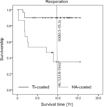
Kaplan-Meier survival curve with failure defined as any reoperation involving the hip. Survivorship curves, as determined by the Kaplan-Meier method, with reoperation as the end point. The 2 lines represent the proportion of hips had not had revision in the 2 comparative groups of patients who had titanium (Ti) coated cup and patients who had hydroxyapatite (HA) coated cup.
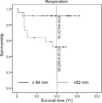
Kaplan-Meier survival curve with failure defined as any reoperation involving the hip. Survivorship curves, as determined by the Kaplan-Meier method, with reoperation as the end point. The 2 lines represent the proportion of hips had not had revision in the 2 comparative groups of patients who above 54 mm cup in diameter and patients who had below 52 mm cup.
We experienced intraoperative greater trochanter fractures in 5 hips (1.1%) which were managed with cerclage wires or permanent sutures. All fractures occurred in patients with localised osteolysis in the proximal femoral area. This fracture was usually a mild or un-displaced fracture. At the last follow-up, fracture fragments had migrated proximally in 2 hips. These patients complained of none or mild discomfort at the fracture site and abduction weakness, but no limping was noted.
Discussion
Generally, removing grossly loose cemented acetabular components is not difficult, however, removal of well-fixed cemented and, especially, cementless components has remained a challenge, often causing significant bone destruction and critical obstacles at subsequent revision procedure. Liner cementation has been frequently used to treat PE wear and localised acetabular osteolysis (18, 19). The technique did not disrupt the stability of the cups and femoral stems which means that potential damage was minimal.
Revision surgery has always been associated with higher dislocation rates than primary total hip arthroplasty due to soft tissue disruption, different components, and the surgical approaches used. In fact, dislocation rates after primary THA range from 0.3 % to 11 %, but revision dislocation rates can be as high as 26% (15). Furthermore, dislocation rates for the posterior approach after revision surgery are significantly higher than those for the anterolateral approach, and the posterior approach has been reported to have a dislocation rate up to 3.2% greater than the direct lateral and/or anterolateral approaches (19). This is likely due to multiple factors and may be related to difficulties encountered with formal repair of the capsule and external rotators, especially true in cases of isolated liner exchange or cementation. Boucher et al (18) reported a 25% incidence in instability following liner exchanges performed using the posterior approach. In our study, we found that dislocation rate was higher with the posterior approach (p = 0.032) compared to the anterolateral approach.
At reoperation after liner cementation failure we frequently found that the liner was posteriorly protruding from the metal shell. This was probably due to surgeons inserting the liner in an elevated position to prevent dislocation. The liner may then have been pushed out by the cement during the curing period. Adequate liner containment within the metal shells and repair of the posterior capsule as well as short external rotators for the posterior approach appear to be critically important in terms of achieving good clinical results (20).
Eccentrically located liners or laterally located PE liners have been reported to show impingement between the rim of the metal shell and neck (21). Contact between a metal neck and a PE liner can have a number of potentially adverse consequences, which include motion and function limitation, increased stress on the liner rim, generation of rim wear and a potentially increased risk of osteolysis (22). We cannot draw any conclusions about the influence of head-neck ratios because our study only included a single skirted neck.
High PE wear is frequently observed after PE liner changes. Another suspected cause is impingement. Retrieval studies have shown that impingement contributes to wear. Yamaguchi et al (23) correlated impingement with linear wear, and found a mean wear rate of 0.33 ± 0.28 mm/yr for impinging liners as compared with 0.19 ± 0.14 mm/yr for liners without impingement (p = 0.009). In this study, we found that cup sizes of less than 54 mm and inclinations >45° were related to wear, loosening and failure (Tab. III). In this study, 3 hips experienced dislocation as well as loosening. We believe that these combined complications are due to initial neck and cup impingement, which causes minimal separation from the socket and subluxation, followed by dislocation at extreme ranges of motion such as those caused by squatting (especially prevalent in Asian people). HA-coated cups showed inreased rates of loosening (p = 0.006). The outer surfaces of HA-coating on the smooth surfaces of metal shells are also a risk factor related to loosening. This finding is supported by the concerns expressed in another report (24). The cause of the high PE wear of HA-coated cups is explained by HA particles separating from the coating, which then lead to increase coated surface disruption from backside of the cup and facilitate wear of the PE (24).
Cox regression analysis to identify significantly affecting factors to reoperation after liner cementation

The cup may be well-fixed, but symptoms of hip pain can occur in association with osteolysis. This is likely related to synovitis and effusion that can develop in response to polyethylene wear debris. Superior and posterior acetabular defects may compromise mechanical support for the acetabular component in the weight bearing region and should be treated surgically. If the shell is well fixed and in a good position, liner exchange or cementation with or without and bone graft for the osteolytic lesions is a better option than cup revision because this does not risk additional bone loss during acetabular component removal. We suggest that the cup size and the state of the coating of the outer metal cups should be considered for this procedure. Less than 54 mm or an HA-coated cup should be considered as a risk factor of liner cementation technique failure.
In conclusion, we conclude that isolated liner cementation appears to be an effective method when strict indication criteria are followed.
Footnotes
Financial support: This paper was supported by Fund of Biomedical Research Institute, Chonbuk National University Hospital and research funds of Chonbuk National University in 2014.
Conflict of interest: None.