






Abstract
Background
Staged revision for periprosthetic infection of the hip is an accepted and widely used technique by many surgeons. However, single-stage exchange of the hip prosthesis remains an attractive option to others because of the advantages of reduced morbidity, shorter treatment time and hospital stay in addition to the reduced cost of treatment.
Hypothesis
Single-stage revision for periprosthetic hip infection can achieve excellent results if a specific protocol for patients’ selection and management is followed.
Methods
52 patients with evidence of periprosthetic infection had preoperative aspiration of the affected hip. The infecting organisms were identified in 33/52 and single-stage revision was performed. The remaining 19 patients had a 2-stage exchange arthroplasty. Patients in the single-stage revision protocol had antibiotic loaded morsellized bone graft, a cemented cup and a long cementless stem.
Results
At an average follow up of 6 (range 4-8) years postoperatively, only 1 case of persistent infection was found in the single-stage group – a 97% rate of eradicating infection was achieved.
Discussion
Single-stage exchange achieves excellent success rate in patients with periprosthetic infection when a specific protocol for patient selection and management is followed.
Introduction
Staged revision for periprosthetic infection of the hip is an accepted and widely used technique by many surgeons (1, 2). However, single-stage exchange of the hip prosthesis remains an attractive option to others because of the advantages of reduced morbidity, shorter treatment time and hospital stay in addition to reduced treatment coasts (3–4–5–6).
There are many contradictory reports on the results of single-stage exchange hip arthroplasty with high success rates in some and unfavorable results in others (7–8–9). This contradiction may be related to the differences in the methods of patients’ selection, technique of implant exchange between cemented and noncemented hips in addition to the intra- and postoperative antibiotic (AB) protocols (9).
The advantages of the staged revision strategy are increased identification of the infecting organisms and double debridement of the infected tissues (10–11–12). This, however, does not rule out the value of performing single-stage exchange in some patients (13), and the question remains of how to recognise the category of patients that would benefit most from each technique.
Manifestations of infection varies from late loosening of implants to acute periprosthetic infection with draining sinuses and septicemia (14). These manifestations are related to the patient's general condition, the infecting organism, local soft tissue condition and bone defects (15).
An active draining sinus, acute septicaemia and inability to identify the infecting organism are seen by many surgeons as contraindications for single-stage revision (16). In this series, a specific protocol for patients’ selection and management was tested in the hope that it would allow for identification of suitable patients who may benefit from excellent outcomes with single-stage exchange in periprosthetic infection of the hip.
Methods
52 patients with chronic periprosthetic infection of the hip have been included in this prospective study that was conducted between August 2006 and September 2011. Approval of the local research committee was obtained as well as patients’ consent for inclusion in the study.
All patients had elevated ESR >30 mm/h and CRP >10 mg/L in addition to radiological evidence of bone resorption and/or periosteal reaction around the infected implant. Patients were subjected to preoperative aspiration of the hip in a sterile theatre under x-ray control and specimens collected were directly cultured on blood agars for 2 weeks in addition to leucocyte count.
The inclusion criteria for single-stage (SS) exchange were: absence of active draining sinus; absence of acute septicaemia; identification of the infecting organism by preoperative aspiration; availability of suitable AB for administration through parenteral route; and finally the presence of a viable soft tissue envelope around the hip at the end of debridement. Patients who did not fulfill the above criteria were excluded from the single-stage exchange protocol.
Preoperative assessment of patients’ general condition was performed and bone defects on the femoral and acetabular sides were graded according to AAOS classification. Harris Hip Scores (HHSs) for hip function were recorded.
The single-stage revision (SS) operative technique
All patients were operated on the lateral decubitus position using the posterior approach described by Gibson-Kocher and an extended trochanteric osteotomy (ETO) was used (17). The length of the osteotomy from the tip of the greater trochanter was determined by the need to remove distally fixed cement mantles. In all cases flipping the greater trochanter allowed removal of distal cement mantles and infected membranes at the bone cement interface. The extensive debridement of all infected bony and soft tissues on the femoral and acetabular sides was performed to leave viable bleeding tissues. On the acetabular side, special attention was given to circumferential removal of the hip capsule. When acetabular erosion had happened, the debridement stops at the medial periosteal cover before entering the extra-peritoneal space. Bleeding surfaces had to be seen before the debridement of sclerotic bone or infected membranes were completed. On the femoral side, meticulous debridement of the medullary canal, calcar area and inner surface of the greater trochanter was performed. At the end of debridement, copious wash of bone surfaces by H2O2 (10%) was performed. This was followed by irrigation using pulsatile lavage systems. 6 litres of normal saline were used at this stage. Once the debridement was completed, intravenous infusion of AB was commenced.
The procedure was performed with the concept of 2 surgeries under the same anaesthesia. Therefore, different sterile set of instruments, drapes and gowns were used before starting the second part of the surgery for reconstruction and implantation of the prostheses.
6 tissue specimens were collected and sent for standard and extended culture (2 weeks) and antibiotic sensitivity tests.
Assessment of acetabular bone defects and impaction bone graft were performed using AB loaded fresh frozen femoral head allograft. Preparation of the graft was performed by removing all attached soft tissues and cartilage then dividing the remaining cancellous bone using power saw and manual rongeurs into bone chips 8-12 mm in size. The bone chips were rinsed with saline. Large doses of AB in powder form (Fig. 1) were mixed to the cancellous chips of the allograft (18–19–20). The bone chips were then impacted progressively in layers into the acetabular defects until fully seated and filled the defects (21, 22). Trial heads from the hip tray were used for the purpose of impaction. No metal mesh was added to the floor or sides of the acetabulae. Instead, direct contact between the bone graft and viable periosteum and bleeding cancellous bone was preferred. However, in cases of severe acetabular defects (grades III & IV) a Burch-Schneider cage was used with superior screw fixation, and seating the inferior lip into the ischium (Zimmer, GmbH, CH-8404 Winterthur, Switzerland) (Fig. 2A, B). The cage was used in 4 cases.
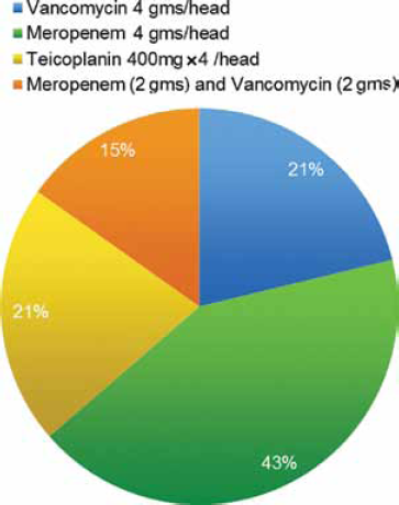
Types of antibiotic powders added to the fresh frozen allograft.
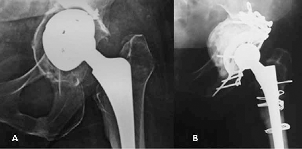
(
All polyethylene cups with an inner diameter 32 mm were cemented in all acetabulae (ZCA, longevity cross linked all poly cup, Zimmer) using standard high fatigue gentamycin loaded bone cement. The powder of 1 vial from the chosen antibiotics was added to each pack of 40 gm bone cement after 1 minute from the beginning of the mixing process (23–24–25).
On the femoral side straight Wagner SL revision cementless stems (Zimmer, GmbH, Sulzerallee 8, 8404 Winterthur, Switzerland) were inserted on all femora having accepted the height and rotation of the trial prosthesis. 32 mm outer diameter cobalt chrome heads were selected in all cases.
Reattachment of the osteotomised trochanter was finally performed using stainless steel (Ortron) doubled wires and soft tissue repair of the external rotators using Ethibond No. 5 trans-osseous sutures.
Postoperative antibiotic protocol
Intravenous infusion of antibiotics according to the preoperative culture results was commenced intraoperatively and continued until tissue biopsy culture results were available (Tab. I). A Combination of IV and oral antibiotics was administered for 4 weeks (Tab. I). After the 4th week, oral antibiotics were continued for another 6-8 weeks.
Data about patients’ comorbidities, organisms identified by preoperative aspiration and through tissue specimens. In addition, antibiotics given intravenously during the first 4 weeks and the following “6-8 weeks” oral antibiotic regimen is provided
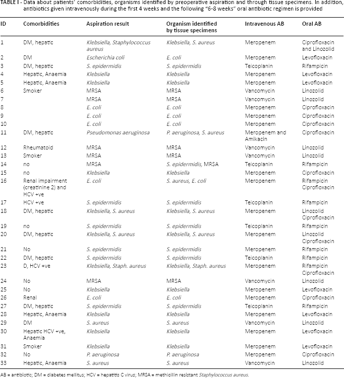
AB = antibiotic; DM = diabetes mellitus; HCV = hepatitis C virus; MRSA = methicillin resistant Staphylococcus aureus.
Patients were allowed touch weight-bearing (WB) mobilisation from the second postoperative day and for 6 weeks. Partial WB gradually progressed from the 6th to the 12th weeks when full WB was permitted.
Laboratory assessment of the patients’ ESR and CRP was conducted on a weekly basis during the first 6 weeks, then every 2 weeks for another 6 weeks and then at 6 and 12 months from surgery. Similarly, x-rays were taken immediately postoperatively then at 3, 6 and 12 months. The HHS was recorded at the end of 6 and 12 months then annually.
Patients who could not attend for checkup after the second postoperative year were contacted by phone and x-rays were annually sent for review.
The Wilcoxon matched-pairs signed-ranks test was used to compare pre- to postoperative results.
Results
52 patients with hip periprosthetic infection were identified. Out of these 52 patients, 33 patients (average age of 61 [range 41-73] years) had a single-stage revision for periprosthetic infection. 13 patients were females and 20 were males. These patients have been followed for an average 6 years (range 4-8 years). None of these patients were lost in the follow-up. 2 patients died 3 and 4 years postoperatively from nonrelated causes. At the latest follow-up, 32 out of the 33 patients (97%) were free of infection. None of these patients was revised or are awaiting revision.
The preoperative aspiration was successful in discovering the organism preoperatively in all 33.
The infecting organisms that were identified by preoperative aspiration were confirmed by tissue specimens collected at time of revision. However, additional microbes were detected in 3 patients (Tab. I).
Bone defects and Impaction grafting was performed in all cases. The grades of acetabular bone defects were variable as shown in (Fig. 3). Radiological assessment of graft incorporation was performed using the criteria used in previous reports (26, 27). No case of graft resorption or lysis was observed in these patients. Incorporation and maturation of the graft within the previous defects were recorded in 29 out of the 33 patients. It was possible to observe trabecular arrangement within the graft even in cases where a metal cage was inserted. Using the criteria described by DeLee and Charnley (28) radiolucent lines were seen in zone II in 3 patients and zone I in 2 patients.
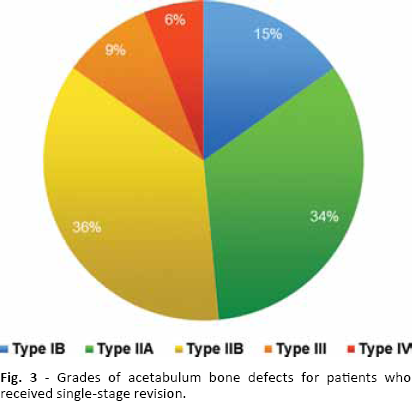
Grades of acetabulum bone defects for patients who received single-stage revision.
Bony union of the ETO occurred in 28/33 patients (Fig. 2). Proximal migration of the trochanter by <10 mm was observed in 5 patients. Trendelenburg gait was recorded in these patients but no hip instability was found.
The HHS has significantly improved from 30 ± 3 pre- to 87 ± 4 postoperative (mean ± STD) (p<0.00001).
Complications
1 patient in this series had a single dislocation that was reduced by closed reduction and continued to be stable afterwards. 1 patient had a draining wound with haematoma formation. This was washed out in theatre and coagulation profile was corrected. The same patient had recurrence of infection 18 months from the index procedure. He continued to use oral antibiotics and opted against further surgery.
2 patients had intraoperative fractures in form of extension of the ETO in 1 and split along the medial wall of the proximal femur in another. All fractures united with the use of circular wires and the period of partial WB was extended to 12 weeks.
Discussion
One of the challenges facing orthopaedic surgeons is to define the best indications and technique for a single-stage exchange in periprosthetic infection (29). Though excellent results of single-stage which are comparable to the 2-stage revision protocols have been published (29), the orthopaedic community needs to see replications of these results from different centres and health systems.
Successful eradication of infection only occurs through adequate debridement of all infected tissues as well as implants. In addition to this, it was equally important to deliver the correct antibiotics in the appropriate doses both locally and systemically (6, 8). Therefore, an ETO was performed in all our cases. The ETO allowed access to the intramedullary canal for removal of bone cement as well as the membranes at the bone cement interface.
On the acetabular side, infected membranes usually tracked through the planes of the original approach. When the approach of the original surgery was the anterolateral approach, identifying the extent of the infected membranes can be achieved by flipping the greater trochanter.
One of the important technical points in these cases is how the surgical approach could be modified during surgery. In many occasions the infected tissues were found after incising the short external rotators or the posterior capsule of the hip joint. However, if caseous materials were found underneath the tensor fascia lata (TFL), the origin of these infected tissues should be identified and included in the debridement.
In their consensus meeting, the musculoskeletal infection society considered patients with draining sinus tracts as a contraindication to single-stage revision. Meanwhile, preoperative identification of the infecting organism and availability of suitable AB were considered prerequisites for considering single-stage exchange (3).
In this series, actively draining sinus tracts or inability to identify the infecting organism preoperatively were considered as exclusion criteria.
The value of preoperative aspiration has been emphasised in many studies (36–37–38–39). This procedure is established in the AAOS paradigm for diagnosis of infection (40). The limitation is still on the ability to identify an organism preoperatively in the cases suspected to be infected. Williams et al (41) reported the overall accuracy of preoperative hip aspiration in defining the infecting organism as 90.1% with 84% sensitivity and 94% specificity. In another study of the same group the positive predictive value of hip aspiration in the radiology department was 74% while the negative predictive value was 94% (42).
In this study, preoperative aspiration has been successful in identifying the organism in 33/52 patients (63%). The specificity of the positive results was 91%, as additional organisms were discovered from the culture results of the specimens collected during surgery in 3 patients. Only 1 of these 3 patients had recurrence of infection at 18 months postoperative. Improving the accuracy and specificity of preoperative aspiration is an important task that may influence decision making and results in the future.
In this series high doses of local antibiotic (vancomycin and meropenem) (Fig. 1), were added to the fresh frozen allograft. Antibiotic impregnated cancellous graft was successfully used in previous studies (43, 44), and was found to locally deliver very high concentration of the antibiotics that can affect not only the planktonic form of bacteria but also the sessile clusters and biofilm colonisation (6).
The most commonly used AB for impregnation of cancellous graft is vancomycin (8, 43). It was found to elute locally from bone graft and deliver very high concentrations that are several folds above the minimum inhibitory concentration of the sensitive bacteria especially the MRSA (18). However, in this series other gram negative bacteria - E. Coli and Klebsiella were identified in many cases preoperatively (Tab. I). The combination of vancomycin and meropenem was found to have a synergistic effect and previously been tested with bone cement (23–24–25–26–27–28–29–30–31–32–33–34–35–36–37–38–39–40–41–42–43–44–45). Therefore, the choice of AB added to cancellous bone graft was according to the preoperative identification of the infecting organism and its sensitivity profile.
The advantages of using antibiotic-loaded bone graft are multiple. Firstly, it allows treatment of bone defects on the acetabular side. It is important to note here that no metal meshes were used to allow direct contact between the viable bleeding tissues and the AB loaded graft. Secondly, achieve better stability for acetabular components and reduce the dead space. Finally, local antibiotics can be delivered in very high doses that cannot be achieved through systemic routes (8, 19).
ABs were infused intravenously (IV) during surgery in the interim period between completion of debridement and insertion of the new implants. These IV antibiotics were continued for 4 weeks postoperatively in combination to other forms of oral antibiotics. Although IV antibiotics were stopped after 4 weeks, the treatment by oral antibiotics was continued for another 6-8 weeks.
Variability in the outcome of single-stage revision for infection is observed in different reports (1, 6). Patients’ selection, quality of debridement and antibiotic protocols varies between centers. The extent of infection into tissue planes and quality of debridement received marked attention in the literature. However, categorisation of the extent of infection into different tissue planes, involvement of muscle layers and bone affection could not be classified. It is logical to think that the ability to excise infected tissues which are confined to the hip space would be more precise and successful than trying to excise infected tissues that are widespread into different muscle compartments or bony sequestrum either femoral or acetabular. Developing a classification that differentiates confined or contained infection from more severe widespread forms of virulent infection would make a step forward in selecting patients for single stage revision and comparison to other studies.
Previous studies that employed the single-stage exchange strategy achieved variable rates of success from 54% (1) to 90% (6) but included patients with draining sinuses and without prior identification of the organism. When a standard protocol for patients’ selection was followed higher rates of success were achieved (3, 13).
The limitations in this study are related to the relatively small number of patients and the lack of a control group. However, it has tested a protocol for patients’ selection and management and reported its outcome without losing any patient from the follow up.
In conclusion, we have shown similar or higher rates of success in eradication of infection when compared to a 2-stage reimplantation protocol. We achieved this by restricting the single-stage revision protocol to patients with no draining sinuses in whom an organism could be identified preoperatively, achieving excellent results at mid-term follow-up.
Footnotes
Financial support: None.
Conflict of interest: None.