






Abstract
Introduction
Gait analysis is one of the poorly understood dimensions of the functional results obtained after periacetabular osteotomy (PAO) due to dysplasia of the hip.
Materials and methods
Spatiotemporal parameters as well as coronal and sagittal plane kinematics and moments were compared among three demographically similar groups of subjects: (i) 23 patients with excellent results according to the Harris Hip Score (HHS) (mean 6.6 years of follow-up); (ii) 12 patients with good and fair results according to the HHS (mean 9.3 years of follow-up); and (iii) 35 asymptomatic controls.
Results
50% of the analysed sagittal plane kinematic parameters were significantly better (p<0.05) in the control group not only for the hip joint but also for the ipsilateral knee and ipsilateral ankle. A vast majority of the other evaluated gait parameters resulted similar among the groups (p>0.05).
Conclusions
In our experience, most of the gait parameters after PAO are close to those observed in control subjects; however, the obtained pattern cannot be classified as a completely normal gait. The excellence in terms of functional results seems not to be determined by the achievement of this theoretical goal.
Introduction
It is well accepted that developmental dysplasia of the hip (DDH) is 1 of the main causes of hip osteoarthritis among young patients (1). Currently, the Bernese periacetabular osteotomy (PAO) (2) is the most popular surgical strategy to treat developmental dysplasia of the hip (DDH) in patients with a mature skeleton and adequate joint congruency to attempt joint preservation, which aims to prolong the survival of the native hip as much as possible without the requirement of a total hip arthroplasty (THA) (3).
Over the last two decades, it has been proved that PAO is not only successful in treating hip symptoms (4) but also can improve quality of life (5), functionality (3) and other important activities of daily living (6). More relevant, it has been demonstrated that it is a cost-effective procedure among patients with no radiographic signs of osteoarthritis (7).
Despite all the clinical and biomechanical research supporting PAO, the gait pattern obtained after the surgery remains as a poorly understood dimension of the functional results. A classical study by Romanò et al (8) showed that patients with residual DDH walk with reduced velocity, shorter steps, and increased stance phase on the non-compromised side. Few articles have described the results of PAO using gait analysis and how the surgery can improve these variables (9-10-11-12). To our knowledge, none of them have differentiated the analysis based on the achievement of excellent functional results.
The aim of this study is to compare the pattern of gait attained after PAO versus asymptomatic controls and to evaluate if the achievement of excellent results correlate with the obtaining of a normal gait pattern.
Patients and methods
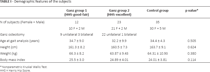
This study was approved by the institutional review board. Medical records of 92 patients who underwent PAO in our institution between 1995 and 2010 were evaluated. All patients were operated using the same osteotomy technique as described by Ganz et al (2) applying the ilioinguinal approach described by Millis (13). 4 patients who had THA during follow-up were excluded. A formal invitation to participate in the study was sent to the rest of the patients. Finally, 35 patients (39 hips) who responded the invitation and volunteered to participate were included in this study. 18 patients had previous surgery in the same hip and 9 received conservative treatment during childhood. Eighteen patients demonstrated limb length discrepancy (range 0.5-3 cm shortening on the affected side). Functional evaluation was performed preoperative and at the latest follow-up using the Harris hip score. For analytic purposes, 3 groups were created. The Group I comprised those patients who underwent PAO and obtained fair and good functional results according to the HHS (>70 to 89 points). The Group II comprised those patients who underwent PAO and obtained excellent results according to the HHS (90 or more points). The control group consisted of 35 healthy, age and gender similar subjects with no history of musculoskeletal conditions (Tab. I).
Demographic features of the subjects
Nonparametric Kruskal Wallis Test.
HHS = Harris Hip Score.
Radiographic analysis included the Tönnis’ and the Wiberg's angles evaluated in a centered, weight bearing anteroposterior view of the pelvis, the Lequesne's angle evaluated in the false profile view. Tönnis osteoarthritis grade was evaluated preoperatively and at the final follow-up.
Gait analyses were performed in Motion Analysis Laboratory at Hacettepe University Medical School, Department of Physical and Rehabilitation Medicine, using Vicon 612 System (Oxford Metrics) with 6 infrared JAI cameras at 50 Hz. The standard Plug-in Gait marker set was used to capture kinematic data. 15 reflective markers were placed bilaterally on the anterior superior iliac spines, mid-lateral thighs, lateral knee joints, lateral cruris, lateral malleolus, second metatarsal heads, and over the posterior calcaneus bilaterally, and one on the sacrum midline between the posterior iliac spines. Two force plates (Bertec Co.) were embedded in the middle of a 10-metres walkway and were used to measure ground reaction forces as well as to calculate kinetic data. Before data collection, each camera and force plates were calibrated. All individuals walked barefoot at their self-selected pace. The joint kinematic and kinetic values were calculated using the average value of 5 good trials (subject's steps landing completely within the force plates). Kinetic data were normalised according to body mass. All data processing was performed using Vicon Workstation software. The obtained values were compared among the groups.
Nonparametric Kruskal-Wallis test and nonparametric Mann-Whitney U-test were used for statistical analysis, p<0.05.
Results
A significant improvement in HHS pain and function scores was noted from a preoperative average of 61 points (range 35-80) to a postoperative average of 90.7 points (rate 73-106) (p<0,001). 12 patients were included in Group I and 23 in the Group II (Tab. II).
Pre-op and follow-up Harris Hip Score

Nonparametric Kruskal Wallis Test.
HHS = Harris Hip Score.
Radiographic analysis revealed significant improvement in Tönnis’ angle from a preoperative average of 37° (range 20°-56°) to a postoperative average of 13.4° (range 4°-35°). The Wiberg's angle improved from 8.9° (range −10°-25°) to 39.8° (range 17°-68°). The Lequesne's angle increased from 4.9° (range −10°-25°) to 35.9° in (range 10°-60°). Preoperative Tönnis grade was grade 0 in 24 joints and grade 1 in 17 joints. At the latest follow-up it was measured as grade 0 in 11 joints, grade 2 in 21 joints and, 3 grade in 1.
Gait analysis
Spatiotemporal parameters
There were no demonstrable statistical differences between the groups in the analysis of cadence (steps/min), limp index, opposite foot contact, opposite foot off, single support, step length, step time, stride length, stride time and walking speed. There was significant increase in double support and foot off in Group I and Group II. Group II also presented significantly higher step width (Tab. III).
Spatiotemporal parameters

Nonparametric Kruskal Wallis Test.
HHS = Harris Hip Score.
Sagittal plane kinematics
Significant differences were found in pelvic tilt range of motion (RoM), maximum hip extension, hip flexion-extension RoM, knee flexion-extension RoM, maximum ankle dorsiflexion, ankle dorsi-plantar flexion RoM (Tab. IV) (Fig. 1).
Sagittal plane kinematics

Nonparametric Kruskal Wallis Test.
HHS = Harris Hip Score.

Hip joint motion in the sagittal plane.
Coronal plane kinematics
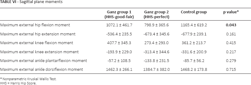
Significant differences were found in pelvic obliquity (Tab. V). There were no significant differences in sagittal plane kinetics moments except for a significant reduction in the mean maximum hip external flexion moment in Group II, demonstrating a possible reduction in the gluteus maximus and hamstring activity (Tab. VI) (Fig. 2).
Coronal plane kinematics

Nonparametric Kruskal Wallis Test
HHS = Harris Hip Score.
Sagittal plane moments
Nonparametric Kruskal Wallis Test.
HHS = Harris Hip Score.

Hip joint motion in the coronal plane.
Discussion
This study demonstrated that the vast majority of the gait parameters obtained after a PAO, including spatiotemporal parameters, coronal plane kinematics and sagittal plane moments, are not significantly different compared to control subjects. The exception in our study was the sagittal plane kinematic parameters, which differed from those observed in control subjects in 50% of the studied variables. Interestingly, these parameters were different not only in the hip joint but also in the ipsilateral knee and ankle when compared to control subjects. Our findings support the fact that the achievement of excellent clinical results based on the HHS seems not to be determined by the restoration of a completely normal gait pattern.
Among the tools that are currently available for the evaluation of results in hip surgery, gait analysis is one of the most objective ones (14). Body kinetics, kinematics and functional electromyography can all be assessed in a reliable way using this approach. Gait analysis has been used previously to evaluate patients with DDH who underwent a PAO. Pedersen at al (9) assessed 9 women before and 18 months after periacetabular osteotomy. They found that all patients walked with an increased knee extension at the standing phase and maintained a reduced flexor moment of the hip but achieved a more erect gait pattern. Sucato et al (10) described gait patterns in a case series of 21 adolescent patients (mean age 16.1 years) who underwent a PAO. Interestingly, and despite significant improvement in the radiographic parameters, none of the patients achieved a HHS >89 points at 1-year follow-up. In this group, the gait analysis revealed that isokinetic abductor strength and abductor impulse at 1-year follow-up was above the preoperative value. Flexion strength and flexion pull-off power were significantly lower compared to the baseline at 6 months. At 1 year, flexion strength was not significantly less than the preoperative value. A more recent study showed that the history of a previous surgery (Salter's, Chiari's or femoral osteotomy) among patients undergoing PAO may influence the hip abductor strength at one year follow-up (11). Additionally, Karam at al (12) have demonstrated that PAO can improve significantly walking velocity and stride length.
Among the possible explanations for the differences observed in the sagittal plane kinematics between PAO patients and controls 2 factors should be considered. The first one is the inherent compromise in gait patterns observed in patients with residual DDH at baseline (8). Adult patients with DDH walk with a reduced velocity, short steps and with a shorter stance phase on the affected site (antalgic gait). They also tend to reduce the rotation of the pelvis. Romanò at al (8) demonstrated that the RoM of the hip, particularly the extension, is reduced. This phenomenon is more evident in subjects who had degenerative joint changes. The ipsilateral knee compensates the reduced hip extension and allows the pelvis to progress forward before toe-off. On heel strike, the ground reaction force is also reduced. These changes were interpreted as an adaptation to reduce the load on the hip joint, hence an adjustment to reduce the pain. It remains unknown if the baseline condition is the main responsible for the complete achievement of a completely normal gait pattern after PAO. With the design of our study, this question cannot be answered. The 2nd factor relates to the surgical technique. Different surgical approaches have been proposed to perform a PAO, including, for instance, a transsartorial approach (15) or a rectus sparing approach (16). Our surgical approach corresponds to the one that modifies the original technique described by Ganz at al (2) applying an ilioinguinal approach to minimise abductor muscle damage (13). It will remain as a subject for future research to demonstrate that the use of the so-called less-invasive approaches for PAO can provide a clinically significant difference in terms of gait patterns.
In conclusion, we are able to show that in our experience, using the classical Ganz PAO technique and ilioinguinal approach, most of the gait parameters are close to those observed in control subjects. The attainment of excellence in terms of clinical results seems not to be determined by the achievement of a completely normal gait pattern in these patients.
Footnotes
Acknowledgement
The authors acknowledge Yıldız Erdoganoglu for data collection, Claudio Diaz-Ledezma for writing and editing, we also appreciate Mumtaz Alpaslan for invaluable logistical support and study administration.
Financial support: None.
Conflict of interest: None.