






Abstract
Introduction:
In the long term the treatment of articular cartilage defects of the hip has the most direct impact on the postoperative outcome and should diminish degenerative changes caused by different pathologies. The purpose of this prospective feasibility study is to describe technical aspects of arthroscopic, injectable autologous chondrocyte implantation in the hip and to report the short-term outcome.
Methods:
Full-thickness cartilage defects of 13 patients were treated arthroscopically with an injectable autologous chondrocyte transplantation product (Novocart Inject, Tetec) in a 2-step surgical procedure. Patient-related outcome was assessed with iHOT 33, EQ-5D and Non Arthritic Hip Score at baseline (day before transplantation), after 6 weeks and 3, 6 and 12 months.
Results:
13 out of 13 patients (all men) with a mean age of 32.7 ± 6.9 years and an average defect size of 1.9 ± 1.0 cm2 were available for follow-up after a mean of 12 months (range 6-24 months). All defects were located on the acetabulum and 11 were associated with a labral lesion of 2.9 hours size. Femoroacetabular impingement (10 cam, 2 combined, 1 pincer) was the cause of all defects. An overall statistically significant improvement was observed for all assessment scores.
Conclusions:
In this study we present the feasibility and short-term data of an arthroscopic injectable autologous chondrocyte transplant as a treatment option for full-thickness cartilage defects of the hip. All patient-administered assessment scores demonstrated an increase in activity level, improvement in quality of life and reduction of pain after a 12-month follow-up. Further randomised controlled trails with long-term follow-up and additional morphological assessment are needed.
Introduction
Besides specific entities such as trauma and mechanical overload, femoroacetabular impingement (FAI) is a -major cause of hip cartilage defects which may subsequently lead to the development of hip osteoarthritis.1–4 In particular, cam-type FAI is associated with large acetabular cartilage defects. 3 As there seems to be a correlation between the stage of cartilage loss and postoperative outcome, the appropriate treatment of articular cartilage defects may have the most direct impact on the postoperative outcome.5–9
Various surgical techniques such as marrow stimulation techniques, osteochondral transplantation and autologous chondrocyte implantation (ACI) have been made to restore articular surfaces. These techniques are well established in the knee; however, at the hip only a few case series have been reported.6,7,10–15
In contrast to the knee joint little has been published on arthroscopic or open autologous chondrocyte transplantation in the hip.16–19 The main reasons for the limited use of ACI procedures on the hip is the restricted space during arthroscopy due to osseous and labral containment and the technically demanding procedure. 16 Additionally, there are high demands on the ACI-System, which should be capable of a purely arthroscopic implantation without further fixation.16,19 The transplant investigated in this study can be applied arthroscopically in the cartilage defect without additional fixation.16,19
The purpose of this study is to show feasibility of the technique and to report short-term data of an injectable, in situ polymerisable matrix associated autologous chondrocyte implantation (ACI) product for the arthroscopic -treatment of cartilage defects of the hip joint. The postoperative outcome is measured by using established scoring systems (iHOT 33, Non Arthritic Hip Score [NAHS], EQ-5D).
Materials and methods
This prospective investigator-initiated trial was conducted according to the principles of the Declaration of Helsinki (World Medical Association). Approval was obtained from the local independent ethics committee. Our institution is authorised for the withdrawal of starting material for donation and procurement according to directive 2004/23/EC.
Participants and trial conduct
In a prospective case series, we report data on the first 13 patients with a focal cartilage defect of the hip treated by an arthroscopically conducted autologous chondrocyte implantation in a two-step procedure. 13 male patients fulfilled the inclusion criteria with an age between 18 and 50 years. Isolated cartilage defects of the hip joint International Cartilage Repair Society (ICRS) grades 3 and 4 with intact subchondral bone lamella and surrounding cartilage were included. Patients with more than one defect, opposing defects or radiographic signs of osteoarthritis higher than grade 2 according to Kellgren and Lawrence were excluded. Patients unable to follow a standardised rehabilitation protocol were excluded.
Preoperative diagnostics included a clinical functional examination and standardised anteroposterior (pelvis centred) and cross-table (hip centred) radiographs. 20
For the detection of cartilage injury, we used magnetic resonance arthrography (MRA) of the hip with radial reconstructions. 21 Only patients with signs of a chondral defect in the MRA were eligible for inclusion.
During arthroscopy, the cartilage defect was classified macroscopically according to the ICRS score, and biopsies and blood samples were taken for the cultivation of the chondrocytes. Only defects classified as grade 3a-3d, according to ICRS were included. On average, 34 days (24/62; min/max) after biopsy and cultivation the ACI was performed in a 2nd surgical procedure.
Instruments for subjective assessments
The following subjective questionnaires were used to evaluate patient outcome on the day before index arthroscopy, which was defined as baseline and at 6 weeks, 3, 6 and 12 months after ACI: (i) Euro-Quol group score (EQ-5D) evaluating the general health with mental and physical components; (ii) International Hip Outcome Tool (iHOT33) assessing pain and functional parameters of daily life and sports; and (iii) the Non Arthritic Hip Score (NAHS) rating symptoms, sports activities and function.23–26
Endpoints
The primary endpoint was the subjective improvement of symptoms and functionality displayed in the NAHS, iHOT 33 and EQ-5D at 12 months after ACT compared to baseline.
Operation technique and treatment with Novocart Inject
After patients provided their informed consent, the indication for treatment with Novocart Inject was verified during arthroscopy. Using the supine approach with ~10 mm joint distraction the defect area with localised cartilage defect was investigated utilising 2 arthroscopic portals (anterolateral and anterior) and classified according to ICRS during index arthroscopy. The treatment of associated pathologies such as labral tears and cam deformity was performed in the 1st operation. After harvesting 2-4 full-depth cartilage cylinders from non-weight-bearing areas of the hip (head-neck junction) during 1st arthroscopy, the specimens were sent together with 10 ml autologous blood to the manufacturer. Patient’s chondrocytes were isolated from the cylinders and expanded as a primary culture in vitro in a Good Manufacturing Practice (GMP) approved facility (TETEC AG). In the 2nd procedure the defect was debrided arthroscopically to produce stable perpendicular margins.
After creating a 3rd posterolateral portal as drainage, the constant fluid irrigation was stopped to keep the defect as dry as possible. The chondral defect was carefully filled with Novocart Inject, a combination of autologous cartilage cells and an in situ polymerisable hydrogel, through the deformable applicator. The injectable hydrogel, a combination of human albumin and hyaluronic acid, together with the autologous chondrocytes is delivered in a special syringe with 2 reservoirs. The fluid of both reservoirs is only then combined when injected through the applicator. It polymerises in 30-60 seconds and bonds immediately to the bottom of the defect. No further fixation had to be applied (Figure 1). The only technical difficulty is to achieve the appropriate mixing-ratio at the beginning of the implantation. Therefore, we recommend discarding the first 10-12 drops of the fluid to achieve an ideal mixing-ratio before implantation. The average operation times for the 2nd procedures were 53 minutes (range 45-68 minutes).
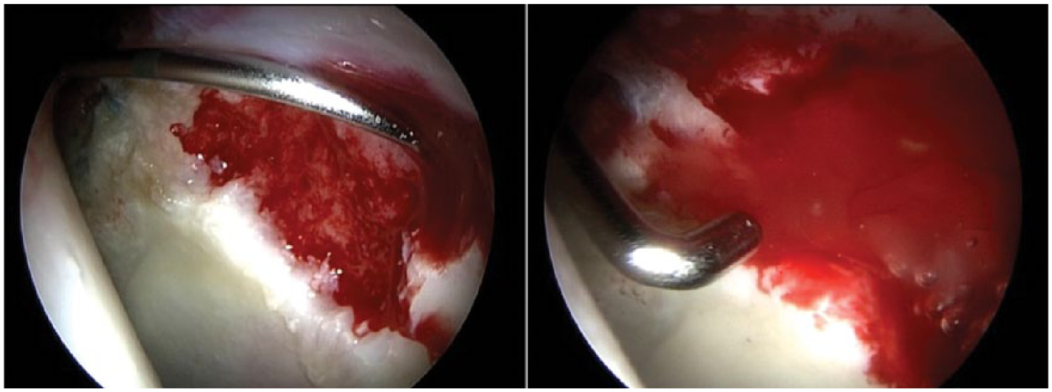
Application of Novocart Inject into the cartilage defect and final testing of transplant stability with a hook.
All patients were reported to have followed the standardised postoperative rehabilitation protocol. Continuous passive motion (CPM) therapy was conducted for 4 weeks with a minimum usage of 6 hours daily. The rehabilitation program included partial weight-bearing for 6 weeks with 10-20 kg. Patients with labral repair were furthermore restricted to a maximum flexion of 80° over 6 weeks. Functional scoring, data concerning tolerability of the treatment and the occurrence of any adverse events (AEs) were gathered. Return to low-impact sports was administered 3 months after transplantation, return to competition sport after 6 months.
Statistics
All statistical calculations were carried out with SAS software, release 9.3 (SAS Institute Inc.).
Quantitative parameters are presented by their mean values together with standard deviations or (for skewed data) by median and range. In order to investigate whether parameters change over time an analysis of variance (ANOVA) for repeated measurements was performed for each outcome parameter using the SAS procedure PROC MIXED (where time was considered as a fixed and patient’s ID as a random factor). In the case of a statistically significant test result Dunnett’s test was conducted as a post hoc test in order to find out if there is a difference in comparison to baseline. Pearson’s or Spearman’s correlation coefficients were calculated in order to investigate an association between variables. The result of a statistical test was considered as significant for p<0.05.
Results
Baseline characteristics
13 patients (all men) aged between 22 and 43 years (average 32.7 years) were included in this investigation. All -patients completed follow-up examinations. The average time of follow-up was 12 months (range 6-24 months). -According to ICRS classification all patients were diagnosed with a full-thickness chondral defect of the acetabulum Grade 3a-3d.
All defects were located on the acetabulum between 12 and 5 o’clock position. The average defect size was 1.9 cm2. 10 defects were caused by cam-type FAI, 1 by Pincer-type FAI and 2 by combined FAI (Table 1). 1 patient had undergone offset reconstruction, acetabulum trimming, labral refixation and microfracturing 16 months before ACI.
Demographic data and baseline characteristics of study population.
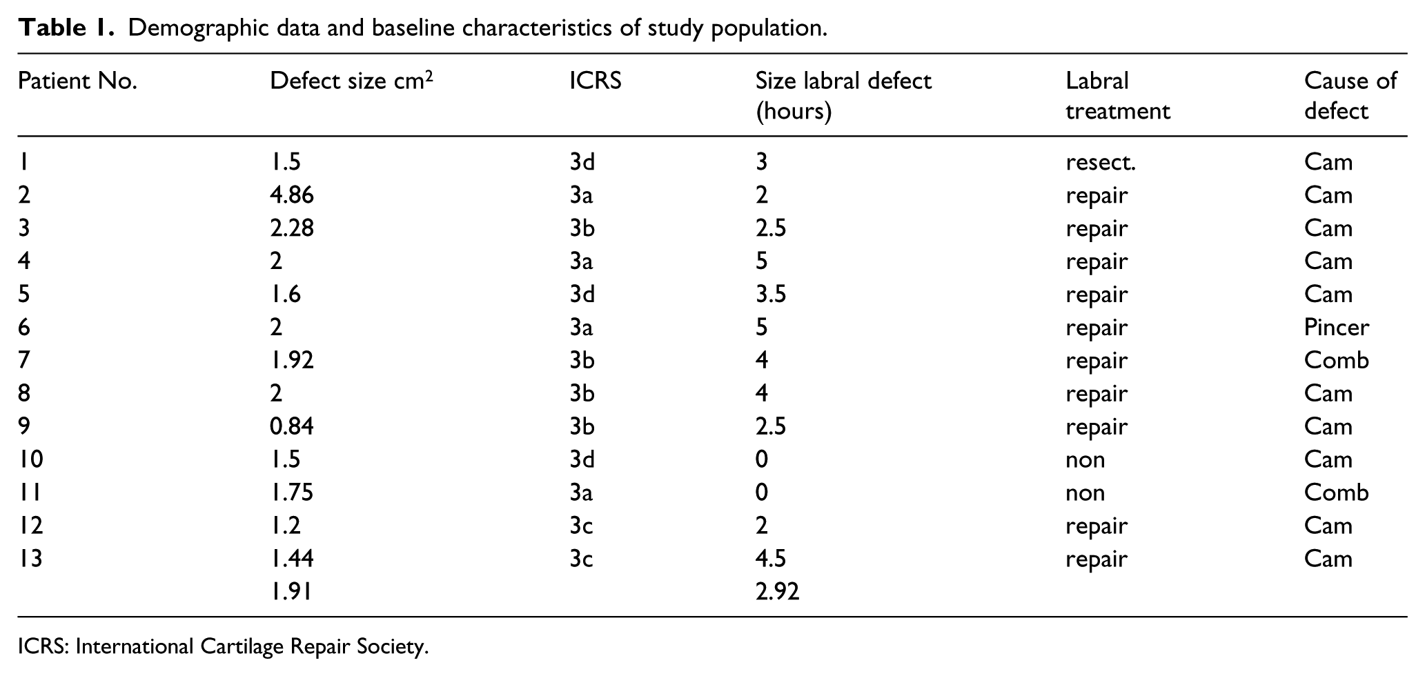
ICRS: International Cartilage Repair Society.
During index arthroscopy, 13 patients received additional treatment on the affected hip: femoral osteoplasty in 13 -patients, of whom 5 received additional acetabuloplasty. 11 patients had labral pathology of whom 10 received labral repair with 1 to 5 anchors and one partial trimming of the labrum. The extension of the labral defects on a clock rating varied from 2 to 5 hours.
Functional outcome evaluation
The clinical efficacy of the ACI-System was evaluated with 3 validated outcome scales; iHOT 33, NAHS and SF-36 Score. Each scale displayed a significant improvement (p<0.05) at 12 months after autologous chondrocyte implantation in comparison to baseline (Figure 2).
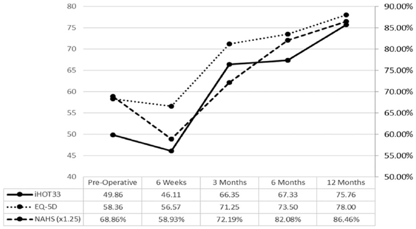
Outcome evaluation of iHOT 33, EQ-5D and NAHS (×1.25).
International Hip Outcome Tool (iHOT 33)
All patients treated with autologous chondrocyte implantation showed an overall improvement according to the iHOT 33 in comparison to baseline (p = 0.0002) (mean score at baseline: 49.8 ± 21.1; 12 months after ACT: 75.8 ± 11.7).
At 3 months significant improvement was seen (p = 0.0048). There was further improvement at 6 (p = 0.0030) and 12 months (p = 0.0003) after surgery.
Non Arthritic Hip Score (NAHS)
Using the ANOVA for repeated measurements significant changes over time could be revealed (p = 0.0014). At 6 (p = 0.0241) and 12 months (p = 0.0175) after surgery the results improved significantly compared to baseline. Also 12 months after autologous chondrocyte implantation patients improved significantly in the NAHS compared to baseline (p = 0.175) (mean score at baseline: 68.9% ± 14.6%; 12 months after ACT: 86.5% ± 7.2%).
Euro-Quol group score (EQ-5D)
In general, changes over time were highly significant (p<0.0001). Patients who underwent ACI of the hip showed an overall improvement according to EQ-5D when compared to baseline data (median baseline: 58.4 ± 16.0; 12 months: 78.0 ± 10.2, p = 0.0010). The subscore for “Mobility” and “Pain” showed that 46% of the patients had “some problems in walking”, 92% had moderate and 8% severe pain and discomfort at baseline. At 12 months none of the patients had mobility problems.
Subgroup analyses
Defect size
The size of the cartilage defect had no significant influence on the functional outcome in the iHOT33 (p = 0.9674) at baseline using the Spearman correlation test. There was no significant correlation of the defect size in either the NAHS (p = 0.5140) or EQ-5D (p = 0.8901) at baseline. 12 months after arthroscopic ACI no influence of the defect size on the functional outcome could be observed (NAHS p = 0.8574; EQ5D p = 0.9147; iHot33 p = 0.6265).
Treatment of labral pathology
A larger tear correlated with significantly poorer results in all assessment scores at baseline (EQ-5D p = 0.2301; iHOT33 p = 0.7095; NAHS p = 0.6604). Considering functional outcome evaluation, the number of anchors (iHOT33 p = 0.5177, NAHS p = 0.68, EQ-5D p = 0.91) and the size of the tear (iHOT33 p = 0.0981, NAHS p = 0.2061, EQ-5D p = 0.1894) did not prove to be of significant influence for functional outcome 12 months after arthroscopic ACI in the Spearman correlation coefficient.
Complications
With regard to postoperative complications, 1 patient had lateral femoral cutaneous nerve neuropraxia and another patient complained about a temporary neuropraxia in both forefeet. In all cases, neuropraxia disappeared within 3 months or less.
Discussion
The aim of the present study was to report the feasibility and the short-term results of 13 patients treated by concomitant impingement surgery and autologous chondrocytes together with polymerisable injectable albumin hyaluronic acid gel in the hip joint. The results were evaluated with respect to mental and physical health, pain and functionality in patients with isolated cartilage defects caused by femoroacetabular impingement. Overall, the final scores after treatment by concomitant impingement surgery and Novocart Inject revealed statistically significant increased levels of activity and quality of life after an average follow-up of 12 months. This indicates that in terms of pain relief and improvement of hip function the combination of impingement surgery and Novocart Inject seems to be an effective treatment method for full-thickness cartilage defects of the hip associated with FAI.
Several authors have reported a correlation of cartilage defects with inferior outcome results in FAI patients. -Philippon et al 26 showed the status of cartilage damage correlates with the subjective outcome. At 2.3 years follow-up, cartilage status classified as mild, moderate and poor had mHHS scores of 87, 79 and 62, respectively. Haviv et al 7 showed at their final follow-up that limited cartilage injury resulted in better outcome in contrast to severe cartilage injury. Singh et al13 concluded that larger defect size might have an inferior postoperative outcome. Other authors concluded that in cases of labral tears a coexisting cartilage defect seems to lead to significantly worse outcome results.5,9,27 This indicates the importance of the appropriate treatment of cartilage defects in the hip.
Arthroscopic access to the hip is difficult as the hip joint is a deep ball and socket joint surrounded by a large amount of muscle. The central compartment of the hip is especially difficult to treat during hip arthroscopy. Additionally, the majority of cartilage defects are localised on the acetabulum. 1 This sets high demands on the ACI System from the technical point of view, as fixation by suture or adhesives are needed. 18
From the preclinical research the components of -Novocart Inject are well known for their biological effects and biocompatibility. 28 The implant is characterised by stabilisation of the chondrocyte phenotype. 29 For the hydrogel an anti–angiogenic and anti-osteogenic effect is published, which inhibits inflammation.30,31 Until now 2 previous studies have displayed the feasibility and technical aspects of a different MACI system (Chondrosphere, co.don AG). In both studies by Fickert et al 16 and Körsmeier et al 19 the patient-administered assessment scores increased in activity level and quality of life in short-term follow-up. A possible advantage of Novocart Inject may be the remarkable bonding capacity of the in situ polymerisable hydrogel.
Fontana et al 18 have reported their 5-year follow-up results of 15 patients on the treatment of chondral defects of the hip with ACI in comparison to a group with simple debridement. They also used a 2-step procedure. However, their polymer-based scaffold was seeded with the expanded chondrocytes after cultivation, as it is an ACI product. From the technical point of view their rigid scaffold could only be used for acetabular defects as further fixation could not be implemented arthroscopically. 18
Similar to this study, Fontana et al 18 excluded patients with osteoarthritis higher than grade 2° as several authors reported that osteoarthritis may be a parameter to predict inferior results after hip arthroscopy in FAI patients.5,6,13,32 We did not include “kissing lesions” as the results in the knee joint have been unsatisfactory. This is in line with the findings of Fontana et al 18 , who reported the worst results in patients with corresponding cartilage defects in the hip. Körsmeier et al 19 in comparison do not specify exclusion criteria.
However, we are not able to comment on the quality of repair tissue or the defect filling after ACI, because MRI or second look surgery with histological or histomorphological analysis is lacking in this study. Körsmeier et al 19 performed second-look arthroscopy in 2 patients due to unsatisfactory range of motion 5 and 8 months after Chondrosphere implantation. They describe a good ingrowth of the transplanted chondrocytes, but further histological examinations have not been performed. 19
Fontana et al 18 reported a significantly better outcome of the ACI group with average defect size of 2.6 cm2 in terms of the Harris Hip Score (HHS) compared to the debridement group at 5 years. These results should be interpreted with care due to a possible selection bias as their control group was -selected out of 144 patients. Fickert et al 16 and Körsmeier et al 19 also reported significant improvement in the outcome results in larger defects (average 3.5 cm2 and 4.52 cm2) in short-term follow-up. Neither study had control groups.
However, the results discussed above are not comparable, as different outcome scores were used. Fontana et al 18 used the HHS, which is limited to pain, movement and daily activities. Among others Körsmeier et al 4 used the -WOMAC score, which is validated for patients with osteoarthritis. These conventional scores do not reflect the expectations and aims of the mainly young and active patients with FAI. Therefore, the iHOT33 was used, which has been developed and validated especially for FAI patients.22,25 We also correlated our results with the general health of the patient (EQ-5D) to evaluate possible influences on the results. 24 Patients in our cohort with larger cartilage defects showed a significantly lower iHOT33 (p = 0.014) score at baseline. We could not see inferior outcomes in correlation to larger defect sizes at 12 months after treatment. As in all cases additional interventions (labral refixation, femoroplasty -acetabuloplasty) were performed, we are not able to distinguish which part of the intervention is most likely to be responsible for the clinical improvement with our study design. However, in the current literature there are no high-level evidence studies dealing with this issue. This illustrates the importance of further trials to discriminate between different therapies.
Evidence is increasing that a repair of a torn labrum rather than a partial excision should be performed.2,26,33–38 The preservation of the labrum seems to be important for the functionality of the hip joint, the lubrication and the protection of the cartilage surfaces. 34 In this study we were able to repair 10 of 11 coexisting labral defects. In 1 patient the labral tear could not be repaired due to the severity of the tear. In the present study no influence of the size of the labral tear and/or number of anchors used for fixation could be seen in regard to the postoperative outcome.
Complications
We monitored temporary neuropraxia in 2 patients. In both patients the symptoms disappeared within 3 months. A comparison with the present literature is difficult as mostly only persistent neuropraxia is mentioned as a complication. Möckel et al 39 reported a 4.5% rate of nerve complications in over 13.000 patients after hip arthroscopy in a 5-year period.
Limitations
The present study only includes a small number of -patients. There is no control group, which does not allow us to make any conclusions, and only a short-term assessment of 12 months was carried out. No 2nd-look arthroscopies, no histomorphological assessment or MRI scans have been performed to improve the value of the presented data. Further limitations are a selection bias during patient inclusion as we only had male FAI patients. Additionally, we are not able to distinguish whether the improved outcome results are only related to the ACI procedure with the current study design.
Conclusion
In this study we present the feasibility and short-term data of an arthroscopic injectable matrix-associated, autologous chondrocyte implant as a treatment option for full-thickness cartilage defects of the hip. All patient-administered assessment scores demonstrated an increase in activity level, improvement in the quality of life and reduction of pain after a 12-month follow-up. However, further randomised controlled trails with additional morphological assessment are needed to approve the results in long-term follow-up.
Footnotes
Declaration of conflicting interests
The author(s) declared the following potential conflicts of interest with respect to the research, authorship, and/or publication of this article:SF had relevant financial activities outside of this work and/or any other relationships or activities that readers could perceive to have influenced, or that give the appearance of potentially influencing, this manuscript (Arthrex- Consultancy for Hip Arthrosocpy; Bauernfeind- Consultancy for strategic orientatation in orthopedics; DePuy- Travel expenses for congress participation).
Funding
The author(s) received no financial support for the research, authorship and/or publication of this article.