






Abstract
Background
Anemia is common in patients with heart failure and is associated with adverse outcomes. Management of anemia in CF-LVAD patients is not well studied. Our purpose is to characterize and identify the etiology of anemia in CF-LVAD patients. Secondary objectives are to describe the effect of CF-LVAD on pre-existing anemia and assess its impact after CF-LVAD support.
Methods
Cross-sectional study from January to July 2015 of ambulatory patients supported with a CF-LVAD for at least 6-months that presented with hemoglobin <12 g/dL and no recent gastrointestinal bleeding. Patients were classified as iron-deficient and non-iron-deficient and compared. Additionally, a retrospective analysis of 116 consecutive patients who underwent CF-LVAD from 2008 to 2013 with reported hemoglobin at 6 months as outpatients were divided into anemic or non-anemic and compared.
Results
In our cross-sectional cohort, iron deficiency was the most common cause of anemia. Notably, 49% of the iron-deficient patients were already on iron supplementation. In our retrospective cohort, 59% of the patients were anemic after 6 months of support. Anemic patients were older, had lower albumin, higher brain natriuretic peptide (BNP), worse renal function and New York Heart Association (NYHA) class. Anemia had a HR of 3.16 (95%CI 1.38–7.26) to predict a composite of 1-year death and HF readmissions, as well as HF-readmissions alone.
Conclusions
The most common cause of anemia in our study was iron-deficiency; almost half of the patients were iron deficient despite treatment, suggesting that oral iron may not be sufficient to reverse anemia. Anemia regardless of etiology was associated with adverse outcomes.
Introduction
The prevalence of anemia and its association with adverse outcomes in patients with chronic heart failure (HF) have been clearly demonstrated (1-3). However, the pathophysiology behind the development of anemia has not been completely elucidated. Anemia is likely a result of multiple underlying mechanisms that involve iron deficiency, renal dysfunction, inflammation, bone marrow dysfunction and hemodilution (4).
The use of durable continuous flow left ventricular assist devices (CF-LVADs) has increased in the past few years as a therapeutic option for patients with end-stage HF (5). There is a paucity of data regarding the prevalence of anemia and its implications in these patients. CF-LVADs have shown to improve HF by decreasing congestion and improving end-organ function, albeit with the trade-off of increased coagulopathy-hemolysis and bleeding (6-8). The impact of these mechanisms on anemia post CF-LVAD is not well characterized.
Few studies have described the prevalence of anemia and its implications in patients with LVADs, though only 1 in CF-LVADs (9, 10). However, the characteristics of anemia and the effect of CF-LVADs on pre-existing anemia remains unknown. We sought to identify the etiology and characteristics of anemia in patients who had been on support for at least 6 months. Our secondary objective was to validate the association of anemia with adverse outcomes.
Methods
Data collection and study design
This study was approved by our Institutional Review Board. The designs for both study arms are shown in Figure 1. We performed a cross-sectional analysis of patients supported with a CF-LVAD for at least 6 months that had hemoglobin <12 g/dL This cutoff represents the mean hemoglobin of the patients in the FAIR-HF trial as well as a previous study of CF-LVADs and anemia (10, 11). We excluded those with recent (3-months) GI-bleeding and/or blood transfusions. Laboratory tests, iron studies, erythropoietin levels and thyroid-stimulating hormone were obtained after at least 6 months of CF-LVAD support. Anemic patients were categorized according to the FAIR-HF trial (11) and characteristics were compared between the groups:
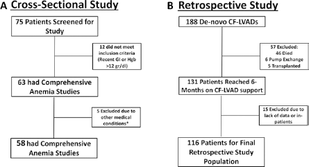
Study design. Flow diagram demonstrating the study design for the cross-sectional arm (A) and the retrospective arm (B). *See text for specific medical conditions excluded.
Absolute iron deficiency: Ferritin less than 100 mg/dL
Functional iron deficiency: Ferritin 100–299 mg/dL plus a transferrin saturation (TSAT) <20%
Non-iron deficiency: Patients that did not meet the above criteria.
Additionally we performed a retrospective analysis of a separate cohort of patients implanted with a CF-LVAD from May 2008 to December 2013 that had a 6-month outpatient visit; all patients, including those with recent GI bleeding were included in this arm of the study. Patients were categorized into anemic and non-anemic using a hemoglobin cutoff of < 12 g/dL (10, 12, 13). Characteristics and outcomes were compared.
Statistical analysis
Analysis was performed using STATA13 (StataCorp LP). A p value <0.05 was considered significant. Continuous variables were analyzed using Student's t-test or Wilcoxon rank sum tests. The proportion of anemic patients before and after CF-LVAD was compared using McNemar's chi-square test. Categorical variables were analyzed using chi-square or Fisher's exact tests.
The follow-up time was calculated from the initial visit (6-months post-CF-LVAD) to 12 months after. Kaplan-Meier curves were estimated to test the association between anemia and a composite of 1-year mortality and 1-year HF readmissions. The components of the composite outcome were also studied separately. Cox proportional hazard regression was used to identify predictors of outcomes.
Results
Cross-sectional study for characterization of anemia in CF-LVAD patients
Out of 75 patients screened, 58 (77%) patients met inclusion criteria; 12 patients were not anemic and 5 had conditions that could influence the nature of anemia (2 patients had myelodysplastic syndrome, 2 were receiving chemotherapy and 1 had hemochromatosis). Baseline characteristics of the patients are presented in Table I. Mean age was 58 years, 76% were male, 59% had ischemic cardiomyopathy and in 65% CF-LVAD was implanted as destination therapy. Mean duration of support was 23 months.
- Baseline characteristics of patients in the cross-sectional study
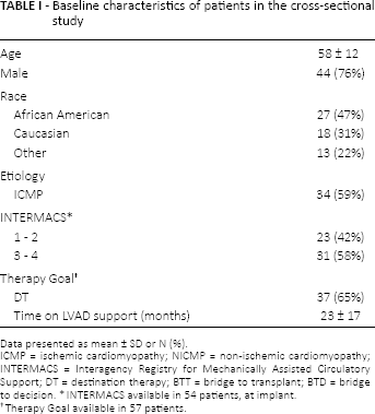
Data presented as mean ± SD or N (%).
ICMP = ischemic cardiomyopathy; NICMP = non-ischemic cardiomyopathy; INTERMACS = Interagency Registry for Mechanically Assisted Circulatory Support; DT = destination therapy; BTT = bridge to transplant; BTD = bridge to decision.
INTERMACS available in 54 patients, at implant.
Therapy Goal available in 57 patients.
Out of 58 patients, 37(64%) were iron deficient, of those 37, 75% had an absolute iron deficiency (ferritin <100 ng/mL) and 25% had functional iron deficiency (ferritin 100–299 ng/mL plus TSAT ≤20% [12]). Comparison of iron-deficient patients versus non-iron-deficient is shown in Table II. There was no significant difference between age, gender, etiology, therapy goal or INTERMACS profile. Iron-deficient patients had higher erythropoietin (67.0 vs. 24.5; p<0.01), lower hemoglobin (10 vs. 11; p = 0.01), mean corpuscular value (MCV) (86 vs. 93; p<0.01) and mean corpuscular hemoglobin (MCH) (26 vs. 29; p<0.01). There was no difference in kidney and liver function or hemolysis between iron-deficient and non-iron-deficient patients. Likewise, B12 and folate levels were no different between the two groups. Patients without iron deficiency had characteristics suggestive of anemia of chronic disease with higher ferritin (302 vs. 74 ng/mL; p<0.01) and a decreased total iron binding capacity (246 vs. 337 ng/mL; p<0.01). There was no difference in NYHA class (p = 0.3) or BNP (p = 0.2).
- Comparison of iron deficient versus non-iron-deficient patients
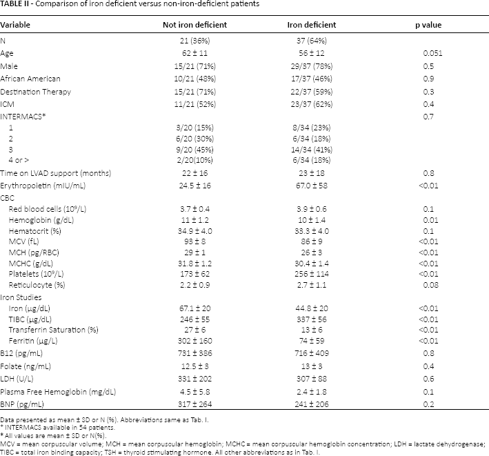
Data presented as mean ± SD or N (%). Abbreviations same as Tab. I.
*INTERMACS available in 54 patients.
*All values are mean ± SD or N(%).
MCV = mean corpuscular volume; MCH = mean corpuscular hemoglobin; MCHC = mean corpuscular hemoglobin concentration; LDH = lactate dehydrogenase; TIBC = total iron binding capacity; TSH = thyroid stimulating hormone. All other abbreviations as in Tab. I.
Impact of oral iron supplementation in iron deficient CF-LVAD patients
The calculated mean total body iron deficit utilizing the Ganzoni formula (14) in the iron-deficient patients was 7,100 mg. Interestingly, 49% (compared to only 18% in the non-iron-deficient group) of the iron deficiency patients were already on oral iron replacement for a median of 230 days (range 9–702; at our institution oral iron replacement therapy is achieved with 325 mg of ferrous sulfate TID with adjuvant ascorbic acid). We evaluated the impact of oral iron therapy over hemoglobin (Fig. 2), the mean hemoglobin when treatment was started was 9.2 g/dL and 1 year later was 10.4 g/dL, while the increase was statistically significant (p = 0.005), the mean improvement in hemoglobin after 365 days was only 1.2 g/dL.
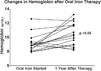
Impact of oral iron therapy on hemoglobin. Changes in hemoglobin after 1 year of oral iron therapy. Mean hemoglobin Δ1.2 ± 0.3 g/dL. Each line represents a different patient. Note: 2 patients are not included in the graph as they did not reach the 1-year follow-up, when included in the analysis; p<0.05.
With regards to anticoagulation, there was no difference in the use of aspirin (57% for both groups; p = 0.9), Coumadin (38% in the iron deficiency group vs. 48% in the non-iron deficient; p = 0.4) or proton pump inhibitors (54% in the iron deficiency group vs. 42% in the non-iron deficient; p = 0.4). The percent of arachidonic acid inhibition was similar between the 2 groups (46 ± 36% vs. 52 ± 37%; p = 0.4) as well as the internationalized normalized ratio (INR) (2 ± 0.4 vs. 1.8 ± 0.5) and partial thromboplastin time (PTT) (41 ± 7 vs. 41 ± 9; p = 0.9).
Retrospective study: patient characteristics
A total of 188 patients were implanted de novo with a CF-LVAD during the retrospective study period (2008–2013). Seventy-two patients were excluded from analysis because of lack of data (15), death (46), transplant (5) and pump exchange (6) before the 6-month visit. Of our final population of 116 patients, 47(41%) had hemoglobin ≥12 g/dL (group A) and 69 (59%) had a hemoglobin <12 g/dL (group B). Patient characteristics are shown in Table III and relevant laboratory data are presented in Table IV. Patients in Group B were older (59 vs. 51 years, p<0.01), females (33% vs. 17%; p = 0.051), with lower body mass index (BMI) (26 vs. 29 kg/m2, p<0.01), ischemic cardiomyopathy as primary diagnosis (65% vs. 47%, p = 0.04), worse kidney function (GFR: 66 vs. 85 mL/min per 1.73 m2; p<0.01. Creatinine: 1.5 vs. 1.1 mg/dL; p<0.01. BUN: 26 vs. 19 mg/dL; p<0.01), and lower albumin (3.9 vs. 4.2 g/L; p<0.01) when compared to those in group A. Group B patients had higher BNP (352 vs. 193 pg/mL; p<0.01) and worse NYHA class (36% were NYHA class III–IV vs. only 2% in group A; p<0.01).
- Baseline characteristics of consecutive patients after 6 months of CF-LVAD support
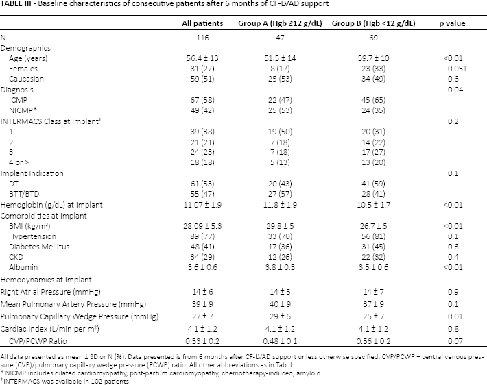
All data presented as mean ± SD or N (%). Data presented is from 6 months after CF-LVAD support unless otherwise specified. CVP/PCWP = central venous pressure (CVP)/pulmonary capillary wedge pressure (PCWP) ratio. All other abbreviations as in Tab. I.
NICMP includes dilated cardiomyopathy, post-partum cardiomyopathy, chemotherapy-induced, amyloid.
INTERMACS was available in 102 patients.
- Selected laboratories of consecutive patients after 6 months of CF-LVAD support
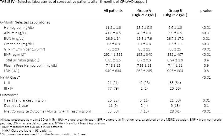
All data presented as mean ± SD or N (%). BUN = blood urea nitrogen; GFR = glomerular filtration rate, calculated by the MDRD equation; BNP = brain natriuretic peptide; LDH = lactate dehydrogenase; NYHA = New York Heart Association.
BMP measurement available in 69 patients.
NYHA Class available in 98 patients.
Outcomes were analyzed from the 6-month visit up to 1 year.
At the time of implantation, the anemic patients were more likely to have a lower BMI and lower albumin levels (3.5 ± 0.6 vs. 3.8 ± 0.5; p<0.01). There were no differences in implant indication or INTERMACS profile. At time of CF-LVAD implant, the pulmonary capillary wedge pressure was higher in the non-anemic group (29 ± 6 vs. 26 ± 7 mmHg; p = 0.01) and there was a trend towards an increased high central venous pressure (CVP)/pulmonary capillary wedge pressure (PCWP) ratio in the anemic group (0.49 ± 0.1 vs. 0.56 ± 0.2, p = 0.07). There were no differences in RA pressures, pulmonary pressures and cardiac index. The mean hemoglobin in group B was 9.9 g/dL versus 13 g/dL in group A (p<0.01). The MCV was higher in group B (89 vs. 85 fL: p<0.01) with a decreased intracellular hemoglobin content as measured by MCHC (31.9 vs. 33 g/dL; p<0.01). Similarly, the RDW was higher in group B (53% vs. 48%; p<0.01). Markers of RBC destruction, bilirubin, potassium, LDH and plasma free hemoglobin were similar in both groups.
Impact of anemia in CF-LVAD patients
The patients in group B had lower hemoglobin values than those in group A (10.5 vs. 11.8 g/dL; p<0.01) before CF-LVAD implant (24 hours before surgery). Furthermore, group B patients' anemia had worsened after CF-LVAD from 10.5 g/ dL pre-implant to 9.9 g/dL after 6 months (~6% decrease; p = 0.02). Similarly, patients in group A were also anemic prior to LVAD implantation, however their anemia improved with CF-LVAD from 11.8 to 13.2 g/dL (~11% increase; p<0.01; Fig. 3). Group B patients had higher transfusion requirements than their non-anemic counterparts. The median number of RBC units group B patients received was 6 (IQR: 4–11), while group A patients received a median of 4 (IQR: 2–7; p = 0.01). There was no difference in the use of platelet products or fresh-frozen plasma.
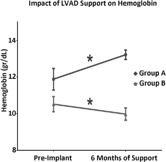
Impact of LVAD support on hemoglobin. Comparison of pre-implant hemoglobin vs. 6 months of support by group. Group A: hemoglobin ≥12 g/dL. Group B: hemoglobin <12 g/dL; *p<0.05 for paired data.
Regarding the composite outcome, the event-free survival at 1 year for those in group B was significantly lower than group A (54% vs. 83%; p<0.01; Fig. 4). Freedom from 1-year HF readmissions alone was lower in group B group compared to those in group A (63% vs. 87%; p = 0.01). One-year mortality was not significant between the 2 groups, suggesting that the composite outcome was driven by heart-failure readmissions.
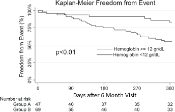
Freedom from composite outcome. Kaplan-Meier curves comparing freedom from a composite of death and heart-failure readmission within 1 year. Group A: hemoglobin ≥12 g/dL. Group B: hemoglobin <12 g/dL.
On univariate analysis, anemia was associated with the highest hazard for the outcome (HR 3.16, 95%CI: 1.38–7.26; p<0.01), followed by albumin (lower, HR: 2.79.95%CI: 1.44–5.4, p<0.01), NYHA class (HR: 2.7, 95%CI: 1.63–4.46, p<0.01) female (HR: 2.22, 95%CI: 1.12–4.33, p = 0.02), and creatinine (HR: 1.36, 95%CI: 1.12–1.73, p<0.01).
While hemoglobin <12 g/dL failed to predict mortality, we did find a significant difference in mortality in those patients with a hemoglobin <10 g/dL (93% vs. 77% p = 0.01).
Discussion
The major findings of our study are as follows.
We have identified iron deficiency defined by either serum ferritin <100 ng/mL (absolute deficiency) or ferritin 100–299 ng/mL plus TSAT ≤20% (functional deficiency) to be the most common cause of anemia in these patients. For the remainder, anemia of chronic disease appeared to be the main etiology.
The use of CF-LVAD had a contrasting effect on hemoglobin with correlation on the patients' pre-operative hemoglobin levels. Patients with low hemoglobin (<12 g/dL) at implant continued to have lower levels after 6 months of support; however, patients with high hemoglobin (≥12 g/dL) prior to implantation improved after CF-LVAD implantation.
We also established that hemoglobin <12 g/dL at 6 months post CF-LVAD is associated with worse outcomes, specifically heart failure readmissions. These results add to what has been previously shown by Jennings et al.
There is limited data about anemia and its etiology in patients with CF-LVADs. One study predominantly had patients with pulsatile LVADs (10) and the other study showed an association with morbidity but did not establish a specific hemoglobin cutoff to identify patients who are at higher risk of adverse outcomes (9). Our study performed on CF-LVAD patients validates the relationship between anemia and adverse outcomes. Patients with hemoglobin <12 g/dL had an increased incidence of HF readmissions and death, as well as HF alone. We also found that patients with hemoglobin <10 g/dL at were at a risk of increased mortality.
The possible mechanisms behind anemia in patients with HF have been widely studied (1, 3), however the impact of CF-LVAD support over these mechanisms remains unknown. It is well known that mechanical support unloads the left ventricle and improves tissue oxygenation. It also leads to decreased venous congestion leading to improved kidney function and gut absorption (15), which, in turn, should improve anemia. The positive effects need to be balanced out by the ongoing degree of hemolysis and microscopic GI bleeding, which can counteract the beneficial effects of improved perfusion. In these patients it may be that improvement in anemia after CF LVAD implantation is dependent on the fine balance between the resolution of HF syndrome and prevalence of ongoing RBC loss.
Effective treatment of anemia requires that the underlying cause be identified; however, this is difficult in patients with CF-LVADs as many different mechanisms may be in effect. Our cross-sectional analysis demonstrated that the vast majority (64%) of patients with CF-LVAD and anemia had iron deficiency. This is consistent with the observations of Nanas et al in the non-CF-LVAD population, where the incidence of iron deficiency in patients with advanced HF and reduced EF was 73% (16).
In the light of these results, 2 questions remain unanswered. First, why are CF-LVAD patients iron deficient? In patients with HF, absolute iron deficiency is defined as ferritin <100 ng/mL Using that definition, absolute iron deficiency was present in 28 (76%) of the anemic patients. Still, when ferritin is between 100–300 ng/mL, diagnosis of iron deficiency is more complicated. Studies have relied on the TSAT, which represents the body's capacity to deliver iron to the target cells. Indeed, patients with ferritin 100–300 ng/mL and a TSAT <20% are also iron deficient, but their deficiency is functional rather than absolute. Using that definition in our study, 9 patients (24%) had functional deficiency (17-19). For both cases, the body is deficient in iron, whether it is due to a lack of iron stores or not enough iron to supply the body's demand.
The underlying cause of iron deficiency (and consequently anemia) in CF-LVAD patients might be explained by the degree of underlying hemolysis during CF-LVAD support as well as GI bleeding, and while our study did not show any difference in overt GI bleeding, occult GI bleeding is well recognized in these patients (20). Likewise, immune activation and HF are now accepted as a common co-occurring phenomena (21). Immune activation is associated with iron sequestration in macrophages and hepcidin-mediated decreased intestinal absorption leading to absolute iron deficiency with or without anemia (1); with CF-LVADs effectively unloading the heart, the effect on inflammation and immune activation might not be as marked (22), leading to a state of chronic inflammation and hence functional iron deficiency with decreased iron utilization in the bone marrow.
Another condition associated with inflammation and heart failure is cardiac cachexia. Inflammatory cytokines that are part of the pathophysiology of cardiac cachexia are also associated with a decreased red blood cell mass, either by direct suppression of the bone marrow and/or disruption of iron handling (functional iron deficiency). Interestingly, in our study anemic patients were more likely to have lower albumin levels both pre-implant and after 6 months of support. These observations suggest that cardiac cachexia could play a role in the development and non-resolution of anemia despite cardiac unloading.
Second, what is the cause of anemia in patients without iron deficiency? While there is no reliable test for identifying anemia of chronic disease, in our study, non-iron- deficient patients had milder anemia, higher MCV (normocytosis) lower erythropoietin production (albeit levels were high), increased ferritin, decreased total iron-binding capacity (TIBC), a higher, yet still low TSAT, and a trend towards a lower reticulocyte production when compared to iron- deficient patients. Patients without iron deficiency were older with no difference in renal function. All these characteristics are suggestive of anemia of chronic disease (23, 24).
It is well recognized that anemia is not just a simple laboratory abnormality and it should always be treated, as our results suggest. It is imperative, then, that the mechanism behind the anemia be identified, whether it is iron deficiency or any other cause. While this might be self-evident to the LVAD clinician, to our knowledge there is no data describing the etiology of anemia in this unique patient population.
Oral iron supplementation is widely used for treating iron deficiency, however, the use of oral supplementation might be ineffective and even detrimental in CF-LVAD patients. In our study, 18 patients (49%) were diagnosed as anemic due to iron deficiency despite being on oral iron. While our results suggest that there is a statistical significant improvement on hemoglobin with oral iron, the clinical impact might be minimal, as mean improvement of hemoglobin was only 1.2 g/dL and this was after 1-year of therapy. The reason for this lack of improvement might be due to various reasons, 1 of them being that oral iron might not be enough to replace iron losses. For example, in our iron-deficient patients the mean iron deficit was 7,100 mg. If a patient receives 325 mg of ferrous sulfate TID (195 mg of elemental iron) only 25 mg will be absorbed and utilized (bioavailability of ~10%) and it would take 284 days to replenish all stores. This is assuming a “perfect world” as it is recognized that patient adherence is hard to achieve with oral iron (60% report side-effects, increased pill burden) (12), and a high proportion of patients are on acid-reducing agents (interfere with absorption).
Furthermore, oral iron is known to increase oxidative stress in the mucosa and hence might place CF-LVAD patients at an even higher risk of GI bleeding. Certainly, intravenous (IV) iron formulations might be a better option, as it will confer a faster and more tolerable strategy to replenish iron. While the risk of infection with IV iron might be of special concern in CF-LVADs, a recent meta-analysis demonstrated that IV iron therapy is not associated with increased risk of infections (25). Likewise, in a randomized trial of chronic kidney disease patients (26), IV iron was more effective than oral in elevating ferritin and hemoglobin and there was no difference in renal, cardiovascular or infectious events (26). Prospective trials to evaluate the safety and efficacy of IV iron in CF-LVADs are required.
Treatment of anemia of chronic disease/inflammation should be focused on treating the underlying disease. Erythropoietin-stimulating agents are commonly used to treat this condition in patients with chronic kidney disease, anemia due to malignancies or autoimmune conditions; however, our study demonstrated that patients supported with CF-LVADs have normal to high erythropoietin. This observation, along with those of the RED-HF trial (27) and the risks of erythropoietin analogues (28) suggest that these medications should be reserved for special situations when the clinician believes the benefits outweighs the risks.
Data on iron supplementation in this cohort of patients is scarce; IV iron infusions have been used in patients with kidney disease undergoing concomitant ESA therapy with good results (29). The efficacy of IV iron alone, in the setting of already elevated erythropoietin in CF-LVAD patients remains to be tested in a prospective clinical trial. Furthermore the FAIR-HF trial demonstrated that treatment with intravenous iron preparation, regardless of anemia status, improved overall morbidity and mortality in patients with chronic HF (11). Whether the effects of iron deficiency and supplementation play a role in patients supported with CF-LVAD should be determined in future studies.
Limitations
The cross-sectional analysis hinders the possibility to identify the potential impact of anemia and iron deficiency in this patient population. Other study limitations include those inherent to all single-center, retrospective studies, with a small sample size, only 1 device type (HeartMate II) and a study population excluding patients that died within 6-months of implant.
Conclusions
Patients supported with a CF-LVAD have a high prevalence of anemia principally due to iron deficiency. The specific causes of iron deficiency and anemia without iron deficiency remain unanswered and further studies are needed to understand the different mechanisms in play during CF-LVAD support. Our results validate the association between anemia and adverse outcomes, and call for the need of protocols to identify and effectively treat anemia in patients supported with CF-LVADs.
Footnotes
Conflict of interest: Dr. Jerry D. Estep and Dr. Arvind Bhimaraj are consultants for St. Jude Medical™ (St. Paul, MN, USA).