





Abstract

Dear Editor,
A 46-year-old man presented to the emergency with severe pain in the right upper limb. Four days earlier, he had undergone percutaneous coronary intervention (PCI) through right radial artery for treatment of his coronary artery disease. However, the coronary intervention was not successful because of difficult manipulation of the catheter through the cannulated radial artery. At that time, it was presumed to be due to spasm of radial artery. The procedure was rescheduled for a different date. During the presentation at the emergency department, no swelling was present on clinical examination. The radial pulse was felt and the capillary refill was normal distally. However, because of the recent cannulation and suggestion of spasm of radial artery, computed tomography angiography (CTA) was considered. CTA showed thin caliber radial artery (measuring around 2-3.5 mm), which was arising from the axillary artery (Fig. 1) and was running superficially along the flexors in the forearm. The ulnar artery and the palmar arch were patent and normal. All the arteries were patent and no thrombus or stenosis was evident.
Brachioradial artery is an uncommon anatomical variation of the upper limb arterial system. It is defined as high origin of the radial artery in the arm above the level of intercondylar line, instead of its usual origin as one of the terminal branches or bifurcation of the brachial artery in the cubital fossa (1, 2). It can arise either from the brachial artery or the axillary artery. It is named as superficial brachioradial artery when it courses superficial to the flexor muscles in the forearm. Brachioradial artery is the commonest anatomical arterial branching variation of upper limb with an incidence of 13.8%, though a superficial brachioradial artery is present in only 1.25-3.12% of normal population.
Embryologically, the artery is derived from its precursor known as superficial brachial artery, which is formed by failure of regression of more than one cervical intersegmental artery (3, 4). Brachioradial artery can be classified into four groups according to its site of origin, namely group one, two, three and four, as the artery arises from distal third, middle third, proximal third of brachial artery or distal infrapectoral portion of axillary artery, respectively (Fig. 2). It courses medially in the proximal arm along with cutaneous nerve of forearm (3). As it reaches the cubital fossa, it runs to the lateral aspect, superficially and across the median nerve. The superficial brachioradial artery runs superficial to the flexor muscles all along the lateral aspect of the forearm to enter the wrist superficial to the flexor retinaculum and end in the first web space by forming deep palmar arch. While in the cubital fossa, it may anastomose with the proper brachial artery at times. This arterial variant can exist with a variant brachial plexus anatomy wherein the medial cutaneous nerve of the forearm and median nerve join to form a common trunk in the arm (3). Another vascular variation of anomalous course of posterior circumflex humeral artery is also known to be associated, where it runs deep to the tendon of latissimus dorsi. The later variation is accompanied by an increased risk of bleeding due to unanticipated injury in surgical procedures involving the surgical neck of humerus.
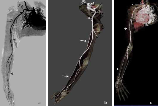
Maximum intensity projection with inversion (a) and volume-rendered images (b and c) of the CT angiography showing the origin of the thin caliber radial artery from the axillary artery.
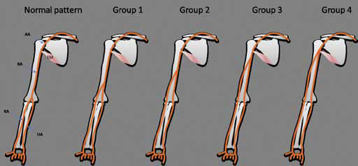
Diagram showing the types of brachioradial artery. AA, axillary artery; BA, brachial artery; RA, radial artery; UA, ulnar artery; TM, teres minor.
As this variant is usually smaller in caliber, it may result in difficult, if not unsuccessful, radial cannulation for diagnostic and interventional procedures, as in our patient (5). In the present angio-interventional practice, especially for coronary arterial interventions, wherein the radial arterial approach is being increasingly preferred over the femoral access, due to reduced local complications and faster patient mobilization, importance of recognizing this variant cannot be overemphasized (6). It is advised not to adopt brachial route for catheterization in cases of abnormal brachial arterial branching pattern in order to avoid complications (7). Superficial location and course of this arterial variant predisposes to increased chances of arterial injury in the setting of trauma and may result in torrential bleeding. Inadvertent puncture of the artery, instead of vein, may result in bleeding, hematoma or pseudoaneurysm formation. It is also relevant in the surgical procedures of the arm and forearm wherein the potential arterial injury can be avoided by prior awareness.
Footnotes
Conflict of interest: The authors have no conflict of interest.