






Abstract
Background
Systemic hypothermia with bilateral antegrade selective cerebral perfusion (ASCP) is the preferred cerebral protective strategy for type A aortic dissection surgery. The optimal ASCP flow rate remains uncertain and the target flow cannot always be reached due to pressure limitations. The aim of this study was to assess the correlation between ASCP flow and regional cerebral oxygen saturation (rSO2).
Methods
A retrospective analysis was performed on 140 patients with acute type A aortic dissection who underwent surgery with moderate hypothermic circulatory arrest and bilateral ASCP between 2015 and 2021. Pearson correlation analysis was performed between ASCP flow and rSO2.
Results
The median circulatory arrest duration was 46.5 (IQR:37.0-61.0) minutes. There was no significant correlation between ASCP flow and rSO2 for both the right (r = -.02, p = .851), and the left hemisphere (r = - .04, p = .618). The rSO2 values for ten patients who received > 10 mL/kg/min flow did not differ significantly from 130 patients who received 10 mL/kg/min or less for both the left hemisphere (p = .135), and the right hemisphere (p = .318). The ASCP flow was 5.1 (IQR:5.0- 6.5) mL/kg/min in five patients with, and 7.2 (IQR:5.8-8.3) mL/kg/min in 135 patients without a watershed infarction (p = .098).
Conclusions
There was no correlation between ASCP flow rate and rSO2 in patients with acute type A aortic dissection. Furthermore, ASCP flow below 10 mL/kg/min was not associated with a reduction in rSO2. Definitive associations between ASCP flow and neurological outcome after type A aortic dissection surgery need further investigation.
Keywords
Introduction
The preferred method for cerebral protection during surgery of the thoracic aorta is moderate hypothermic circulatory arrest (MHCA) with antegrade selective cerebral perfusion (ASCP). A target cerebral blood flow of 10 mL/kg/min for ASCP at 25°C with cerebral perfusion pressures between 40 and 70 mmHg is most commonly used, although flow rates vary between clinical centers ranging from 8 to 15 mL/kg/min.1–4 The ASCP flow rate that provides optimal cerebral protection is still controversial.5,6 Animal studies have shown an increase in ischemic markers when ASCP flow was below 6 mL/kg/min, while ‘luxury’ perfusion of 18 mL/kg/min was associated with delayed neurobehavioral recovery.7,8 The upper and lower safety limits of ASCP flow are not well-defined. Despite advances in operative techniques and intra-operative monitoring the incidence of stroke after type A dissection surgery is still high, ranging between 5 and 15%.9–11 Although the majority of strokes after type A dissection surgery are embolic in nature, optimizing ASCP parameters to prevent watershed infarction is of critical importance due to its significant associated morbidity.12–15 Near-infrared spectroscopy (NIRS) is commonly employed for monitoring regional cerebral oxygenation (rSO2) during type A dissection surgery. It has been demonstrated, that a sustained decrease in rSO2 is associated with postoperative neurological events.16,17 The aim of this study was to investigate the correlation between ASCP flow and rSO2 to determine the optimal ASCP flow for cerebral oxygenation in patients undergoing surgery for type A aortic dissection. Additionally, ASCP flow rates and rSO2 data in patients with and without watershed infarction were compared.
Materials
Patients and methods
Between January 2015 and December 2021, 180 patients underwent surgery for (acute) type A dissection with ASCP in our institution. Patients were excluded from analysis if NIRS data were incomplete (n = 14), if stroke was present on admission at the hospital (n = 1), if unilateral ASCP or deep hypothermic circulatory arrest was used (n = 4), or if patients died before postoperative neurological assessment was performed (n = 21). Data from the remaining 140 patients were retrospectively analyzed. The diagnosis of type A aortic dissection was confirmed by computed tomography and transesophageal echocardiography. The primary outcome variable was postoperative watershed infarction. Patients were considered to have had postoperative watershed infarction if they exhibited new neurologic dysfunction during intensive care unit (ICU) admission after the surgical intervention, confirmed by computed tomography scanning or magnetic resonance imaging. The distinction between watershed and non-watershed stroke was made by a radiologist. Approval for this study was received from the local ethics committee, that waived the need for an informed consent (nWMO-2022.003).
Procedure
Extracorporeal circulation (ECC) was established by median sternotomy, femoral, subclavian or direct aortic arterial cannulation and cavo-atrial cannulation for venous return. The left side of the heart was vented through the right superior pulmonary vein. Cardiac arrest was achieved using St Thomas cardioplegia or histidine-tryptophan-ketoglutarate cardioplegia (Dr Franz Köhler Chemie, Bensheim, Germany) injected directly into the coronary ostia. The aorta was opened, and the proximal anastomosis was performed. The aortic valve and or root was repaired or replaced when necessary. Patients were cooled until a bladder temperature of 25°C was achieved before arresting systemic circulation. Cooling was performed with a maximal gradient of 10°C between the venous inflow and arterial outlet of the oxygenator. For ASCP, a 15 french retrograde coronary sinus cannula (Medtronic Inc., Kerkrade, The Netherlands) was inserted into the brachiocephalic artery, while a 13 french cannula of the same manufacturer was inserted into the left common carotid artery through the aortic lumen. In case of selective cannulation, the flow over both cannulas was controlled using roller pumps. Alternatively, subclavian artery cannulation using a vascular graft prothesis was used for ASCP of the right hemisphere and a 13 French retrograde coronary sinus cannula was inserted into the left common carotid artery. Cerebral perfusion flow was initiated at a rate of 10 mL/min/kg and the flow was adjusted to maintain a maximal pressure of 70 mmHg in the corresponding artery, measured at the tip of the cannula. The aortic arch. was inspected and partly or fully replaced when nessecary. In case of femoral artery cannulation, the arterial line of the extracorporeal circuit was connected to a side branch of the vascular prosthesis using a 3/8-inch tubing connector upon completion of ASCP to ensure antegrade perfusion flow after repair.
Equipment
The procedures were performed with a Livanova custom pack containing an Inspire 6FM oxygenator and a Revolution centrifugal pump (Livanova, Mirandola, Italy). The circuit was primed with 1000 mL sodium chloride, 500 mL Voluven (8%), 200 mL mannitol (15%), 40 mL sodium bicarbonate (8.4%), 7500 IU heparin, and 500 mg tranexamic acid. ASCP flow was achieved with two 85 mm mast roller pumps (Livanova, Munich, Germany) and occlusion was set before each case. Near-Infrared Spectroscopy (NIRS) oximetry was measured with the Foresight Elite system (Edwards Lifesciences, Irvine, United States).
Statistical analysis
The data were analyzed by SPSS version 26 (SPSS Inc. Chicago, USA). The normality of distribution of continuous variables was tested by the Shapiro-Wilk test. Categorical variables were represented as absolute numbers (n) and relative frequencies (%). Continuous (numeric) variables were expressed as the mean ± standard deviation (SD) or as median ± interquartile ranges [IQR;25th and 75th percentile], depending on normality. Differences were compared with the independent t test if data were normally distributed, or with the Mann-Whitney U test if data were not normally distributed. For all comparisons p-values were presented, and statistical significance was set at p < .05. The frequencies of categorical data were compared by Chi Square test or by Fisher Exact test if frequencies of less than five occurred in the contingency table. The correlation between ASCP flow and rSO2 was assessed using both the Pearson correlation coefficient and linear regression analysis. Correlation coefficients between 0 and 0.3 were considered very weak; those between 0.31 and 0.5 were regarded as weak; those between 0.51 and 0.7 were deemed moderate; those between 0.71 and 0.9 were seen as strong; and those between 0.91 and one were classified as very strong. 18 Baseline rSO2 was defined as the mean rSO2 from start of cardiopulmonary bypass until circulatory arrest. ASCP rSO2 was defined as the mean rSO2 value from start to stop of ASCP.
Results
Study cohort
Demographic parameters and indication for surgery.
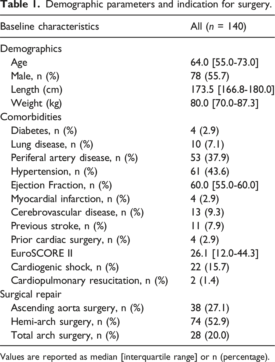
Values are reported as median [interquartile range] or n (percentage).
Antegrade selective cerebral perfusion flow in relation to cerebral oximetry
The mean flow in the brachiocephalic artery was 348.5 ± 94.4 mL/min, while the mean flow in the left common carotid artery was 219.9 ± 55.6 mL/min. As shown in Figure 1, there was no significant correlation between ASCP flow in the brachiocephalic artery and rSO2 for the right hemisphere (r = −0.02, p = .851), or for ASCP flow in the left common carotid artery and rSO2 for the left hemisphere (r = −0.04, p = .618). The study cohort was divided into two subgroups based on the mean ASCP flow received during surgery. In 10 patients the ASCP flow was >10 mL/kg/min (high-flow group), with a median flow rate of 10.8 (IQR: 10.5 - 11.9) mL/kg/min. In 130 patients the ASCP flow was 10 mL/kg/min or less (low-flow group), with a median flow rate of 7.0 (IQR: 5.6 - 8.2) mL/kg/min. The rSO2 in the low-flow group was not significantly different compared to the high-flow group in both the left hemisphere: 68.0 (IQR: 63.5 - 72.9) versus 73.0 (IQR: 68.1 - 73.9); p = .135), and the right hemisphere: 66.7 (IQR: 62.4 - 70.9) versus 68.2 (IQR: 66.6 - 69.6); p = .318), respectively. Patients in the low-flow group had rSO2 values below 55% for 0 (IQR: 0 - 1.2) minutes while patients in the high-flow group had rSO2 values below 55% for 0 (IQR: 0 -0) minutes (p = .252).

Scatterplots of ASCP flow in the left common carotid artery and oxygen saturation of the left hemisphere (a), and ASCP flow in the brachiocephalic artery and oxygen saturation of the right hemisphere (b). Red markers indicate patients with a postoperative watershed infarction. In each subplot, a linear regression best fit line is included for trend visualization, along with corresponding R-squared values and p-values. ASCP: antegrade selective cerebral perfusion; rSO2: regional cerebral oxygen saturation.
Antegrade selective cerebral perfusion flow and clinical outcome
Intraoperative perfusion parameters.
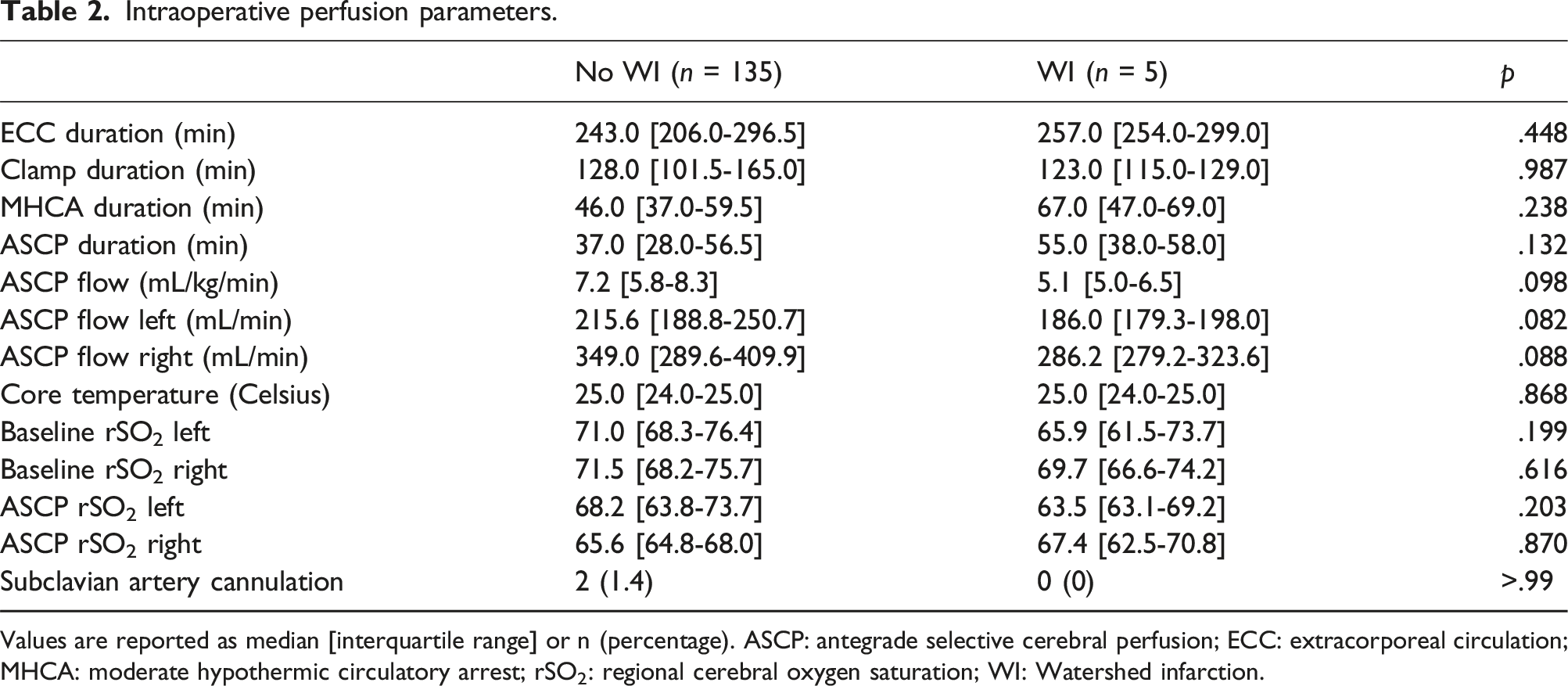
Values are reported as median [interquartile range] or n (percentage). ASCP: antegrade selective cerebral perfusion; ECC: extracorporeal circulation; MHCA: moderate hypothermic circulatory arrest; rSO2: regional cerebral oxygen saturation; WI: Watershed infarction.
Discussion
In this retrospective study, the correlation between ASCP flow and rSO2 was reviewed for 140 patients who underwent surgery for acute Type A dissection. The main finding of the study is that ASCP flow below 10 mL/kg/min was not associated with lower cerebral oxygenation. Additionally, there was no significant difference in ASCP flow or rSO2 between patients with or without postoperative watershed infarctions. Follow-up studies with larger cohorts may be warranted for developing a more patient-tailored cerebral perfusion strategy.
ASCP protocols vary among surgical centers regarding target ASCP flow, perfusion pressure, and usage of alpha stat or pH stat. While a target flow of 10 mL/kg/min at 25°C with cerebral perfusion pressures between 40 and 70 mmHg is commonly used, alternative protocols have also shown excellent outcomes.2,6,19 There is still no gold standard protocol for ASCP, and while some authors speculate 10 mL/kg/min may be excessive, others suggest it may be insufficient.5,20–22 It is important to note that most studies regarding outcomes after acute type A aortic dissection do not report the achieved ASCP flow and its relation to neurological outcomes, even among the articles proposing specific ASCP flow rates.4,23,24 To achieve an ASCP flow of 10 mL/kg/min, in our opinion, often necessitates perfusion pressures that have been reported as unsafe in experimental studies.7,21 In this scenario a challenging decision has to be made: to aim for the target flow with potentially excessive perfusion pressures, or keep the perfusion pressure within the advised range while accepting lower flow. The best course of action in this scenario to optimize patient outcome is unclear.
The findings of this study indicate that flow below 10 mL/kg/min does not lead to decreased cerebral oxygenation. In this study, no significant difference in rSO2 was observed between patients who received a median flow rate of 10.8 (IQR: 10.5 - 11.9) and those who received 7.0 (IQR: 5.6 - 8.2) mL/kg/min. This notion is supported by a recent study by Friess. et al who conducted detailed individualized assessments with multiple flow and rSO2 measurements per person. They found no increase in rSO2 after ASCP flow above 8 mL/kg/min. 22 However, Friess et al. calculated ASCP flow based on ideal body weight, whereas this study calculated ASCP flow based on actual body weight, following current recommendations. 4 This distinction becomes more significant due to the inverse relationship between cerebral blood flow and body mass index.25–27 Consequently, our approach may have led to high target flow rates for patients with higher body mass indices which resulted in lower flow without neurological complications or cerebral desaturation.
A sustained drop in rSO2 below 55% has been associated with neurological events. 16 Harrier et al. proposed an algorithm in which rSO2 levels below 55% serve as a trigger to convert from unilateral to bilateral ASCP. 28 In the current study, the absence of significant periods below 55% rSO2 in patients who received less than 10 mL/kg/min suggests that a target flow of 10 mL/kg/min may be higher than necessary.
Contrary to the findings of the present study, another investigation reported a significant correlation between ASCP flow rates and postoperative neurologic complications, however the authors did not correct for other stroke risk factors. 20 As the mean ASCP flow was even higher than in the present study, other variables, such a thromboembolic stroke, may have contributed to adverse neurologic outcome. As stroke is most commonly embolic in origin in type A dissection surgery, it is of crucial importance to exclude these patients from analysis when the relation between stroke and ASCP is assessed.12–15 Also in the present study, patients with watershed infarctions had a higher incidence of preoperative cardiogenic shock and cardiopulmonary resuscitation. As preoperative cardiogenic shock represents an important risk factor for stroke, the possibility that neurological damage occurred before initiation of ASCP cannot be ruled out. 11 Furthermore, NIRS has limited power in detecting (micro-)embolic events, and it is possible that (micro-)embolisms played a role in the onset of watershed infarctions for patients in this study. 16 Studies with larger cohorts are necessary to validate such findings.
Considering the large number of patients in our study who received less than 10 mL/kg/min ASCP flow without experiencing postoperative neurological complications, we believe these findings warrant further investigation of optimal ASCP flow. Current recommendations may need to be reconsidered to instead use metrics like ideal bodyweight, age, or total brain volume to calculate ASCP flow which may offer more tailored flow rates that better align with the individual needs of patients.23,29,30 Furthermore, future study designs should focus on perfusion strategies for the individual hemispheres, as anatomical variations of the circle of Willis are present in 50% of cases. 31 However, guidelines to guide such strategies are currently lacking. Data from this study implies there may be a flow threshold for each hemisphere below which the odds of infarctions increase. In this study no watershed infarctions were observed when ASCP flow rates were above 350 mL/min in the brachiocephalic artery and 200 mL/min in the left common carotid artery. However, a cut-off value for minimal ASCP flow could not be calculated due to the limited number of events. If individual roller pumps are used to control the flow to each hemisphere, the flow of the pump perfusing the brachiocephalic artery should be set to a higher flow rate to account for run-off into the right subclavian artery. A final recommendation is to integrate ASCP data into a comprehensive type A aortic dissection database. This would facilitate further studies into optimal ASCP flow parameters, neurological outcomes, and the influence of various patient characteristics.
Study limitations
Our current study has some limitations. Unfortunately, ASCP perfusion pressures could not be retrospectively obtained for a number of patients and were not included in the analysis. Furthermore, the limited number of events in our study reduced its statistical power, also preventing us from correcting for other stroke risk factors.
Conclusion
In this single-center study, no significant correlation was found between ASCP flow rate and cerebral oxygenation. ASCP flow below 10 mL/kg/min provided sufficient cerebral oxygenation, suggesting its safety within the studied range. Although statistical significance was not attained, our observations suggest hemisphere-specific perfusion requirements. These findings underscore the necessity of re-evaluating how to determine the target ASCP flow to ensure optimal cerebral protection during type A dissection surgery.
Footnotes
Declaration of conflicting interests
The authors received no financial support for the research, authorship and/or publication of this article.
Funding
The author(s) received no financial support for the research, authorship, and/or publication of this article.
