





Abstract
Background: The aim of anterior cruciate ligament reconstruction is to reduce excess joint laxity, hoping to restore normal tibiofemoral kinematics and therefore improve joint stability. It remains unclear if successful ACL reconstruction restores normal tibiofemoral kinematics and whether it is this that is associated with a good result.
Study: Case series.
Purpose: To assess the kinematics of the anterior cruciate ligament-reconstructed knee using open-access MRI.
Methods: Tibiofemoral motion was assessed using open-access MRI, weightbearing through the arc of flexion from 0° to 90° in 10 patients with isolated reconstruction of the anterior cruciate ligament (hamstring autograft) in one knee and a normal contralateral knee. Midmedial and midlateral sagittal images were analyzed in all positions of flexion in both knees to assess the tibiofemoral relationship. Sagittal laxity was also assessed by performing the Lachman test while the knees were scanned dynamically using open-access MRI.
Results: The amount of excursion between the tibial and femoral joint surfaces was similar between the normal and reconstructed knees, but the relationship of tibia to femur was always different for each position of knee flexion assessed—the lateral tibia being about 5 mm more anterior in the anterior cruciate ligament-reconstructed knees. This anterior tibial position is statistically significantly different at 0° (P < .0006), 20° (P = .0004), 45° (P = .002), and 90° of flexion (P < .006). Anteroposterior laxity was similar between normal and anterior cruciate ligament-reconstructed knees.
Conclusion: Anterior cruciate ligament reconstruction reduces sagittal laxity to within normal limits but does not restore normal tibiofemoral kinematics despite a successful outcome.
The aim of surgical reconstruction of the anterior cruciate ligament (ACL) is to reduce excess joint laxity, hoping to restore normal tibiofemoral kinematics and therefore improve joint stability.15,17 The technique has become increasingly reliable. § However, recent cadaveric studies have called into question the ideal placement of the femoral tunnel for ACL reconstruction with the traditional 11 o'clock position (to replicate the insertion of the antero-medial bundle of the ACL) being less favorable at resisting combined anterior and rotational loading compared to the 10 o'clock position of the posterolateral ACL bundle. 33 It remains unclear if successful ACL reconstruction restores normal tibiofemoral kinematics and whether it is this that is associated with a good result.
References 1, 10-12, 20, 22, 28, 31, 34, 36, 38, 39, 45.
Several cadaveric studies of ACL reconstruction have suggested that normal kinematics are restored.3,19 These studies cannot reproduce normal neuromuscular activity around the knee and are limited by their use of plain radiography and the difficulty of interpretation of bony landmarks on lateral knee radiographs. Dynamic magnetic resonance imaging (MRI) using MR tracking resolves this issue allowing the medial and lateral compartments of the weightbearing knee to be visualized separately and simultaneously. This study employs analysis of ACL- reconstructed knees in living subjects using weightbearing and stress MR scanning.
Materials and Methods
Patients
Consent was obtained from 10 patients (all male) with isolated unilateral rupture of the ACL who had undergone arthroscopically assisted single-incision four-strand hamstring autograft reconstruction (Pinczewski technique) 44 performed by an experienced consultant orthopaedic surgeon with a subspeciality interest in reconstructive knee surgery. Patients were selected after successful review at 6 months clinic follow-up and were contacted on a voluntary basis. The mean age of the patients was 32 years (range, 25-42 years) with the mean time from injury to performing the ACL reconstruction being 18 months (range, 1 month- 6 years). Three patients sustained their injury playing football, 4 were skiing, and the remaining 3 were rugby injuries. The indication for reconstruction in all patients was symptomatic instability. All patients were scanned at least 9 months after the ACL reconstruction (range, 9-18 months) and had resumed normal daily and sporting activities. All the reconstructions were symptomatically stable. The patients filled out a modified Lysholm score sheet.
Patients were excluded from the study if they had had an associated meniscal tear, any other ligamentous injuries, a symptomatic contralateral knee, pain at the time of clinical examination, or symptoms in the ipsilateral hip, ankle, or foot.
Anterior tibial translation in normal knees has been shown in numerous trials to vary widely between individuals but to show very little difference between the knees of the same subjects (95% of normal subjects the difference is less than 2 mm).13,46 Hence, for this study the contralateral normal knee was used as the control for direct comparison.
Surgical Technique. 44
The four-strand hamstring (gracilis and semitendinosus) autograft was sutured with a “whip stitch” of nonabsorbable braided suture material distally and with absorbable braided suture proximally. The tibial tunnel was positioned to allow the graft to pass through the center of the native ACL stump. The femoral tunnel was placed at the 11 o'clock position for the right knee (1 o'clock position for left knees). The graft was fixed proximally and distally with titanium interference screws with the graft tensioned in near extension by a maximum manual pull during insertion of the tibial interference screw.
Rehabilitation Protocol
All patients followed the same postoperative rehabilitation protocol. Full weightbearing was commenced within 24 hours, with range of motion of 0° to 90° achieved by 2 weeks. Closed kinetic chain exercises were used for muscle strengthening and proprioceptive work. Patients were allowed to return to normal sporting activities 6 months after surgery.
MR Scanning
All patients were scanned using the “open” configuration Signa SP MR Imaging System (General Electric Medical Systems, Milwaukee, Wis). This consists of a 0.5 tesla superconducting magnet, whose coils are housed in separate but communicating cryostats. The vertical open “double- doughnut” configuration of the scanner allows the patients to stand squat in increments to 90° flexion (Figure 1). Sagittal fast spoiled gradient echo T1 images (5-mm thick, 0-mm spacing) across the entire knee articulation (approximately 15 images) were taken with the patient weight- bearing at the following extensions: 0°, 20°, 45°, and 90° of flexion (angle measured with a goniometer).
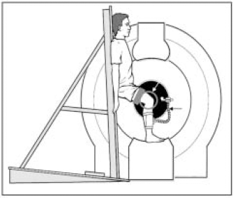
Weightbearing dynamic MRI using an openaccess scanner. White arrow, MR coil; black arrow, MR tracker.
The open magnet also allows the patient to sit and provide access for the examiner to assess the anteroposterior laxity of the knee by performing a Lachman and a “reverse” Lachman test (Figure 2). Sagittal fast spoiled gradient echo T1 images (5-mm thick, 0-mm spacing) across the entire tibiofemoral articulation (approximately 15 images) were taken with the patient sitting and the knee scanned at 20° (angle measured with a goniometer). The first measurement (measurement 1: neutral reference point) was obtained with the foot and ankle supported in a neutral rotation and the quadriceps relaxed. The second measurement was a reverse Lachman stress applied to displace the tibia posteriorly relative to the femur. The third measurement was taken when a standard Lachman stress was applied displacing the tibia anteriorly relative to the femur (Figure 3). We have previously shown the reproducibility and validity of the Lachman test when compared to the more objective “radiological Lachman.” 31 The patients’ contralateral normal knees were also scanned in all positions as a control. All clinical tests were performed by the same examiner.

Lachman test in the open-access scanner.

Measurement method after Iwaki et al. 26 FFC, flexion facet center; d, distance measured to ipsilateral posterior tibial cortex.
MR Tracking
Assessment of joint motion using MR tracking was first described in 1999 by Pearle et al. 40 As well as providing the space for access to the patients, the scanner has a tracking device that maintains the same plane of scanning despite changes in knee position between individual scans. The MR signal in an MR tracking procedure is detected by a small radiofrequency coil that is applied around the leg, just below the knee.
Measurements
The position of the posterior femoral condyles relative to the tibia was measured in the sagittal plane at midmedial and midlateral positions of the knee according to the method of Iwaki et al. 26 The centers of the posterior circular surfaces of the femoral condyles seen on sagittal MRI images—the flexion facet centers (FFCs)—are reliable femoral reference points. These were identified by placing acetate overlays with circles of varying diameters over the femoral condyles of midmedial and midlateral images. The distance between this center and a vertical line drawn from the ipsilateral posterior tibial cortex was measured for each position with a Vernier caliper and corrected for magnification (Figure 2). The accuracy of this technique has been shown to be ± 1.5 mm for each measurement.23,26,35
Statistical Analysis
The results were normally distributed. A power analysis was performed post hoc on the means (±SD) of the non- weightbearing and the weightbearing data between the normal and ACL-reconstructed knees. A power value of 0.8 was regarded as sufficient. This showed that n = 10 was sufficient for comparison, and the paired Student's t test was used. A P value of less than .05 was regarded as statistically significant. The reconstructions were performed by two surgeons using the same technique (seven by one author and three by another). The data were analyzed for surgeon-specific effects; there were none, with all patients having persistent anterior subluxation of the lateral tibial plateau throughout the are of motion.
Results
The patients’ average postoperative Lysholm score was 98 (range, 89-100). Clinical examination of the ACL-recon- structed knees prior to MRI scanning showed that all patients had full range of motion, a firm endpoint on the Lachman test (range, 0-1+), and a negative pivot shift. There were no abnormalities detected on clinical examination of the contralateral asymptomatic normal knees.
Anteroposterior Laxity
Table 1 shows the anteroposterior laxity in the ACL- reconstructed knees. There is no statistically significant difference in the anterior or posterior movement in the medial compartment compared to the contralateral normal knee (P < .36 and P < .98, respectively) (see Table 2). There is also no statistically significant difference in the anterior or posterior movement in the lateral compartment compared to the contralateral normal knee (P < .8 and P < .51, respectively). As with previous studies, the Lachman test internally rotates the tibia as it moves anteriorly. 31 The reverse Lachman test similarly externally rotates the tibia as it moves posteriorly. The anteroposterior laxity observed in the normal knees was very similar to previously published work using stress MRI. 31
Data for Anteroposterior Laxity in ACL-Reconstructed Knees a

NWB 20, nonweightbearing at 20°. Mean posterior tibial translation (neutral-reverse Lachman): medially = 0.7 mm ± 0.8, laterally = 2.3 mm ± 1.2; mean anterior tibial translation (neutral- Lachman): medially = 2.1 mm ± 1.2, laterally = 3.9 mm ± 2.
Data for Anteroposterior Laxity in Contralateral Normal Knees a

NWB 20, nonweightbearing at 20°. Mean posterior tibial translation (neutral-reverse Lachman): medially = 0.7 mm ± 0.9, laterally = 2.6 mm ± 1.6; mean anterior tibial translation (neutral- Lachman): medially = 1.6 mm ± 1.3, laterally = 3.7 mm ± 2.1.
Weightbearing Tibiofemoral Motion
Figures 4 and 5 show the clear difference in relative tibiofemoral position in the lateral compartment of the ACL-reconstructed knees compared to the contralateral normal knee observed throughout the flexion arc 0° to 90°. Figures 6 and 7 show the mean position of the FFCs of the femoral condyles in the ACL-reconstructed and contralateral normal knees, respectively. The lateral femoral condyle moved 10 mm posteriorly from 0° to 90° in the ACL-reconstructed knees compared to 10 mm in the normal knees (Tables 3 and 4, Figures 6 and 7).
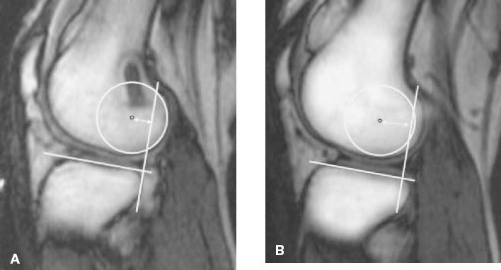
Lateral compartment of (A) ACL-reconstructed knee and (B) contralateral normal knee at 20° flexion.
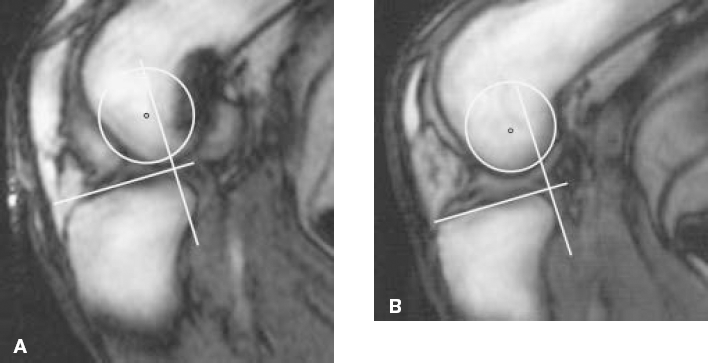
Lateral compartment of (A) ACL-reconstructed knee and (B) contralateral normal knee at 90° flexion.
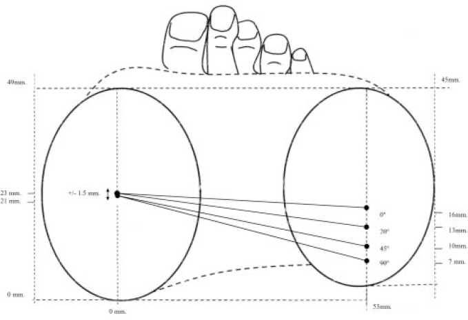
The mean positions of the femoral condyles as given by lines connecting their flexion facet centers at 0° to 90° in the ACL-reconstructed knees.
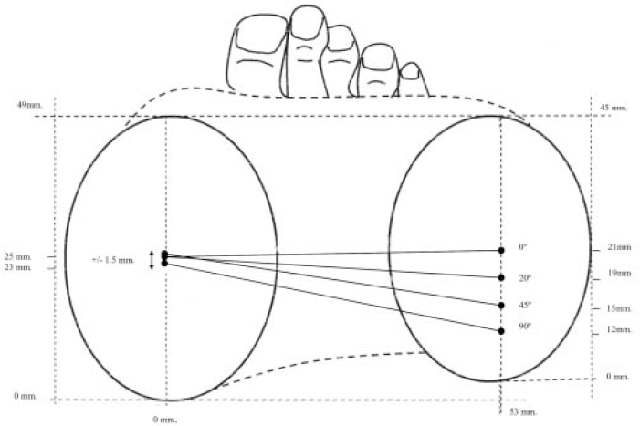
The mean positions of the femoral condyles as given by lines connecting their flexion facet centers at 0° to 90° in the contralateral normal knees.
Weightbearing Data for Contralateral Normal Knees

Weightbearing Data for ACL-Reconstructed Knees

Looking at the data presented in Tables 3 and 4 and Figures 6 and 7, there is a persistent anterior subluxation of the lateral tibial plateau/posterior subluxation of the lateral femoral condyle of approximately 5 mm throughout the flexion arc 0° to 90° in the ACL-reconstructed knees. This anterior tibial position is statistically significantly different at 0° (P < .0006), 20° (P = .0004), 45° (P = .002), and 90° of flexion (P < .006). In summary, the amount of excursion between the tibial and femoral joint surfaces was similar between the normal and reconstructed knees, but the relationship of tibia to femur was always different for each position of knee flexion assessed—the lateral tibia being about 5 mm more anterior in the ACL-reconstructed knees.
The medial femoral condyle movement relative to the tibia was minimal—from 0° to 90° in both ACL-reconstructed and normal knees with no evidence of “rollback.” The mean positions of the medial tibial condyles in the ACL-reconstructed knees were slightly anterior compared to the normal knees, but this only reached statistical significance at 90° of flexion (P < .04)
Discussion
Our research unit has previously published on the kinematics of the weightbearing normal knee using MRI.23,26 We have shown that knee flexion is accompanied by internal tibial rotation due to a differential relative motion of femur on tibia between the medial and lateral compartments. The medial femoral condyle has minimal movement anteroposteriorly with flexion to 120° and does not roll back as was previously thought,23,26,41 whereas the lateral femoral condyle moves posteriorly on the lateral tibia.
In this study, the contralateral normal knees flexed from 0° to 90° as per the normal knees previously studied.23,26 The ACL-reconstructed knees also showed tibial internal rotation with flexion due to posterior movement of the lateral femoral condyle; however, the lateral tibial plateau was consistently displaced anteriorly throughout the active flexion arc by approximately 5 mm. Following ACL reconstruction, there is a normal magnitude of relative joint surface excursion laterally, but the relative position of the tibia and femur is changed with persistent anterior subluxation of the tibia relative to the femur. Therefore, in the lateral compartment of the ACL reconstructed at full extension, the tibiofemoral position is equivalent to 45° flexion in the normal knee, and at 45° flexion it is equivalent to more than 90° flexion in the normal knee. In fact, the posterior subluxation of the lateral femoral condyle observed after ACL reconstruction is identical to the ACL- deficient knee, 32 suggesting that reconstruction does not improve the weightbearing kinematics of the lateral compartment of the knee. The squat was performed by a double-leg squat. We have previously investigated the potential differences between a monopodal and bipedal stance in patients with ACL deficiency (personal observations) and could find none. Due to the limited space inside the MR scanner, by positioning the knee to be scanned in the isocenter of the magnet, if anything more weight would be taken through that knee during scanning. This would be the same for both the ACL-deficient knees and the control contralateral knees.
It may be viewed that to avoid roof impingement ACL reconstruction using autograft aims to replace the posterolateral ACL bundle tibial insertion by placing the tibial tunnel as posteriorly as possible,21,25 but the well- documented femoral attachment of 11 o'clock and 1 o'clock positions (for the right and left knee, respectively) aims to replicate the origin of the anteromedial bundle of the ACL. 33 Perhaps this anatomical “deficit” explains our findings. A recent MRI study has revealed that successful ACL autografts do not recreate the normal sagittal obliquity of the ACL having instead a more vertical orientation. 4 It could be argued that a double-bundle anatomical graft technique may be more likely to reproduce more natural kinematics. 49 Current methods of ACL graft fixation do not replicate the normal twisting of the fibers of the ACL as they insert onto the tibia, nor do they replicate the tibial and femoral insertions noted to be more than three times larger than the midsubstance cross-sectional area of the ACL.4,21
Several recent in vivo studies using gait analysis, 7 radiostereometry,8,9 and arthrometry 2 have all shown no improvement in postoperative ACL reconstruction kinematics compared to the preoperative ACL-deficient knee. Gait analysis and arthrometry measure bony movement indirectly, lack accuracy due to soft tissue interposition, and are limited by their inability to differentiate the relative contributions of the medial and lateral compartments to joint motion.7,18 Radiostereometry studies are limited by the exposure to radiation and the invasive nature of the technique with insertion of markers into the distal femur and proximal tibia. A recent study by Kanisawa et al using an in vivo weightbearing fluoroscopy-based three-dimensional technique to measure tibiofemoral motion from 0° to 70° of flexion showed no difference in kinematics between normal and ACL-reconstructed knees. 30 The impact of Kanisawa et al's study was reduced by the fact that all the patients had concomitant meniscal surgery that could affect knee kinematics and also by the difficulty of interpretation of bony landmarks on lateral knee radiographs.
In this present MRI study, we can see that ACL reconstruction reduces passive anteroposterior tibial translation to within normal limits similar to the findings of Bull et al. 10
Several studies have shown that proprioception is improved following ACL reconstruction.6,27,42 Reinnervation of autografts has been demonstrated histologically using both animal models and human patients5,37,47 and also in vivo by detection of somatosensory-evoked potentials after direct mechanical stimulation of ACL autografts. 38 Using an invasive electromyographic technique, Tsuda et al have recently shown that ACL autografts in vivo can restore the ACL-hamstring reflex arc. 48 The relative contribution of the mechanical impact of ACL reconstruction versus altered neuromuscular control in restoring joint stability is unknown.
Current methods of ACL reconstruction have been successful at restoring joint stability by reducing excess sagittal anteroposterior laxity and perhaps by restoring more normal proprioception, but they do not, however, restore normal tibiofemoral kinematics. Despite this, all patients in this study had resumed sporting activities with no episodes of giving way. The average postoperative Lysholm score was 98 (range, 89-100), emphasizing the success of this procedure. In other words, although the knee has not been restored to normal, the subject clearly copes well with it compared to preoperative symptoms. All of the patients scanned have abnormal tibiofemoral kinematics in the lateral compartment, which may help to explain, at least in part, why ACL reconstruction may not reduce the incidence of osteoarthritis.14,16,24,29,43
An obvious limitation of this study is that there are no kinematic data on these patients prior to reconstruction of the ACL. This is currently being addressed in a prospective study using the same assessment of knees described here for ACL-deficient knees before and after reconstruction. A further limitation of our study was that we assessed only the kinematics of the knee in the sagittal plane and thus were unable to determine the three-dimensional, six degrees freedom of motion of the knee. More complex motions such as pivoting were not assessed.
This study confirms that ACL reconstruction reduces anterior tibial laxity to within normal limits but does not restore normal tibiofemoral kinematics despite a successful outcome.