
Abstract
Rotary blood pumps (RBPs), used as Ventricular assist devices (VADs), feature a suspended impeller (rotor) within a housing (stator). Titanium alloys are mostly used for those parts, but their limited wear resistance in critical contact areas, leading to scratches and promoting thrombus formation. Therefore, hard material coatings (HMCs) can be applied to increase wear resistance and bonding agents ensure stable coatings on the bulk material. Still, coating damage may occur and expose the material to blood, requiring hemocompatibility assessment. Therefore, their hemocompatibility must be evaluated as well as that of the HMCs. Platelet adhesion as thrombus formation indicator was investigated using an in vitro flow chamber and fluorescence microscopy for the following materials: Silicon diamond-like carbon (SiDLC), titanium nitride with and without droplets (TiN_D, TiN), bonding agents chrome (Cr), chrome nitride (CrN), uncoated Ti6Al4V (Ti), and aluminum (Alu) as positive control. CFD simulations determined wall shear rates, averaging 5730.5 1/s on the evaluated area. The normalized percentage of the covered surface (NCSA) area was statistically evaluated (p-values, Wilcoxon effect size). NCSA analysis showed that Alu had the highest value (8.7), significantly exceeding CrN and SiDLC (both 0.3, p < 0.05). Cr (3.6) exhibited significantly more platelets than CrN with a medium effect compared to CrN, Ti (0.2), TiN (0.3), and SiDLC, and a weak effect compared to TiN_D (0.4) and Alu. No significant differences were observed among HMCs, Ti, and CrN. This study highlights Cr’s elevated thrombogenicity, whereas the other surfaces (except Alu) showed hemocompatibility comparable to Ti, supporting their use in VADs.
Keywords
Introduction
Heart failure is known as a pathophysiological state, in which the heart can no longer pump blood at a sufficient rate. This condition is associated with a severe influence on the quality of life 1 and with high morbidity and mortality rates. Heart transplantation is considered as the gold standard for end-stage heart failure patients, with post-transplant survival rates showing a significant improvement over recent years. 2 However, due to the limited availability of donor organs and surgical as well as technical advances, 2 rotary blood pumps (RBPs), such as the HeartMate 3 (Abbott GmbH, Chicago, IL, USA), are implanted as ventricular assist devices (VADs) to provide mechanical circulatory support. 3
In clinical practice, VADs play a pivotal role in bridging patients to recovery or transplantation. The number of patients on the waiting list is approximately doubling the number of available transplants annually and there is increasing proof that patients on the waiting list with a VAD have a higher survival rate. 4 VADs have become indispensable in modern heart failure management. Nevertheless, frequent hemocompatibility-related adverse events such as thrombus formation can occur. It is a major complication associated with VAD therapy, for example, when the thrombi is flushed into the circulatory system, causing strokes or peripheral thromboembolisms. 5
In RBPs, human whole blood is exposed to a broad range of shear rates. Further, blood comes into contact with foreign surfaces, triggering the subsequent deposition of plasma proteins and cells, mediated by the surface characteristics.6,7 Titanium alloys are widely used as biomaterials. Compared to pure titanium, the titanium alloy Ti6Al4V (medical grade) is particularly known for its higher strength and fatigue resistance and is also used for VADs.8,9 On the metal’s surface, a titanium oxide layer is naturally present, which spontaneously forms at room temperature, mainly contributing to its hemocompatible properties. 7
Second- and third-generation continuous-flow VADs feature an impeller (rotor) within a housing (stator). Although frictional contact between the rotor blades and stator is undesirable, it may still occur under certain conditions. Titanium alloys are well known for their low wear resistance, making them unsuitable for those contact areas. Scratches can damage the titanium oxide layer, exposing the blood to the metal ions underneath the oxide layer and a changed surface topography, subsequently increasing the risk of thrombus formation. To minimize this risk, these surfaces must possess excellent wear resistance. Moreover, while titanium exhibits a high level of hemocompatibility (due to the oxide layer), there remains scope for further optimization, as anti-coagulation drugs are still indispensable for VAD therapy.
More wear- and scratch-resistant coatings like titanium nitride (TiN) and diamond-like carbon (DLC) are being researched for these regions.3,10–13 To ensure stable adhesion of these coatings to the substrate, bonding agents such as chromium (Cr) or chromium nitride (CrN) are commonly used. It should be noted that damage (e.g., scratches) to the coating often occur and in this case, expose bonding agents to the bloodstream. Chrome and chromium nitride are among the most established PVD coating adhesion promoters. A key reason for their selection is their excellent adhesion to titanium alloys.
While the hemocompatibility of established coatings like TiN and DLC has been well-documented, there is a notable lack of data on Cr and CrN, especially when they are directly exposed to blood due to coating damage. In the current literature, Cr/CrN are mentioned almost exclusively as components of multilayer systems or alloys, but not as sole contact surfaces in blood. Particularly with regard to platelet adhesion there’s a notable lack of studies specifically investigating Cr or CrN coatings on Ti6Al4V under VAD-like conditions. To prevent adverse reactions, their hemocompatibility must be evaluated to ensure VAD system safety and durability.
We evaluated different hard material coatings (HMCs) and bonding agents for platelet binding affinity under flow. Wall shear rates (WSR) are determined by CFD simulations. To the best of our knowledge, the present study represents one of the first investigations evaluating Cr and CrN as individual layers on a commonly used implant substrate (Ti6Al4V) in terms of hemocompatibility. By addressing this knowledge gap, we contribute to a fundamental understanding of whether Cr and CrN can be safely employed as bonding layers in blood pump-like applications, or whether they pose a thrombogenic risk.
Methods
Tested surfaces
Three hard material coatings
(1) Silicon-incorporated diamond-like carbon (SiDLC).
(2) Titanium nitride with droplets (TiN_D).
(3) Titanium nitride without droplets (TiN) and two bonding agents.
(4) Chrome (Cr).
(5) Chrome nitride (CrN).
were included. Aluminum (Alu) served as positive control and uncoated titanium grade 5 (Ti6Al4V) as the reference. All test samples (diameter of 10 mm), except of the positive control, were polished before coating with arithmetic average roughness of Ra = 0.1 µm. They were cleaned for 3 min in an ultrasonic bath with a protein-dissolving cleaning agent (Mediclean HomeCareService GmbH, Zwenkau, Germany) and rinsed with PBS before each experiment.
Blood collection and fluorescent labelling of the platelets
Human blood samples of five different donors (see Supplemental Table 1) were taken by venipuncture into 10 mL blood collection tubes (Sarstedt S-Monovette Citrat 3.2%, SARSTEDT AG & Co. KG, Nümbrecht, Germany). The blood samples were either used immediately or stored at 4°C until the start of the experiment within less than 1 h. Mepacrine (quinacrine dihydrochloride, Sigma-Aldrich Chemie GmbH, Taufkirchen, Germany), which served as fluorescent marker for the platelets, was mixed with PBS (0.5 μg Mepacrine per 1 mL PBS). Immediately before the experiment, the blood samples were incubated with this mepacrine-PBS-mixture (ratio of 10:1) and warmed to 37°C. The study was approved by the ethics committee of the Charité – Universitätsmedizin Berlin (EA4/279/20).
Experimental setup and flow chamber
The experimental setup (Figures 1 and 2) was in accordance with Esslinger et al. 11 with minor modifications. For each experiment (N = 5), all test surfaces were included using human blood of the same blood draw. The experiment took place in a water basin (37°C) and 8 mL of the prepared blood sample was used per surface sample. A volume flow of 400 mL/h was chosen to have an average wall shear rate of 5730.5 1/s in the flow channel. The flow direction was reversed after each 3 mL of infused or withdrawn volume (approximately every 27 s), over a total duration of 10 min (syringe pump: PHD ULTRA, Item 70-3007, Harvard Apparatus, MA, USA). The remaining 5 mL of blood were priming volume of the system. Afterwards, the system was perfused with 15 mL of PBS (120 mL/h) to gently wash away the non-adhered cells.
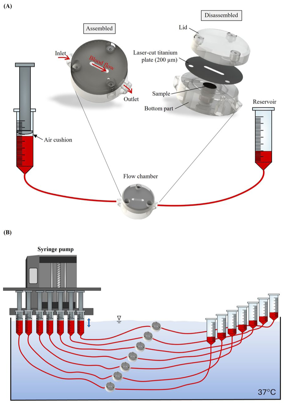
Experimental setup: (a) exemplarily presented for one flow chamber including the assembled and disassembled flow chamber; (b) For one experiment, five flow chambers are connected in a water basin to the same syringe pump. Blood samples for each surface are not pooled.
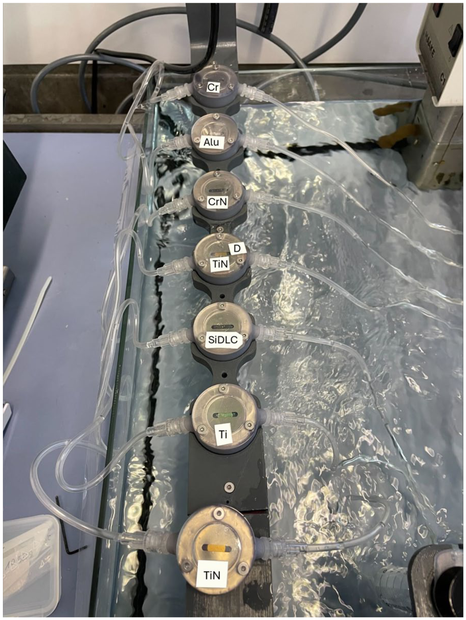
Exemplary picture of the samples in the flow chambers in the in-vitro setup before blood perfusion.
CFD simulations of wall shear rates on sample surfaces in the flow chamber
Computational fluid dynamics (CFD) simulations were conducted using Simcenter STAR-CCM+ (Version 19.04.009, Siemens Digital Industries Software, Plano, TX, USA). The geometry was created in SolidWorks (Version 2024 SP4.0, Dassault Systèmes, Vélizy-Villacoublay, France) and discretized using a finite volume approach with polyhedral and prism elements. A 33.0 µm base size was used for meshing (surfaces and flow volume) after quantifying the discretization error per Roache’s method 14 (see Supplemental Material). At no-slip walls, 10 prismatic layers were extruded from the surface mesh to resolve the boundary layer more accurately. An inflow and outflow area of prismatic cells with a length of 15 mm was connected through a conformal interface with the flow chamber geometry to minimize the impact of the boundary conditions. The meshing parameters produced 4,782,176 cells in the flow domain.
The inlet flow was set to 400 mL/h, with the outlet at environmental pressure. All other surfaces were treated as no-slip boundaries. Blood was modelled using the Carreau-Yasuda viscosity model with model parameters from Abraham et al. 15 With a 200 µm chamber height and 0.0035 Pa * s infinite shear viscosity, the flow yields a Reynolds number of ~11, indicating a laminar regime. The SIMPLE algorithm was used as a pressure-velocity coupling algorithm and the convective flux was discretized using a second-order upwind scheme.
The steady simulation was declared as converged following a reduction of the normalized residuals below the value of 1e-5.
A scalar wall shear rate


Microscopic analysis of adherent platelets
Platelet adhesion on the samples was visualized via fluorescence microscopy (Axio Observer Z1, Carl Zeiss AG, Oberkochen, Germany; Figure 3(a)). Analysis of the fluorescence images was performed using MATLAB (The MathWorks, Inc., Natick, USA, version R2021a). Only the center part of the flow channel area (14.88 mm2, Figure 3(b)) on the sample was evaluated to avoid near wall and inlet/outlet artefacts. The covered surface area was calculated by dividing attached platelet pixels (white in Figure 3(b)) by the total pixels of the binary image. Additional details and exemplary images can be found in the Supplemental material.
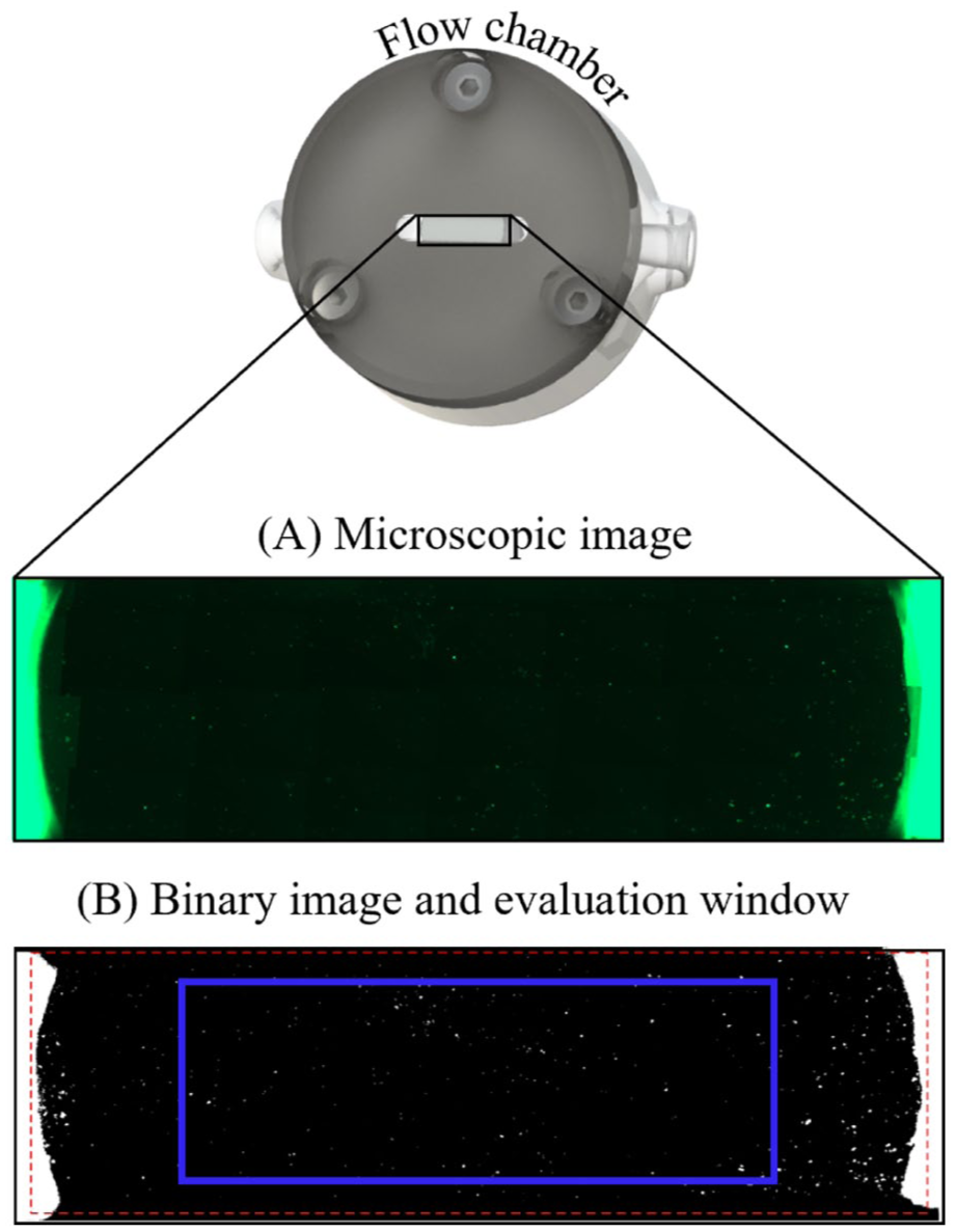
(A) Microscopic image after the evaluation with the fluorescence microscope; (B) Generated binary image and evaluation window (blue box) using MATLAB.
Statistical evaluation of adherent platelets
The normalized percentage of the covered surface area (NCSA) was calculated by dividing the percentage of the covered surface area by the platelet count of the respective blood sample to enhance comparability between different blood samples. For visualization purposes of the results, this value was then multiplied by 105. Statistical analysis was conducted using SPSS (IBM Deutschland GmbH, Ehningen, Germany). p-values below 0.05 were considered significant. Given the small sample size inherent to human blood experiments, classical significance testing has limited explanatory power. Therefore, the Wilcoxon test for paired data was applied to better capture differences between conditions despite the limited data. The Wilcoxon test effect size was calculated to assess the magnitude of differences between the groups, effect sizes between 0.1 and 0.3 were considered weak, between 0.3 and 0.5 were considered medium, and >0.5 were considered strong.
Results
CFD wall shear rate simulation
The wall shear rates on the sample surface in the channel is shown in Figure 4. The simulation reveals little variation of the WSR over most of the channel wall. However, the WSR drops sharply at the sides of the channel close to the wall due to the lower velocities in this region. Additionally, the WSR varies close to the inlet and outlet of the channel, but this effect is limited due to the low Reynolds number flow.
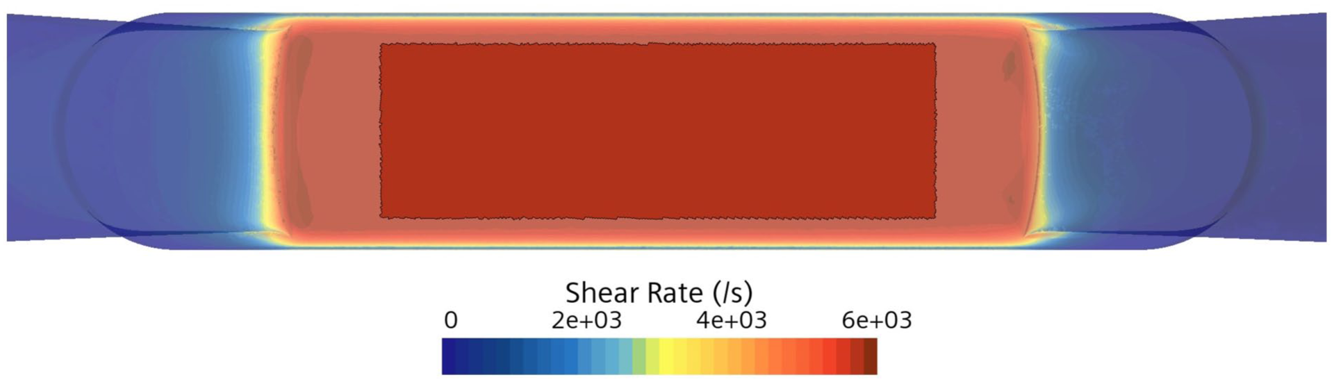
Wall shear rate on the sample surface in the area of interest for the flow rate of 400 mL/h.
We defined an area of interest (highlighted in Figure 4), where the WSR varies little. Here the WSR has a mean value of 5732.2 1/s, with a standard deviation of only ±12.1 1/s. This defined area was then used for the experimental evaluation to ensure comparable conditions for adhesion.
Platelet adhesion experiments
The boxplots in Figure 5 show the normalized percentage of the covered surface area (NCSA), including the mean value and the effect size for the pairwise comparison with the Wilcoxon test. p-values < 0.05 are indicated with asterisks. Two samples (TiN in experiment 1 and Ti in experiment 2) could not be analyzed due to excessive thrombi formation during the experiments. Alu shows the highest value (NCSA = 8.7), followed by chrome (NCSA = 3.6). Alu exhibits a significantly higher NCSA (p < 0.05) compared to CrN and SiDLC (both NCSA = 0.3) and the Wilcoxon effect size for Alu shows a medium effect compared to all HMCs, Ti (NCSA = 0.2) and CrN, and a weak effect compared to Cr. Cr shows significant higher platelet adhesion (p < 0.05) compared to CrN and a medium effect compared to CrN, Ti, and the coatings TiN (NCSA = 0.3) and SiDLC, and a weak effect compared to TiN_D (NCSA = 0.4) and Alu. There are no significant differences between the different HMCs, the reference Ti, and CrN.
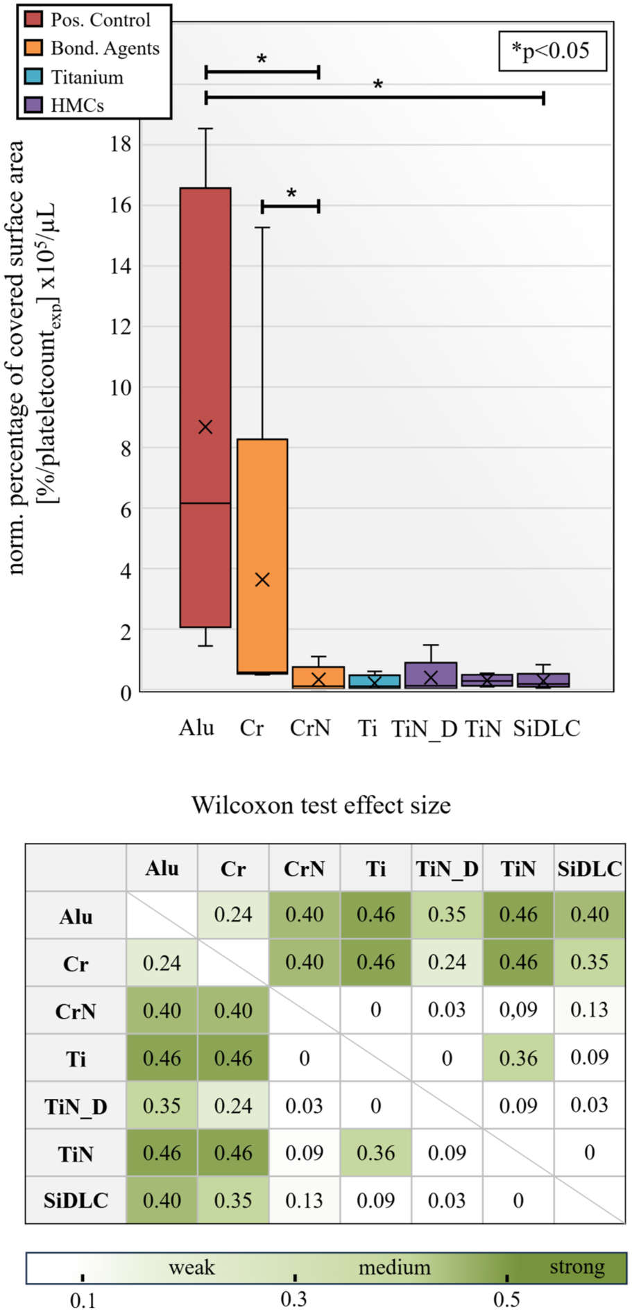
Boxplots for the percentage of the covered surface area for all surfaces normalized by the platelet count of each donation including the mean value (✕) and the p-values and effect size for the pairwise comparison with the Wilcoxon test.
Discussion
With our setup, it was possible to investigate different opaque surfaces in flow chambers with defined wall shear rate conditions, focusing on platelet binding affinity to estimate hemocompatibility in VADs in a dynamic shear setting.
Shear rates in VADs can exceed physiological values, particularly in areas prone to wear, such as the gap between the impeller and housing. The in vitro setup was designed with consideration for both the duration of the experiment, necessary to observe sufficient platelet adhesion, and the efficient use of the valuable resource of human whole blood. Pathologically high shear rates exceeding 5.000 s−1, known to trigger platelet thrombosis 17 guided our choice of experimental conditions. For the flow rate of 400 mL/h, a mean WSR of 5730.5 1/s was calculated using CFD simulations and the evaluable surface area was defined. As a result, the flow conditions represented a compromise between realistic in vitro replication and promoting platelet adhesion under controlled circumstances.
Alu served as suitable positive control since it had the highest amount of adherent platelets with significant differences compared to CrN and SiDLC and medium Wilcoxon effect sizes compared to the HMCs, Ti, and CrN. The results indicate that the chrome surface has a high risk of thrombosis due to a high platelet adhesion during the experiments, especially compared to CrN (p < 0.05). It showed weak to medium effect sizes compared to all HMCs, Ti, and CrN. Therefore, using Cr as bonding agent should be avoided, since eventual scratching of the HMC may expose the bonding agent to the blood and increase thrombosis risk. According to this study, CrN is more suitable since it had significant lower platelet adhesion than Cr, medium effect sizes compared to Alu and Cr, and no significant higher adhesion than the reference Ti. Also, regarding the effect sizes, there was no effect compared to Ti.
In addition to their use as bonding agents, chromium alloys are also employed as biomaterials for implants. 18 To the best of our knowledge, there is no in vitro study so far that investigated chrome apart from for example alloys like CoCrMo19,20 steal alloys like CrNiMo (316L) or FeCrMnMoN (BIOSSN4).20,21 Moreover, no studies were found that investigated the behavior of CrN coatings with respect to thrombogenicity.
Overall, no HMC showed significantly higher potential for the formation of thrombi compared to Ti. Moreover, only TiN showed a medium effect compared to Ti, indicating a negative influence of the process related droplets on the TiN_D surface in terms of platelet adhesion compared to when the droplets are being removed afterwards. Our findings are consistent with previous studies investigating inorganic hard material coatings for blood-contacting applications, 11 where none of the investigated hard material coatings (among them titanium nitride and diamond-like carbon) showed higher platelet adhesion than Ti in a similar in vitro flow chamber setup. Notably, TiN even demonstrated significantly lower platelet coverage compared to Ti, supporting our observation that TiN does not increase thrombogenicity and is a suitable wear-resistant coating. Other studies have also emphasized the hemocompatibility of DLC-based coatings for cardiovascular applications (e.g., heart valves, 22 stents, 23 and VADs12,24), and DLC coatings were stated to be superior to TiN coatings. 12 However, the in our study investigated DLC surface additionally includes Si and the results show no evidence of SiDLC being inferior to TiN in terms of platelet adhesion. This is consistent with literature, as for example, the groups of Ong et al. 25 or Roy et al. 26 showed good hemocompatibility of SiDLC surfaces.
The agreement between our results and the literature reinforces that these HMCs represent promising options for blood-contacting components without increasing thrombotic risk.
By including Cr-based surfaces in this study, we highlight a potential thrombogenic hazard not addressed in previous investigations, underscoring the importance of careful material selection, especially regarding underlying bonding agents. Overall, these results contribute valuable comparative data on the thrombogenicity of HMCs, supporting their further investigation and potential implementation in medical devices.
It should be noted that blood samples for in vitro experiments must always be anticoagulated, as they would otherwise clot within minutes and experiments would not be possible. Sodium citrate acts by chelating ionized calcium, a critical cofactor for coagulation, thereby effectively preventing coagulation. 27 While this limits the reproduction of full in vivo conditions, particularly with respect to thrombin and fibrin formation, platelet function remains largely intact. 28 Citrated blood allows reliable investigation of primary platelet-surface interactions, which are central to the early stages of thrombus formation.
Due to considerable effort and ethical challenges involved in conducting human whole blood experiments, it was not feasible to perform additional experiments within the scope of this study. However, we anticipate that increasing the number of experiments would amplify the observed effects (resulting in higher Wilcoxon effect sizes) while maintaining similar trends. It is important to note that, despite the relatively small sample size in this study, the current results already reveal differences, even if not fully captured by p-values. We want to highlight the importance of considering effect sizes, particularly in studies where sample size is limited due to resource constraints.
Conclusion
Our study evaluated the hemocompatibility of various hard material coatings and bonding agents for the use in VADs by assessing the platelet binding affinity under defined shear rate conditions. Wall shear rate conditions were simulated using CFD and had a mean wall shear rate of 5730.5 1/s on the evaluated surface area. Chrome showed a higher thrombosis risk and should be avoided, while chrome nitride demonstrated lower platelet adhesion and performed similarly to the reference titanium. The tested HMCs silicon-incorporated diamond-like carbon, titanium nitride with droplets and titanium nitride without droplets showed comparable compatibility to Ti, supporting their use in VADs.
Supplemental Material
sj-jpg-4-jao-10.1177_03913988251398845 – Supplemental material for Platelet adhesion on hard material coatings and bonding agents for ventricular assist devices in a flow chamber
Supplemental material, sj-jpg-4-jao-10.1177_03913988251398845 for Platelet adhesion on hard material coatings and bonding agents for ventricular assist devices in a flow chamber by Isabell Esslinger, Henri Wolff, Tim Bierewirtz, Michael Lommel and Ulrich Kertzscher in The International Journal of Artificial Organs
Supplemental Material
sj-pdf-1-jao-10.1177_03913988251398845 – Supplemental material for Platelet adhesion on hard material coatings and bonding agents for ventricular assist devices in a flow chamber
Supplemental material, sj-pdf-1-jao-10.1177_03913988251398845 for Platelet adhesion on hard material coatings and bonding agents for ventricular assist devices in a flow chamber by Isabell Esslinger, Henri Wolff, Tim Bierewirtz, Michael Lommel and Ulrich Kertzscher in The International Journal of Artificial Organs
Supplemental Material
sj-pdf-2-jao-10.1177_03913988251398845 – Supplemental material for Platelet adhesion on hard material coatings and bonding agents for ventricular assist devices in a flow chamber
Supplemental material, sj-pdf-2-jao-10.1177_03913988251398845 for Platelet adhesion on hard material coatings and bonding agents for ventricular assist devices in a flow chamber by Isabell Esslinger, Henri Wolff, Tim Bierewirtz, Michael Lommel and Ulrich Kertzscher in The International Journal of Artificial Organs
Supplemental Material
sj-pdf-3-jao-10.1177_03913988251398845 – Supplemental material for Platelet adhesion on hard material coatings and bonding agents for ventricular assist devices in a flow chamber
Supplemental material, sj-pdf-3-jao-10.1177_03913988251398845 for Platelet adhesion on hard material coatings and bonding agents for ventricular assist devices in a flow chamber by Isabell Esslinger, Henri Wolff, Tim Bierewirtz, Michael Lommel and Ulrich Kertzscher in The International Journal of Artificial Organs
Footnotes
Acknowledgements
We would like to thank Shayan Essam for his efforts improving the MATLAB script.
Declaration of conflicting interests
The author(s) disclosed receipt of the following financial support for the research, authorship, and/or publication of this article: This study was conducted at the request and with the financial support of Berlin Heart GmbH. The study’s design, data collection, analysis, and interpretation were performed independently by the research team, ensuring the integrity and objectivity of the results. All findings and conclusions presented in this paper are solely those of the authors.
Funding
The authors received no financial support for the research, authorship, and/or publication of this article.
Supplemental material
Supplemental material for this article is available online.
References
Supplementary Material
Please find the following supplemental material available below.
For Open Access articles published under a Creative Commons License, all supplemental material carries the same license as the article it is associated with.
For non-Open Access articles published, all supplemental material carries a non-exclusive license, and permission requests for re-use of supplemental material or any part of supplemental material shall be sent directly to the copyright owner as specified in the copyright notice associated with the article.