
Abstract
Development of controlled, targeted drug delivery systems represents one of the frontier areas of biomaterials science, where a multidisciplinary approach is of direct benefit to human healthcare. We demonstrate herein the potential of sol–gel derived phosphate-based glass for use in drug delivery applications. Our low-temperature sol–gel synthesis of phosphate-based glasses has made it possible to incorporate relatively unstable functional molecules for controlled release. We demonstrate the potential of this approach by incorporating the chemotherapy agent cisplatin in a CaO–Na2O–P2O5 glass. X-ray absorption spectroscopy is used to show that the chlorine ligands of cisplatin undergo exchange with oxygen during the synthesis, consistent with binding to the phosphate groups of the sol–gel. UV–visible spectroscopy reveals the subsequent release of cisplatin into an aqueous medium.
INTRODUCTION
Controlled drug delivery offers numerous advantages compared to conventional dosage methods; these include improved efficacy, reduced patient toxicity, and improved patient comfort. Many materials have been employed as carriers in drug delivery applications. These include polymeric micelles [1,2], liposomes [3], hydrogels [4], biodegradable polymers [5–7], magnetic nanoparticles [8], and carbon nanotubes [7,9]. The size range of drug carriers investigated spans three orders of magnitude from nanoparticles that can cross the cell membrane [5] to microspheres for interstitial injection [10] or chemoembolization [11,12]. Recent key areas of advancement include functionalizing the surface of carrier particles for improved uptake by specific tissue or for improved biocompatibility [8,9,13], the use of magnetic nanoparticles so that drug carriers can be externally directed by a magnetic field or their position monitored externally [8,14], the use of pH- or thermo-sensitive materials so that the drug release can be tuned to a particular physiological environment [1,15], and the use of self-assembled prodrugs for enzymatically triggered delivery [15]. Here we present a new material for potential use in drug delivery devices.
Phosphate-based glass containing Ca2+ and Na+ ions has desirable properties for drug delivery applications. For several years, such glass has been used as passive host materials for the controlled release of metal ions for a variety of applications including veterinary treatments [16] and as antibacterial materials [17]. It is both biocompatible and bioresorbable [18], has near linear dissolution rate in aqueous media, and the rate of degradation can be finely tuned through subtle variations in composition [19]. Linear dissolution is particularly important because one of the common problems associated polymer-based drug delivery devices is an ‘initial burst’ of release that is followed by a much lower release rate [20]. Biocompatibility may also offer a significant advantage over the current polymer-based systems where their degradation can result in polymer fragments with heterogeneous chain-lengths which could lead to toxicity [15]. Until now the use phosphate-based glass in drug delivery devices has been limited by the high temperatures required in their preparation, by quenching a melt consisting of oxide precursors, which restricts the types of molecules that can be incorporated. Use of the low-temperature sol–gel route largely circumvents this problem.
In order to demonstrate the potential of sol–gel phosphate-based glass for use in drug delivery devices we introduced the chemotherapy drug cisplatin into our sol–gel reaction. We chose cisplatin as our test drug because it is a widely used and effective cytotoxic agent in the treatment of malignancies of the lung, head and neck, and ovarian cancers [21] and its effectiveness is expected to be significantly enhanced by targeted delivery. The major limitation in the clinical application of cisplatin is the development of resistance in tumors: to achieve an effective intracellular concentration, very high systemic doses of cisplatin are necessary. Unfortunately, such high doses result in severe systemic toxicity and poor patient compliance.
We introduced cisplatin into our sol–gel synthesis of CaO–Na2O–P2O5 glass [22]. After processing, drying, and modest heat treatment the product was a glass of nominal composition (CaO)0.3(Na2O)0.2(P2O5)0.5 containing 1 wt% cisplatin. To verify that cisplatin is successfully encapsulated by the sol–gel matrix, we studied the platinum environment using Pt LIII-edge extended X-ray absorption fine structure (EXAFS) spectroscopy. Having established successful encapsulation of cisplatin within the phosphate glass matrix, we used UV–visible spectroscopy to monitor its release into an aqueous medium.
MATERIALS AND METHODS
Sample Preparation
The sol–gel reaction used to prepare the phosphate-based glass is described elsewhere [22] and outlined schematically in Figure 1. The calcium methoxyethoxide used in the reaction was freshly prepared by the reaction of calcium with 2-methoxyethanol [23]. The cisplatin was dissolved in dimethylformamide (DMF) and added to the sol–gel before the final gelation. The resultant gel was dried at 60°C for 1 week and 120°C for 1 week before heating to 200°C to consolidate the structure and form the glass. This temperature was chosen on the basis of our previous structural study that showed that consolidation of the sol-gel network structure occurs above 200°C [22].
Schematic of the sol–gel synthesis of cisplatin-loaded phosphate-based glass.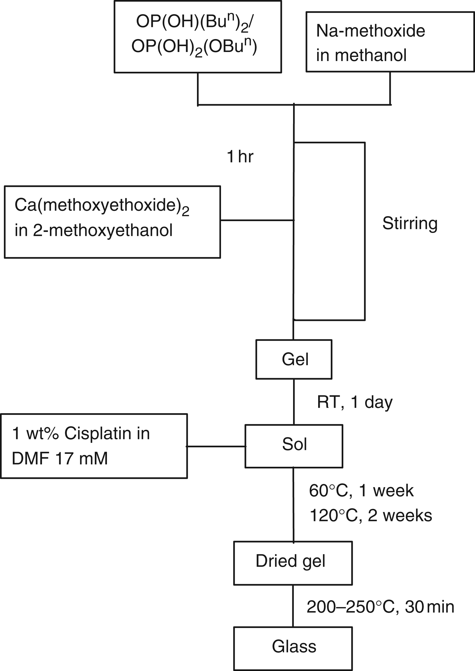
Characterization
The Pt LIII-edge EXAFS measurements were recorded on Station 9.3 at the SRS, Daresbury Laboratory, UK. Finely ground samples were mixed with polyethylene and pressed into pellets to allow easy mounting in the X-ray beam; in the case of the pure cisplatin sample this dilution also reduces self-absorption of the signal. Data were collected in fluorescence mode and analyzed using the software EXCALIB, EXSPLINE, and EXCURV98 [24]. As well as data from the cisplatin-loaded dried gel and glass, reference data were also collected from pure cisplatin.
The UV–visible data were collected on a Cary 400 spectrophotometer: 0.1 g of the cisplatin-loaded glass was placed in 3 mL of aqueous 0.9 wt% NaCl solution and the absorbance of the liquor above the sample monitored at 301 nm as a function of time. A sodium chloride solution was used because cisplatin is more stable in 0.9 wt% NaCl solution than in water [25]. In the reference beam was placed a sample of glass containing no cisplatin in saline solution (0.1 g in 3 mL of 0.9 wt% NaCl(aq)). Each time point is the average of at least two measurements with errors calculated on the basis of statistical variation between results.
RESULTS AND DISCUSSION
EXAFS reveals the local environment of a target atom by analysis of the oscillations superimposed on its X-ray absorption edge. These oscillations are due to modulation of the absorption cross-section caused by interference between outgoing photoelectron waves and those back-scattered from surrounding atoms. Modeling of EXAFS data using curved-wave theory returns the distances between the target atom and those surrounding it as well as the number of atoms in each coordination sphere observed and a measure of the disorder amongst the bond distances (given by the Debye–Waller factor). Figure 2 shows the EXAFS data collected from our drug-loaded samples together with data from a sample of cisplatin for comparison. The structural parameters obtained by modeling the EXAFS data are given in Table 1.
Pt LIII-edge EXAFS fluorescence data: (top) cisplatin, (middle) dried cisplatin-loaded phosphate gel and (bottom) cisplatin-loaded phosphate-based glass. (a) k3-weighted EXAFS data and (b) their Fourier transforms. The experimental data are depicted by solid lines and the fits by dashed lines. Structural parameters obtained from the EXAFS data. N = coordination number, R = atomic separation, A = Debye–Waller factor, and Rfit = measurement of the agreement between the fit and the data. Note that parameters in italics have been fixed during the refinement.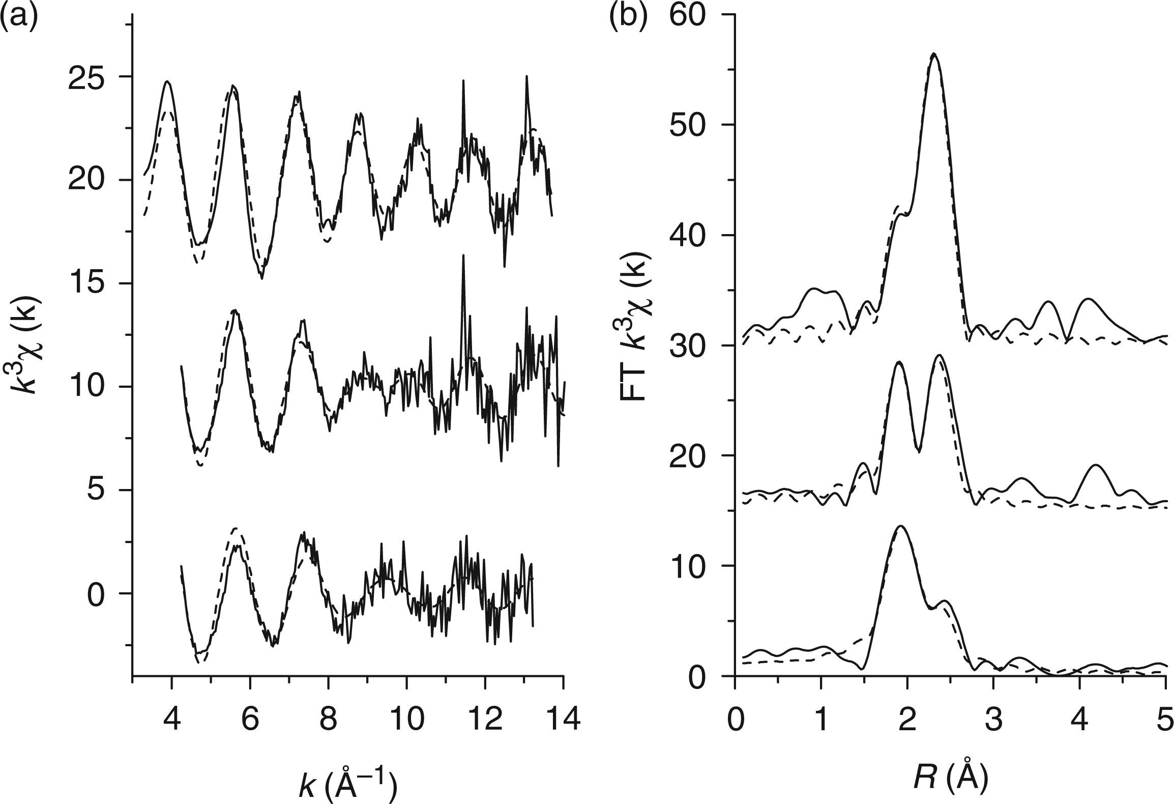
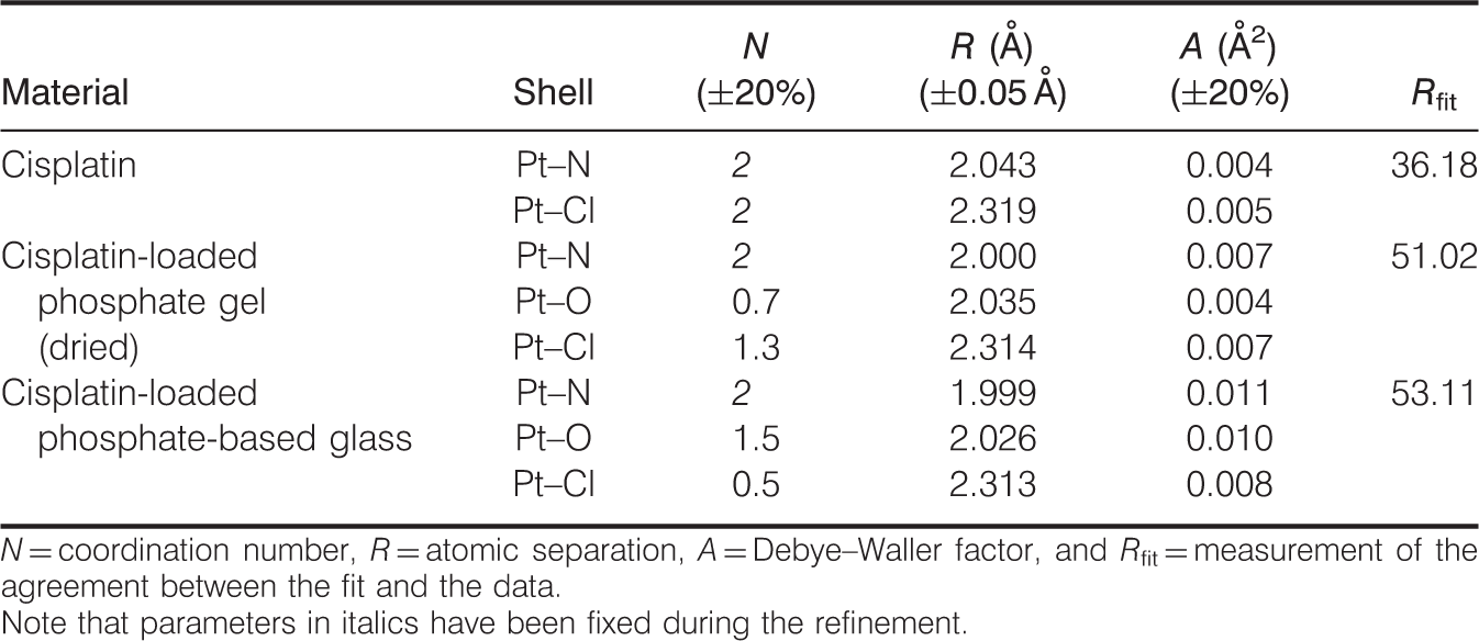
Cisplatin is a square-planar Pt2+ complex with two chlorine and two ammonia ligands arranged in the cis configuration as depicted by Figure 3. The Fourier transform of the EXAFS data from the cisplatin sample exhibits two partially overlapping peaks which correspond to the Pt–N and Pt–Cl distances within the cisplatin molecule. Consistent with the known structure of cisplatin, the data have been modeled with the Pt–N and Pt–Cl coordination numbers fixed at 2. Refinement of the remaining structural parameters returned Pt–N and Pt–Cl distances of 2.04 and 2.32 Å, respectively, consistent with the crystallographic values of 2.00 and 2.33 Å [26].
(a) Cisplatin and (b) carboplatin molecules.
Inspection of the EXAFS data from our sol–gel samples shows that the intensity of the peak due to Pt–Cl bonds is reduced relative to that due to the Pt–N bonds compared to the data from pure cisplatin. This can be rationalized if we consider the effect of substituting some of the chlorine ligands for oxygen ligands. To do this, we need to take note of two points: firstly, because the atomic number of oxygen is much lower than that of chlorine, oxygen atoms are much weaker and back-scatterers of photoelectrons; secondly, Pt–O bonds in square-planar Pt2+ complexes are only slightly longer than Pt–N bonds (in the carboplatin complex depicted in Figure 3, the Pt–N bond-length is 2.02 Å and the Pt–O bond-length is 2.03 Å) [26]. Thus, substitution of the chlorine ligands by oxygen is expected to reduce the amplitude of the Pt–Cl peak and broaden the Pt–N peak due to the extra contribution from Pt–O at a slightly longer distance; this is what is observed experimentally in Figure 2. The results suggest greater substitution of the chlorine ligands by oxygen in the heat-treated sol–gel glass compared to the dried gel. The degree of ligand exchange can be quantified by modeling the EXAFS data in Figure 2. For the data from the cisplatin doped sol–gel, three platinum correlations have been used: Pt–N, Pt–O, and Pt–Cl. In this case, the Pt–N coordination number, and the sum of the Pt–O and Pt–Cl coordination numbers have been fixed at 2; thus constraining the geometry of the platinum environment to square planer. Refinement of the structural parameters yields Pt–N, Pt–O, and Pt–Cl distances of 2.00 A, 2.03 A and 2.31 Å, respectively, consistent with a mixed Pt2+ environment. Refinement of the linked Pt–O and Pt–Cl coordination numbers confirms that the extent of substitution of the chlorine ligands by oxygen is greater in the heat-treated sol–gel. From these coordination numbers we can calculate the fraction of chlorine ligands that undergo exchange with oxygen during the process of encapsulating in the sol–gel. In the dried gel 35 ± 7% of the chlorine ligands are exchanged whereas in the heat-treated glass 75 ± 15% are exchanged. This observation of chlorine ligand exchange is not surprising since it is well known that the initial step in the mechanism of action of cisplatin is exchange of its chlorine ligands with water to form reactive aquated forms [27]. Furthermore, displacement of the chlorine ligands by phosphate ions has been noted [27], and one study describing the adsorption of cisplatin by hydroxyapatite has proposed the formation of Pt–phosphate complexes as one of the binding interactions [28]. Therefore the results presented here are consistent with cisplatin binding to the sol–gel via exchange of its chlorine ligands with oxygen from the phosphate network that forms the backbone of the sol–gel structure. The results also show that the degree of exchange is greater in the heat-treated glass sample.
Following methodology reported elsewhere [29], we measured absorbance values at 301 nm, the maximum absorbance of cisplatin, as a function of time. The results plotted in Figure 4 show sustained release of cisplatin over a period of several days: the curve fitted to the time points suggests that the release follows logarithmic growth. The results here compare favorable with those from studies of the release of cisplatin from silica-based sol–gels [29,30] where a two-stage release mechanism was observed with a high dose of cisplatin released in the first few hours followed by a much slower release. In fact, in those samples over 50% of the total amount of cisplatin released is released in the first 3 h and 90% of the total released within 25 h.
Release of cisplatin into saline solution: plot of absorbance of the solution at 301 nm against time. The error bars represent two standard deviations from the mean absorbance at each time point.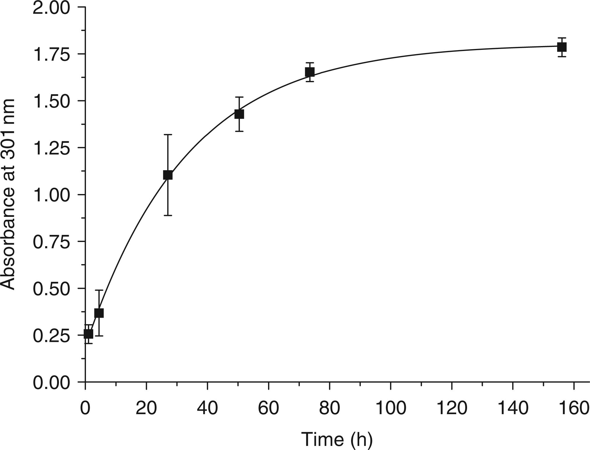
These results demonstrate the potential of sol–gel phosphate-based glass to be used in cisplatin delivery devices that can be implanted in the vicinity of tumors offering particular opportunities for the treatment of head and neck cancer where the tumors tend to be readily accessible. Such an approach would involve preparing this material as microspheres designed to be injected directly into a tumor such that they become physically trapped within the smaller blood vessels (chemoembolization), and sustain the action of cisplatin through controlled release – thereby improving efficacy and reducing systemic toxicity. Additional benefits may arise from venous occlusion, reducing the supply of blood to the tumor.
Furthermore, as a proof-of-principle this work suggests that sol–gel phosphate-based glasses may be suitable carriers for many other drugs and active molecules. Also there is potential to incorporate beneficial co-dopants within the glass matrix such as gallium which has potent antibacterial qualities [23] and has displayed anti-tumor activity [31].
CONCLUSIONS
The data presented here demonstrate that cisplatin can be successfully incorporated in a sol–gel phosphate-based glass and subsequently released into an aqueous medium. Sustained in vitro release was observed over a 4-day period. The results demonstrate the potential of sol–gel phosphate-based glass as a drug delivery vehicle.
Footnotes
ACKNOWLEDGMENTS
The authors wish to acknowledge funding from the EPSRC (EP/C000714, EP/C000633, and GR/T21080). We thank STFC for access to the synchrotron at Daresbury Laboratory. This work was supported in part by WCU Program through the National Research Foundation of Korea (NRF) funded by the Ministry of Education, Science, and Technology (No. R31-10069).