






Abstract
Vital pulp therapy (VPT) and direct pulp capping (DPC) are procedures regularly performed in dogs for the management of acute tooth fractures and as part of management for traumatic malocclusions. The purpose of this review is to apply an evidence-based medicine approach to systematically review and evaluate the scientific literature evaluating the efficacy of mineral trioxide aggregate (MTA) to other commercially available materials used for VPT in the permanent teeth of dogs. The 9 studies meeting inclusion criteria were reviewed and each studies evidence was classified using a grading system modified from the Oxford Centre for Evidence-Based Medicine. For the studies meeting inclusion criteria, MTA consistently performed as well or better than other commercially available products in terms of calcific barrier formation and biocompatibility. This review found a lack of consistency between the studies making a direct comparison of the results unreliable. Future studies would benefit from the implementation of a standard scoring system for histology, equivalent and longer study duration times and the correlation of histological and radiographic data.
Keywords
Introduction
The purpose of vital pulp therapy (VPT) is to preserve the vitality and function of the dental pulp. 1,2 This is achieved by covering the exposed pulp with a biocompatible material that ideally enables and promotes the formation of a calcific barrier, leaving the apical portion of the pulp free from inflammation. 3 In veterinary dentistry, frequently performed VPT procedures include direct pulp capping (DPC) and partial coronal pulpectomy or pulpotomy following recent complicated crown fracture (tooth fracture with pulp exposure); crown-height reduction to eliminate trauma in cases of malocclusion; and pulp exposures that occur during restorative preparation. 4,5 VPT is less frequently employed as a treatment for caries in dogs as compared with people, with only an estimated 5.3% of domestic dogs affected by this condition. 6
The American Veterinary Dental College (AVDC) defines VPT as a procedure performed on a vital tooth with pulp exposure, involving partial pulpectomy, DPC and access or fracture site restoration. 7 The AVDC further defines DPC as a procedure performed as part of VPT and involving the placement of a medicated material over an area of pulp exposure. 7 This definition of direct pulp capping is similar to that used by the American Association of Endodontists (AAE). 8 The AAE do not have a broad definition for VPT, instead, they define pulp capping as the treatment of an exposed vital pulp by sealing the pulp wound with a dental material such as calcium hydroxide (CH) or mineral trioxide aggregate (MTA) to facilitate the formation of reparative dentin and maintenance of a vital pulp. 8 The studies included in this review refer to both DPC and pulpotomy as separate procedures. Pulpotomy is defined by the AAE as the removal of the coronal portion of a vital pulp as a means of preserving the vitality of the remaining radicular portion. 8 In the studies included in this review, DPC is itself considered a procedure where a material is placed directly on a pulp exposure without specific removal of pulp. For the purpose of this review, we will follow these definitions when referring to VPT, DPC and pulpotomy.
Many materials have been explored as DPC agents including, but not limited to, CH, bioceramics, MTA and dental adhesive resins (DAR). Long considered the gold standard in DPC in humans, 9 CH first became widely known as a successful DPC agent through the work of Hermann 10,11 who demonstrated the formation of secondary dentin in the amputation sites of vital pulps capped with CH. CH has excellent antibacterial properties due to its high pH, with a long track record of success in the human literature. 12 -14 There are, however, multiple disadvantages associated with CH, including irritation and necrosis of the pulp directly beneath the capping material; high solubility in oral fluids; dissolution over time; lack of inherent adhesive properties; and the presence of tunnel defects within the calcific barrier leading to a failure to produce a hermetic seal to the underlying pulp against bacteria due to microleakage. 15 -18 Additionally, there is great variability in the available formulations and properties of different commercially available CH preparations as demonstrated in a review by Fava and Saunders. 19 While CH has long been considered by some to be the gold standard pulp capping material, no one formulation has ever been referenced consistently as the control product against which others are measured. Fava and Saunders 19 demonstrated the great variability in non-setting CH pastes, broadly classifying products into aqueous, viscous and oily types, each with slightly different suggested indications. This does not consider that CH is also available in numerous powder and cement formulations. Different forms of CH have produced marked differences when applied to a pulp exposure, 20 -22 and this is likely a factor that has led to great variability in the reported success of CH. 23
The problems associated with CH have led to the development of several new materials. MTA has gained popularity in recent years and it has been recommended as a suitable replacement for CH for VPT. 24 MTA was first described in 1993 as a cement used in repairing lateral root perforations. 25 ProRoot® MTAa (PMTA) was the first commercial MTA and was under patent, which delayed release of competing products. 26 For this reason, PMTA was chosen as the control material in this review. Since its invention, MTA has been evaluated extensively under laboratory conditions, in animal studies and clinical trials. 23,27,28 These studies have demonstrated that MTA has excellent biocompatibility, bioactivity and sealing ability, as well as being associated with desirable clinical outcomes when used for VPT. 23,27,28 MTA possesses many favorable characteristics as a DPC agent. The ability of MTA to set is not affected by the presence of blood or serum. 29 Similar to CH it has a high pH, resulting in extraction of growth factors from dentin thought to promote calcific barrier formation. 30 MTA has also demonstrated good sealing ability with the capacity to bond to dentin, the exact mechanism of which is unclear however, an initial mechanical bond between MTA and dentin forms with a chemical bond being produced over time. 31,32 Once set, MTA is nonabsorbable, unlike CH which can degrade and dissolve, allowing potential ingress of microorganisms and bacterial contamination of the pulp through tunnel defects in the calcific barrier. 33
However, MTA is not without potential drawbacks, including a long setting time, tooth discoloration, high cost and difficult handling characteristics. 23,27,28,34,35
With continued attempts to develop a material without the drawbacks of MTA, MTA-like cements have emerged with descriptors such as “bioceramics” 36 and “bioactive endodontic cements.” 23 A review of properties of MTA and MTA-like cements by Ha et al 37 indicated that despite the differences in terminology, these cements are similar in their elemental compositions to MTA and, hence, fall within the description of MTA.
In the time since MTA became commercially available it has gained popularity, replacing calcium hydroxide in many veterinary dentists’ armementarium. 38 Within the veterinary literature, only one peer-reviewed study evaluating the success of VPT in dogs with MTA has been published. 5 This clinical retrospective study found that MTA was an effective option for use in crown reduction to treat malocclusion and for the treatment of recent crown fractures in immature or mature permanent teeth. 5 A systematic review of the use of MTA in VPT, specifically relating to dogs, has not been published. The lack of published literature in veterinary journals supporting MTA as the gold standard material in dogs suggests that the recent shift toward the use of MTA may be based on data extrapolated from studies performed in species other than dogs.
Despite the lack of veterinary studies focusing on VPT in dogs, the popularity of this species as a model for human dental research means that several studies published in the human literature contain data relevant to veterinarians performing VPT procedures in this species. The purpose of this review was to apply an evidence-based medicine approach to the use of MTA as a DPC material in dogs and systematically review and evaluate the scientific literature reporting the outcome of a single commercially available MTA product as a control, compared with other commercially available DPC materials.
Materials and Methods
The clinical question was formulated using the PICO process, a technique used in evidence-based practice to frame and answer a clinical or healthcare related question. 39,40 The PICO acronym stands for patient or problem, intervention, comparison intervention and outcomes. 40 The focused question formulated using this process which this systematic review aimed to answer, was: “Does PMTA result in superior ongoing pulpal health following DPC of recently pulp-exposed, permanent dogs’ teeth compared to other commercially available DPC agents?” A “recently exposed pulp” was defined as a pulp exposure that occurred no greater than 48 hours prior to treatment. 41
A systematic literature search of 3 online databases (PubMed, Web of Science and Medline) was performed on April 29th, 2018. The 3 online databases were searched using the following terms: [(pulpotomy OR direct pulp cap OR vital pulp therapy OR partial pulpectomy) AND (dog OR dogs OR canine OR canines) AND (mineral trioxide aggregate OR MTA OR calcium hydroxide OR bioceramic OR calcium silicate)]. Additionally, potential manuscripts were searched by hand in the bibliography sections of textbooks and manuscripts.
Inclusion and exclusion criteria for manuscript selection were predetermined to reduce bias. Studies were included in this systematic review if they were published in peer-reviewed journals, available in the English language, examined the histological response of permanent dog teeth following DPC, and compared PMTA (the first commercially available MTA and used as the control MTA product) to other commercially available materials. The studies were required to provide original data with procedures performed on vital teeth with a pulp exposure time no greater than 48 hours with a minimum follow-up period of 28 days. No maximum follow-up period was set.
Studies were excluded if they were not published in the English language, if non-commercially available materials or experimental techniques were applied or if an indirect pulp capping procedure was performed. Studies were also excluded if the patient had an underlying medical condition or outside pharmacological influences were applied (e.g. use of immunosuppressant medication). Articles that did not contribute new experimental data, those that were purely in vitro studies or those where DPC was performed but the result of this procedure was not the focus of the article were also excluded from this review. Reports in book chapters, abstracts, and seminar proceedings that were not peer reviewed were excluded from the study.
One study that met inclusion criteria was subsequently excluded due to discrepancies in the data throughout the article. 42
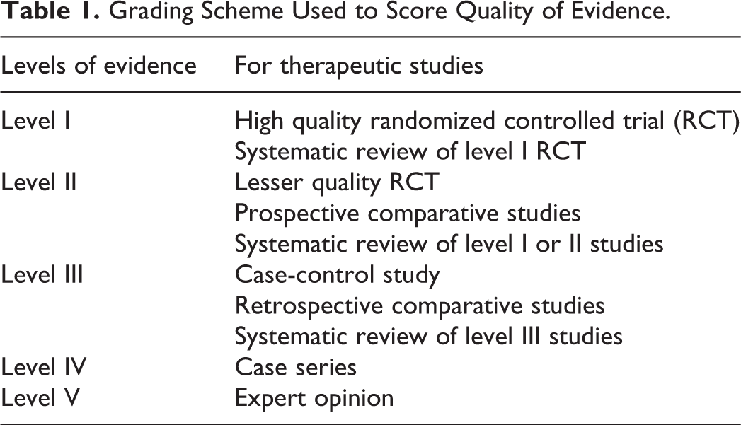
Following the database searches, the authors read the titles, abstracts or full text of the publications to determine the eligibility of the study. Publications that met inclusion criteria were reviewed and each study’s evidence was classified using a scoring system of strongest (I) to weakest (V) evidence modified from a grading system published by the Oxford Centre for Evidence-Based Medicine (Table 1). 43
Grading Scheme Used to Score Quality of Evidence.
The following data was extracted from each article included in this study: the first or only author name, the publication year, the study type, materials used, the type of pulp capping procedure, the number of teeth tested per material, the purpose of the study, duration of the follow-up, key results and outcomes, level of evidence and the funding source, if disclosed.
Results
Literature searches revealed 307 manuscripts for review; with 10 meeting the inclusion criteria and 1 subsequently eliminated for reasons described above. The “Preferred Reporting Items for Systematic Reviews and Meta-Analyses” (PRISMA) guidelines were utilized to show how the search results were narrowed to the 9 manuscripts included in this systematic review. 44,45 A flowchart modified from these PRISMA guidelines is provided in Figure 1. 44
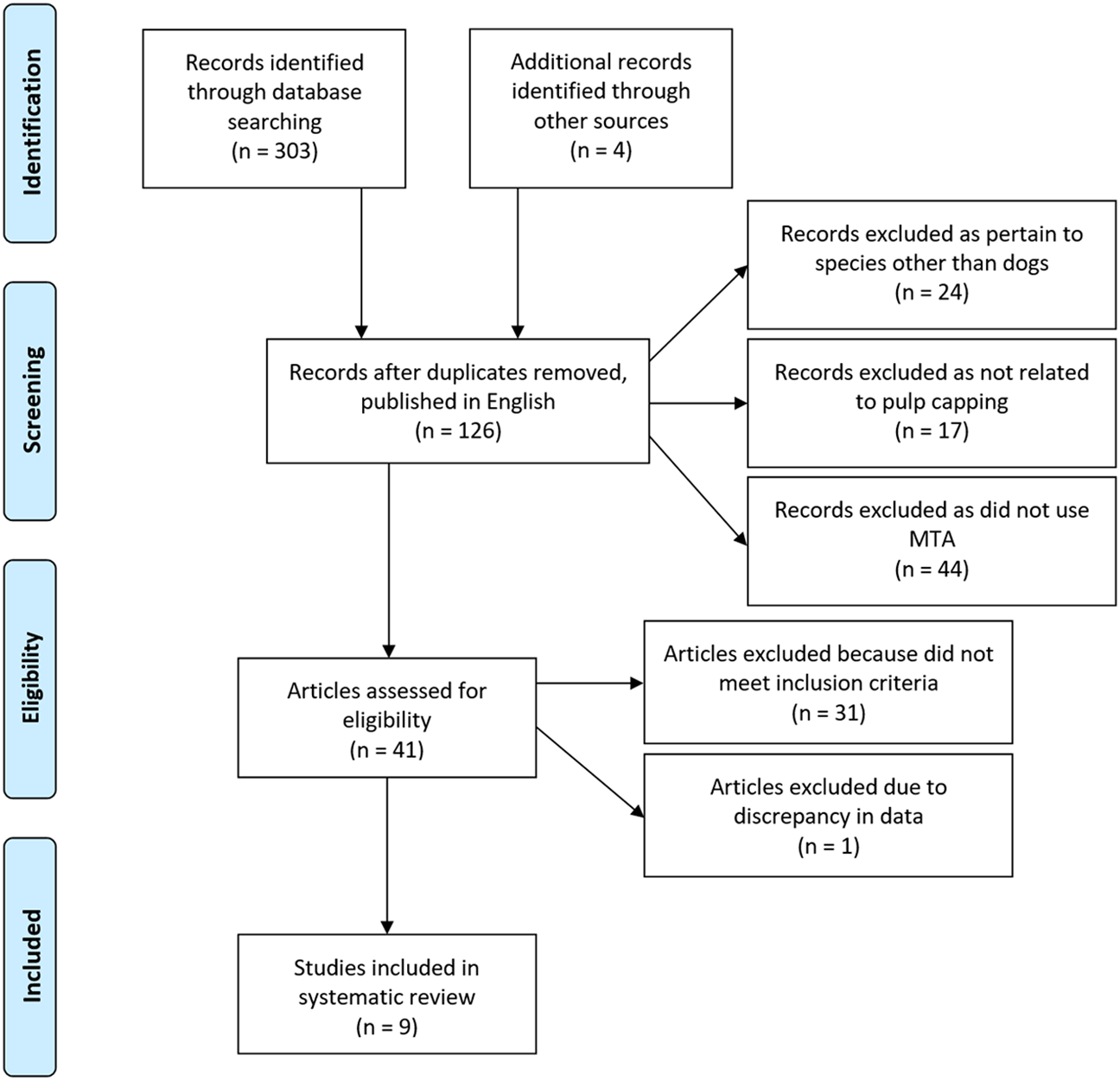
Flowchart modified from the PRISMA 44 guidelines, demonstrating the process by which the search results were narrowed to the 9 articles included in this systematic review.
The most common reason for exclusion of manuscripts was due to commercially available MTA (i.e. experimental MTA products) not being employed as a material in the study, articles focusing on species other than dogs or those not related to a DPC procedure. Of the studies meeting inclusion criteria, all were prospective comparative studies providing level II evidence relative to the study question.
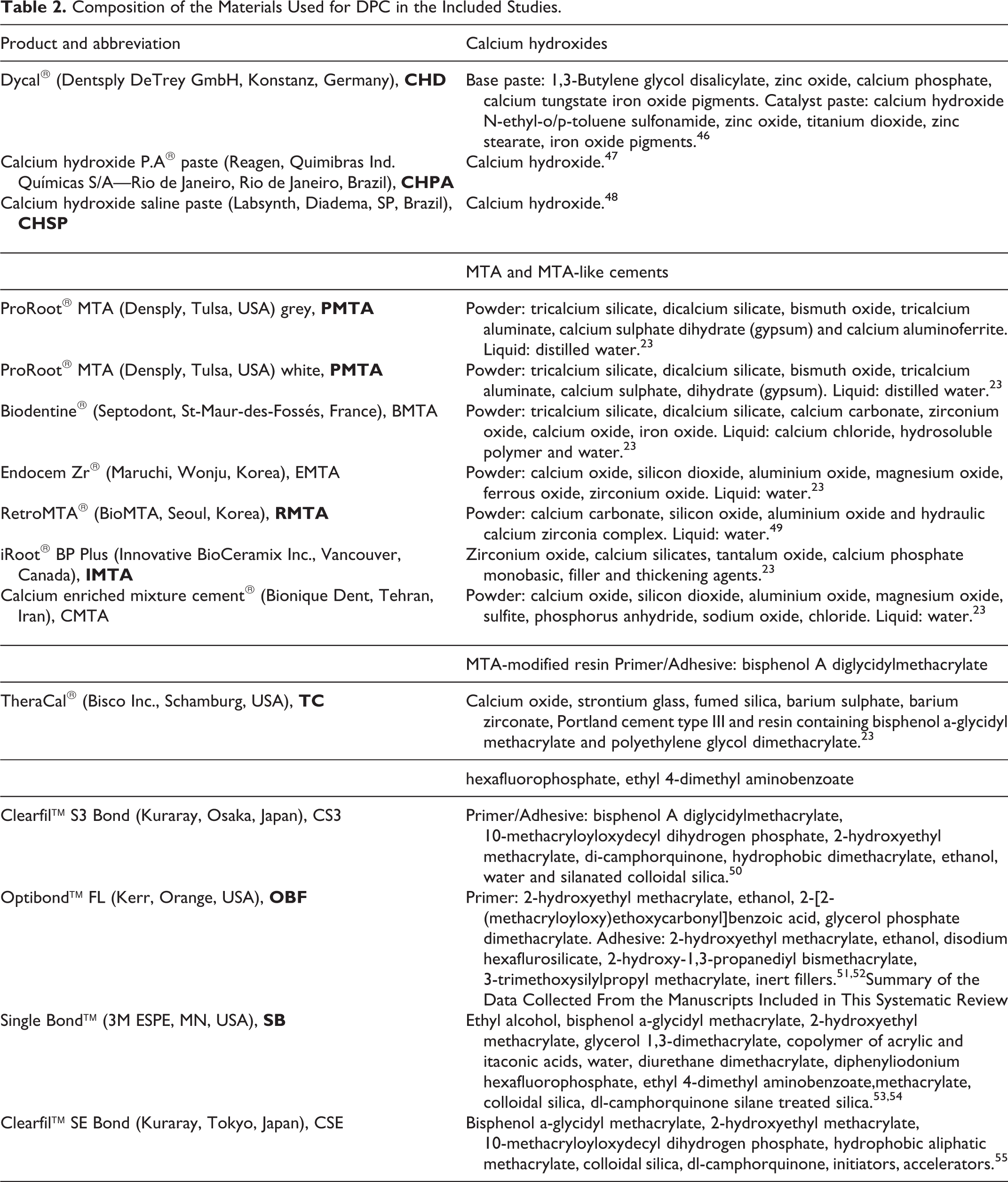
The DPC materials used in the included studies and their chemical composition are detailed in Table 2.
Composition of the Materials Used for DPC in the Included Studies.
The results of the included studies are detailed in Table 3.
Summary of the Data Collected From the Manuscripts Included in This Systematic Review.
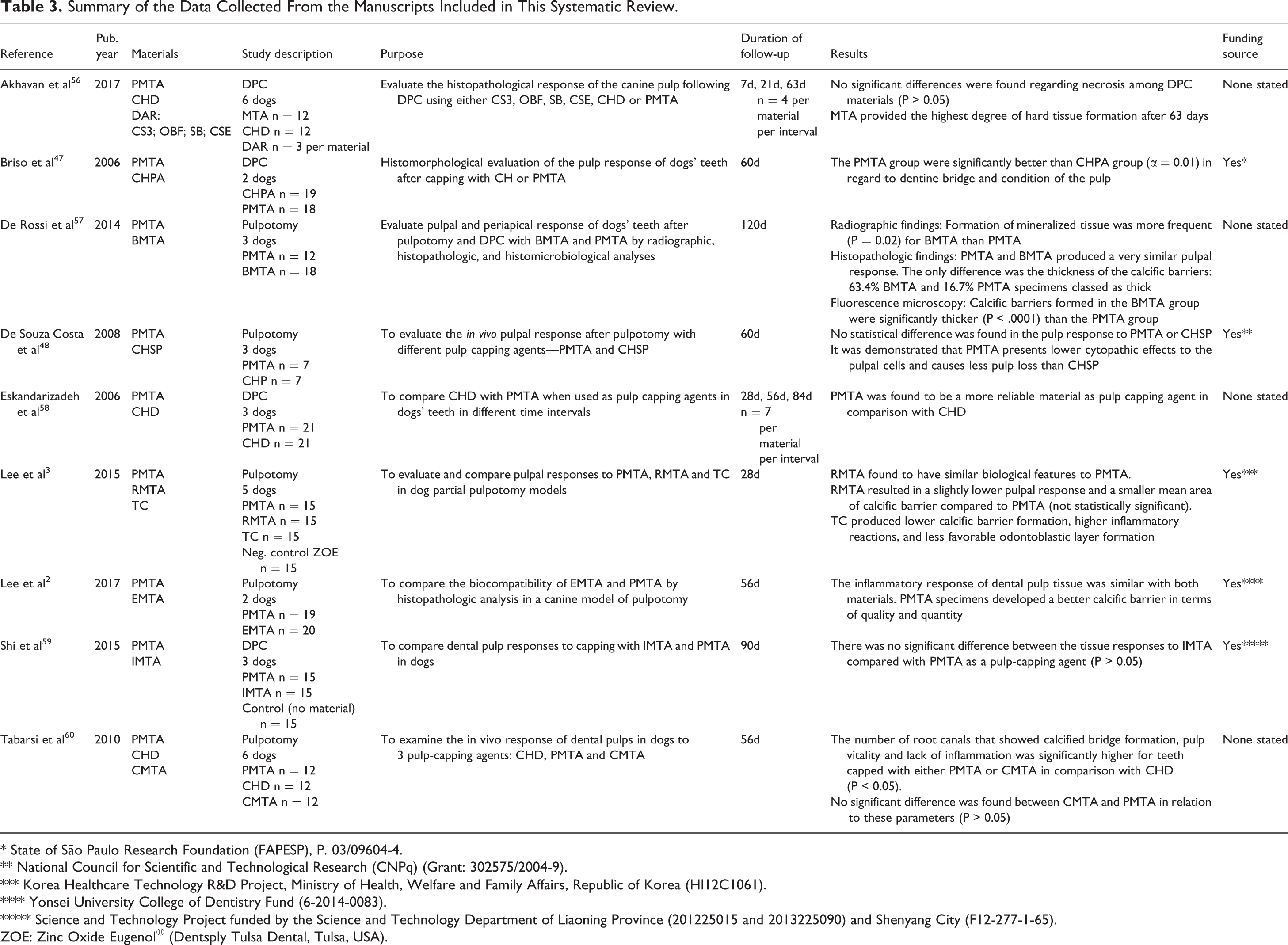
* State of São Paulo Research Foundation (FAPESP), P. 03/09604-4.
** National Council for Scientific and Technological Research (CNPq) (Grant: 302575/2004-9).
*** Korea Healthcare Technology R&D Project, Ministry of Health, Welfare and Family Affairs, Republic of Korea (HI12C1061).
**** Yonsei University College of Dentistry Fund (6-2014-0083).
***** Science and Technology Project funded by the Science and Technology Department of Liaoning Province (201225015 and 2013225090) and Shenyang City (F12-277-1-65).
ZOE: Zinc Oxide Eugenol® (Dentsply Tulsa Dental, Tulsa, USA).
A total of 15 DPC materials were used in the studies. DPC alone with no or minimal pulp amputation was performed in 4 studies. 47,56,58,59 Pulpotomy with amputation of the coronal pulp followed by DPC was performed in 5 studies. 2,3,48,57,60 The long-term follow-up time varied from 28-120 days. 3,57
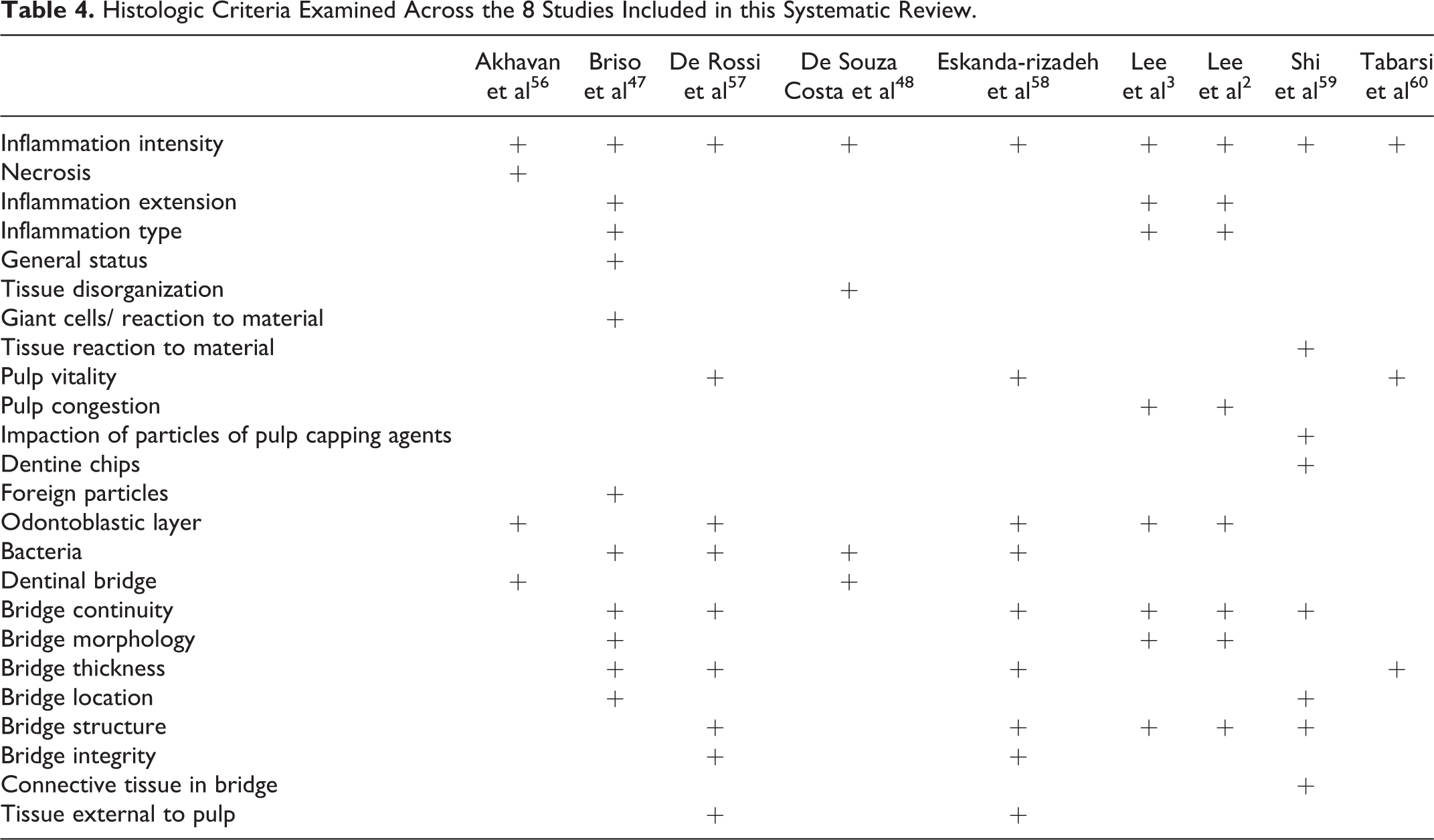
All articles assessed for the presence of a calcific or hard tissue barrier and pulpal inflammation. However, there was no standardized method within these studies for evaluating histological response to DPC. A combination of qualitative and quantitative assessment of the calcific barrier and pulp were performed in each study. The level of detail provided, and exact factors assessed varied between studies as can be observed in Table 4.
Histologic Criteria Examined Across the 8 Studies Included in this Systematic Review.
Discussion
Since its introduction, MTA has gained popularity as a pulp capping material, yet there is minimal published evidence in the veterinary literature that MTA is superior to other commercially available pulp capping materials in dogs.
This systematic review shows in DPC or pulpotomy procedures in dogs, MTA performed as well or better than other commercially available products in terms of the calcific barrier formation as well as biocompatibility.
Three of the studies included in this review demonstrated that PMTA was superior to CH regarding calcific barrier formation and degree of pulpal inflammation. 47,58,60 One study found that PMTA resulted in lower levels of inflammation and higher amounts of hard tissue formation at 63 days compared to CH Dycal®b. 56 Another study found no significant difference in pulpal response to CH saline pastec and PMTA however, the authors did find PMTA was more biocompatible and caused less pulp loss than CH saline paste. 48 It must be noted, however, that the very small sample sizes in these studies makes it difficult for any reliable statistical inferences to be drawn.
The more predictable formation of calcific barriers subjacent to MTA as compared to CH is likely due to a combination of different factors. CH is a soluble material that cannot seal against microleakage, 61 whereas MTA is relatively insoluble and possesses excellent sealing ability, 62 -65 most likely due to a physical bond between MTA and dentin. 32 This seal reduces bacterial penetration to the pulp amputation site. 66,67 A study 17 found 89% of teeth capped with CH contained tunnel defects, most of which appeared to be patent, and these findings were supported by several other studies. 61,68,69 These patent tunnels fail to provide a hermetic seal to the underlying pulp against infection due to microleakage. In contrast, MTA produces more histomorphologically favorable calcific barriers than CH, with earlier formation of hard tissue bridges, larger daily dentin increases and few tunnel defects. 28,70 -73
PMTA was found to be superior to the CH formulations. PMTA also performed well against other MTA-like cements, with few significant differences noted. This may be because they are of similar elemental compositions and possess bioactivity as a common property. 23
One notable difference between PMTA and the MTA-like cements was that Biodentin®d (BMTA) formed significantly thicker calcific barriers than PMTA. 57 The significantly thicker calcific barriers may be explained by BMTA having a greater release of calcium and hydroxyl ions during the initial setting of the material, with reduced ion release over time creating more favorable conditions for pulp repair. 74 While it is widely accepted that the release of calcium hydroxide from pulp capping materials induces a beneficial inflammatory reaction contributing to the formation of the calcific barrier, 74 prolonged release of calcium and hydroxyl ions may have a negative impact due to chronic pulpal inflammation. 57
A second significant difference between PMTA and the other MTA-like endodontic cements was also reported, with Endocem Zr®e (EMTA) treated specimens demonstrating a reduced quantity and quality of calcific barriers with a greater inflammatory response when compared with those treated with PMTA.
EMTA contains a lower percentage of calcium silicate than PMTA due to the quantity of zirconium required to achieve adequate radiopacity. 75 This theoretically has a negative effect on calcium ion release and has been demonstrated with Portland cement. 76,77 EMTA has been demonstrated to have a lower calcium ion concentration than PMTA. 75 EMTA’s less favorable sealing ability and initial transient cytotoxicity, 78,79 are proposed mechanisms for its poorer performance as compared to PMTA. 2
TheraCal®f (TC) performed poorly when compared to PMTA in relation to the frequency of pulpal inflammation and calcific barrier formation. TC is a light-cured, resin-modified calcium silicate marketed for both indirect and DPC and as a protective base/liner. 80 An in vivo study by Cannon et al 81 found that TC treated teeth have hard tissue formation comparable to pure Portland cement, the key ingredient of MTA, and better hard tissue formation than Prisma® VLC Dycal,g a light-cured calcium hydroxide base/liner. 81 However, this study mixed Portland cement with chlorhexidine instead of water, and chlorhexidine is a known retardant of the setting process of Portland cement, 80 which may affect the formation of a calcific barrier. Furthermore, the light-curable calcium hydroxide is different in composition to those listed in this review. Lee et al 3 postulated the poorer response may be due to the reduced biocompatibility of TC compared to PMTA. TC contains Bisphenol A-glycidyl methacrylate, an acrylic monomer that is cytopathic to cultured cells. 82 -84 The authors proposed that the curing of the material may be reduced in a partial pulpotomy model as compared to the DPC model utilized in the study by Cannon et al 81 with leaching of the uncured acrylic monomer into the pulp and dentinal tubules inducing greater pulpal inflammation and reduced calcific barrier formation. A further disparity exists as to the ability of TC to release calcium. Gandolfi et al 85,86 found that calcium was released by TC in lab-based studies at higher rates than the CH-based biomaterials it was tested against and performed well against other calcium silicate-based biomaterials. Whether the material was light-cured prior to testing was not specified. Camilleri 87,88 demonstrated that TC did not leach CH due to limited moisture diffusion within the material. Calcium ions were released but at a reduced rate compared to BMTA, likely due to the resin matrix modifying the setting mechanism and calcium ion leaching of TC. The resin component of TC must also be questioned as to whether it contributes to microleakage secondary to polymerization shrinkage.
Finally, the use of DAR as a pulp capping material remains controversial, with their poor performance highlighted in this review. Several factors, such as acid etching, toxicity of the leached components, microleakage due to polymerization shrinkage and nanoleakage, sensitization and sudden temperature rise during setting may be harmful to the pulp. 89 In a clinical setting, the effective application of these materials may be made more challenging by the difficulty in maintaining hemostasis during their application.
Data regarding the histopathological response of pulpal tissue to both TC and DAR is limited, contradictory and not reliable. Further research is required before making recommendations for their use in VPT in dogs’ teeth.
A meta-analysis was not performed as part of this systematic review due to variation in histological data collected among studies, variation in sample sizes and heterogeneity in the duration of the studies. Consequently, the results of each study were reported individually. All studies presented level II evidence in the form of prospective comparative studies. Based on the criteria established in this systematic review, a search of the current scientific literature did not reveal any studies presenting level I evidence comparing the use of PMTA to other pulp capping materials in a canine model. Evidence-based medicine has become a mainstay of human medicine and more recently its value has been recognized by the veterinary profession with the formation of the Evidence-based Veterinary Medicine Association. It is recognized that several barriers exist to performing high-quality patient-based studies in veterinary science including a lack of external funding, cost, interference with daily practice, inadequate infrastructure as well as ethical concerns from practitioners, the public, animal societies and authorities. 90 Despite this, with the ongoing push to gain higher quality evidence, prospective randomized control studies should be performed to further evaluate the use of PMTA and other pulp capping materials in VPT.
This review identified a lack of a standardization in several important experimental design parameters in the studies presented. Firstly, the criteria used for histological assessment of calcific barrier formation and the pulpal response to pulp capping procedures varied widely across studies. Only 2 articles used the same scoring system, 2,3 while the remaining 7 articles modeled their scoring systems from several different previously described scoring systems. 47,56 -60 Secondly, there was a lack of standardization in the duration of the studies, which ranged from 28 days to 120 days. 3,57 Thirdly, there was wide variation in the restorative materials used to cover the pulp capping materials. In 4 of the articles, glass ionomer cements were placed over the pulp capping material, 2,3,57,59 amalgam was placed in 3 studies, 48,58,60 a resin-modified glass ionomer was placed in 2 studies, 47,56 with the DAR group in one of these studies having a nano-filled composite placed directly over the pulp capping material. 56 With bacterial leakage around restorative margins considered to be a key contributing factor to chronic pulpal inflammation following VPT, 91 -93 uniformity as to the type of restorative material placed in these studies needs to be adopted. Finally, only 3 studies evaluated the presence of bacteria in their histological scoring systems, a finding that would contribute significant information about the relative roles of microleakage and product biocompatibility in the development of pulpal inflammation.
The variation in age of the dogs used in studies included in this review should also be considered. The ages ranged from 8-24 months with one study simply stating that mature dogs were selected. 2,3,47,48,56 -60 A common belief has been that mature teeth (closed apex) will not respond favorably to VPT. A number of veterinary studies have failed to find a statistically significant difference in the maintenance of vitality and patient age following VPT procedures. 4,5,41 This is further supported by recent studies in human subjects with the success of vital pulp therapy being very high when appropriate case selection is followed. 33,94,95 While there may be some variability in results related to age, current evidence suggests this may not be as an important factor as previously considered.
The poor standardization of study design is a significant impediment to the comparison of existing results and for the planning of future studies. Assessment of the successful outcome of VPT is best determined by histopathology, as the state of pulp health or pathology cannot be determined by clinical signs, symptoms or radiological appearance. 15 All studies should adopt the same histological scoring system with a focus on quantitative rather than qualitative measurements. Less heterogeneity in study duration and assessment times, as well as standardization of factors that are influential in the long-term success of these procedures such as the choice of restorative material, would be ideal.
There are many commercially available MTA and MTA-like cements. 23 Although some of these products possess similar composition and characteristics, 37 this review highlights that their performance as DPC materials is not identical. As MTA continues to gain popularity and is now considered by some to be the new material of choice for VPT procedures, 70,96,97 it would seem imperative that a standard control product is selected for future studies.
There are several considerations and limitations that could potentially affect the results and conclusions of this review. Although a thorough electronic and hand search of reference lists was performed as part of this systematic review, it is possible that some manuscripts have been inadvertently excluded because the author did not include a relevant search term. Inclusion and exclusion criteria were pre-defined to reduce bias; however, these criteria resulted in the exclusion of potentially significant articles. For example, 3 articles were excluded as they did not specifically state that PMTA was the MTA product used in their studies, despite PMTA being the likely material of the study. 72,98,99 MTA performed favorably in these 3 excluded studies demonstrating superior performance to CH in terms of calcific barrier formation and pulpal response in 2 articles. 72,99 In the third article, no differences were noted between CH and MTA-treated teeth in terms of calcific barrier formation and pulpal response with all teeth responding favorably to treatment. 98
Another potential limitation is that our review article only included peer-reviewed manuscripts, and this may have resulted in underrepresentation of studies with negative findings secondary to publication bias. The restriction to papers published only in English may also have led to the exclusion of important data. Blinding for histopathology scoring was only implemented in 4 studies included in this review, 2,3,57,60 leaving the remainder open to potential bias. The small sample sizes in the studies included in this review article must also be considered and have contributed to limiting the breadth and scope of the paper. The studies were all of short duration and, as such, no conclusions may be drawn as to the long-term success of MTA as a DPC material from the studies included in this review. Finally, the data included in this review were collected in controlled, experimental conditions and there may be differences in the response of pulp tissues undergoing pulp capping procedures as a treatment for clinical traumatic injuries.
When considering DPC materials for VPT veterinarians should adopt an evidence-based approach to their selection of materials. In clinical practice, veterinarians are limited to radiographic studies as a determinant of pulp vitality following VPT. In studies included in this systematic review, the inflammatory response and calcific barrier formation were determined by histology as this is considered the best way to assess pulp status. 15 There is merit in assessing both histologic and radiographic outcomes in studies in the manner that one article included in this review has done. 57 Studies with positive radiographic results may have more effect on which pulp capping materials veterinarians choose in practice.
Conclusion
In the studies included in this systematic review, PMTA outperformed CH, TC and DAR. PMTA performed as well or better than other MTA-like cements in terms of the calcific barrier formation and biocompatibility. With the continued emergence of new products for VPT, evidence-based assessment of their merits for use in canine VPT requires standardized studies with a consistent control material such as PMTA, implementation of a standard scoring system for histology, equivalent and longer study duration and the correlation of histological and radiographic data.
Footnotes
Materials
ProRoot® MTA (Dentsply) Dycal® (Dentsply DeTrey GmbH) Calcium hydroxide saline paste (Labsynth) Biodentine® (Septodont) Endocem Zr® (Maruchi) TheraCal® (Bisco Inc) Prisma® VLC Dycal (Dentsply)
Declaration of Conflicting Interests
The author(s) declared no potential conflicts of interest with respect to the research, authorship, and/or publication of this article.
Funding
The author(s) received no financial support for the research, authorship, and/or publication of this article.
