
Abstract
Antemortem domestic pig (Sus scrofa domesticus) dental pathology literature is sparse. This observational descriptive study evaluated 23 client-owned pigs that while sedated/anesthetized for routine annual care had intraoral dental radiographs and an oral examination performed. Age, gender, weight, and breed for each pig were recorded. Oral examination and radiographic findings were reviewed to create a comprehensive list of dental abnormalities identified. Descriptive statistics were performed to summarize the data. The study population included 14 castrated males and 20 Vietnamese pot-bellied mini-pigs. The median age was 3 years (range 2-12 years), and the median weight was 39 kg (range 11-140 kg). The most common finding was missing teeth (21/23 pigs); the first premolar tooth was the most likely to be absent (64/106 missing teeth). Periodontal disease was common (20/23 pigs). Advanced stages primarily affected the first molar teeth frequently in the form of a mucogingival defect. Supernumerary roots were discovered on the maxillary canine teeth in female pigs only (10/25 teeth with supernumerary roots). The most common persistent deciduous tooth was the maxillary second incisor (15/19 persistent deciduous teeth). Non-age or gender related open apices were most likely associated with mandibular first and second incisor teeth (26/96 teeth with open apices). Tooth resorption was also identified (7/23 pigs). The study findings prove that pet pigs commonly have dental pathology; therefore, thorough oral examinations with intraoral radiographs should be included in porcine routine health care regimens.
Introduction
Research into the area of porcine dental health and disease is limited in the veterinary literature. With the surge in popularity of domestic small pigs as companion animals, there is an increasing need for veterinarians and veterinary dentists to become more familiar with the porcine oral cavity. Pigs (Sus scrofa domesticus) are omnivores; therefore, they have a diphyodont, heterodont, and brachydont dentition with the exception of male canine teeth (tusks), which are aradicular, hypsodont, and elodont in nature. 1,2 Female canine tooth apices have been reported to close around 2-3 years of age. 2 -4 Therefore, these teeth could be referred to as radicular, hypsodont teeth, but the literature is vague on this point. 2 -4 The porcine deciduous dental formula is 2(i 3/3, c 1/1, p 3/3). The porcine permanent dental formula is 2(I 3/3, C 1/1, P 4/4, M 3/3), and it includes the first premolar and molar teeth absent in the deciduous dentition. 1 -3,5
Dental eruption dates for pigs have been previously reported for European wild hogs, domestic white pigs, and Clawn strain mini-pigs. 4,6,7 Despite some discrepancies between the papers, all provide a similar age for complete eruption and root development, which is 29-30 months (2.5 years). 4,6,7 In healthy domestic pigs, apical closure of permanent tooth roots occurred at the following times: incisor 1 at 16 months, incisor 2 at 24 months, incisor 3 at 12-16 months, canine teeth at 16 months, premolar 1 at 12 months, premolar 2 and 3 at 16 months, premolar 4 at 12-16 months, molar 1 at 8 months, and molars 2 and 3 at 30 months. 4 This data, however, did not specify if pigs were male or female; therefore, apical closure of the canines could not be interpreted. Similar data was found in Göttingen mini-pigs with complete development of all permanent dentition by 23 months (2 years). 8 To the authors’ knowledge, eruption dates have not been established for Vietnamese pot-bellied pigs.
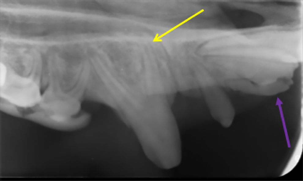
Finally, tooth root number has been described for domestic pigs. Incisors, canines, and first premolar teeth have only 1 root, while premolars typically have 2 roots. 1 -3 Molar teeth in pigs are particularly unique in that they have long, slender, often overlapping roots. Molar teeth 1 and 2 have 4 roots, while molar tooth 3 has 5-6 roots. 2,5,7 The remaining structures of the skull in pigs are relatively similar to dogs and cats except for the extremely long, slender maxilla and mandible, thick layer of adipose tissue, restricted oral aperture, and prominent parotid papillae. The parotid papillae are also purplish in color and can easily be mistaken for bruising or an oral mass by untrained eyes (Figure 1).

Normal parotid duct papilla in a pig (yellow arrow). The periodontal probe is in a deep periodontal pocket associated with the left maxillary first molar tooth.
A handful of studies have attempted to describe dental pathology within the wild and domestic pig populations. 7,9 -13 Dental abnormalities identified in these papers included: hypodontia (decreased number of teeth), supernumerary teeth and roots, dental malocclusion, tooth resorption, dental caries, periodontal disease (PD), dental fractures, attrition/abrasion, and enamel defects. 7,9 -13 All studies evaluated post-mortem specimens, and no study utilized intraoral dental radiography or detailed dental charting techniques. As a result, a study comparing wild boar and domestic pig dental health revealed similar dental anomalies and pathology between the populations despite the “unnatural diet and eating habits” of domestic pigs, and no PD was identified in either population. 9 In contrast, a study focusing on feral and domestic pig PD, which lacked intraoral radiographs but utilized a PD scoring system based solely on intraoral examination, revealed that 24% of feral and 23% of domestic pigs had evidence of PD. 10 Both these articles elucidated the need for an antemortem study utilizing intraoral dental radiography and full mouth dental charting to accurately record dental pathology, anomalies, and to stage PD.
Therefore, the authors designed an in vivo observational descriptive study of anesthetized/sedated client-owned domestic pigs that included intraoral dental radiographs and detailed dental charting. The authors hypothesized that a study of this nature would uncover more dental anomalies and pathology than previously reported. The first aim of the study was to record all dental anomalies and pathology identified upon anesthetized/sedated oral examination with detailed dental charting and intraoral radiography in a client-owned pig population. The second aim of the study was to stage PD found within the population utilizing periodontal probing measurements and intraoral radiography.
Materials and Methods
Data for the study was acquired from institutional medical records, dental charts, and intraoral radiographs of client-owned pet domestic pigs that were presented to Colorado State University Veterinary Teaching Hospital for routine veterinary care between January 2015 and September 2018. All animals were treated in accordance with national and international rules regulating animal welfare. All clients signed a photo release form and paperwork indicating that hospital medical records and diagnostic testing could be used for research purposes. Routine porcine care offered at the institution included: physical examination, ear/eye cleaning, hoof trimming, vaccination, bathing, oral examination with dental charting, full-mouth intraoral radiography, dental prophylaxis, tusk trimming, dental treatment planning for future visits, and any other ancillary service(s) required for individual health conditions. All services listed above were performed under general anesthesia or heavy sedation. Clients determined which services their pet received at each visit based on doctor recommendations, personal desires, and cost estimates.
Every animal included in the study needed to have a complete medical record, detailed dental chart with periodontal probing, and a full-mouth set of intraoral radiographs. Exclusion criteria included: animals under 1 year of age, animals that were undernourished, systemically ill or being treated for chronic systemic health conditions (e.g. chronic kidney disease, liver failure, neoplasia), and animals that had received a dental cleaning or treatment within the previous 4 months.
Data collected for each pig included age, gender, weight, breed, sedation or anesthesia protocol, and dental anomalies/pathology recorded within the medical record, dental chart, or found upon review of the dental radiographs. Oral examination and detailed dental charting had been performed by 1 of 2 individuals, and examination was conducted with a surgical head lamp and loupes(Loupes Micro LED™; SurgiTel, Ann Arbor, MI, USA), Minnesota retractor(Minnesota retractor 14 cm; iM3®, Vancouver, WA, USA), periodontal probe, and dental explorer(Hu-Friedy Dental Williams Explorer Probe UNC 15/23, Chicago, IL, USA). The 2 examiners consisted of 1 faculty member (JR, American Veterinary Dental College (AVDC) Diplomate) and 1 resident (MS, Dentistry and Oral Surgery resident), who had been trained by the faculty member in porcine oral examination and dental radiography.
All oral and dental findings had been recorded on the Dentistry and Oral Surgery Service’s porcine dental chart. The modified triadan system was used for tooth identification, and AVDC abbreviations and nomenclature were used to denote anomalies or pathology. 14,15 Teeth had been periodontally probed at 3 locations on the lingual/palatal and buccal/labial aspects of each tooth. Gingivitis was recorded using a standard 0-3 scoring system and attachment loss was measured in mm. 16
Radiographs were obtained using either a mobile(iRay D3 Handheld X-ray System, CA, USA) or mounted(Heliodent Plus Dental X-ray machine by Dentsply Sirona Imaging, PA, USA) x-ray generator and size 4 phosphor plates(Carestream CS7600 Smart Phosphor X-ray Dental Plate Size 4 Bitewing, Atlanta, GA, USA) processed by a commercially available CR dental radiograph system(Carestream CS7600 CR system, Atlanta, GA, USA). All images were reviewed from the institution’s PACS system on a general use monitor(IntelliSpace PACS Radiology; Philips Healthcare Informatics, CA, USA, HP Z220 computer; Hewlett Packard Enterprise, CA, USA). Radiographs and intraoral findings were compared and used to stage periodontal disease. Periodontal disease was scored on a 0-4 scale beginning with gingivitis as stage 1 and progressing to different percentages of attachment loss in the consecutive stages. Attachment loss is defined as gingival recession, periodontal pocketing, and bone loss noted radiographically or by furcation exposure on oral exam. 17,18 Stage 2 periodontal disease is defined as <25% attachment loss, Stage 3 as 25-50% attachment loss, and Stage 4 is >50% attachment loss. 17
Descriptive statistics were performed for all data collected. Co-variable analysis could not be performed to determine if age or weight was associated with PD, missing teeth, unerupted teeth, persistent deciduous teeth, supernumerary roots, and partially erupted teeth given the small sample size. Continuous variables were represented by mean and standard deviation or median and range. Categorical variables were represented by percentages. Statistical software SAS v. 9.4 (Statistical Analysis Software(Statistical Analysis Software, NC, USA)) was used for all statistical analyses. P-values of < 0.05 were considered statistically significant.
Results
Study Population
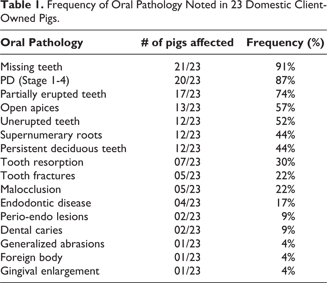
The medical records, dental charts, and intraoral radiographs of 23 pigs were evaluated for inclusion in the study. There were 14 castrated males (14/23; 60.87%), 7 spayed females (7/23; 30.43%), and 2 intact females (2/23; 8.70%) included in the study. The median age was 3 years, ranging from 2-12 years. The breeds included were 20 Vietnamese Pot-bellied mini-pigs (20/23; 86.96%), 1 Vietnamese Pot-Bellied pig (1/23; 4.35%), and 2 domestic mixed breed pigs (2/23; 8.70%). The median weight of the pigs was 39.3 kg ranging from 11.6 kg to 140 kg. All dental anomalies and pathology discovered were recorded (Table 1).
Frequency of Oral Pathology Noted in 23 Domestic Client-Owned Pigs.
Periodontal Disease
Overall, 20 pigs (20/23; 87%) had evidence of PD. The mean weight for pigs with PD was 46.6 kg (± 26.5 kg) while pigs without PD had a mean weight of 23.3 kg (± 6.76 kg). The mean age of pigs with PD was 4.7 years (± 2.9) while pigs without PD had a mean age of 2 years (± 0). No statistical significance could be drawn regarding age or weight when comparing to PD due to the small sample size.
Many pigs had multiple stages of PD associated with different teeth within the oral cavity. Stage 1 PD affected 19 pigs (19/20; 95%), Stage 2 PD affected 15 pigs (15/20; 75%), Stage 3 affected 6 pigs (6/20; 30%), and Stage 4 PD affected 3 pigs (3/20; 15%). Teeth most commonly affected by stage 3 and 4 PD are represented in Figure 2 by tooth number. Stage 3 and 4 PD led to severe mucogingival defects in 6 pigs (6/9; 67%) for a total of 12 defects. Of these 12 severe mucogingival defects, 9 defects (9/12; 75%) involved the first molar teeth, 2 defects (2/12; 17%) involved fourth premolar teeth, and 1 defect (1/12; 8%) involved the second molar tooth (Figures 3 and 4).
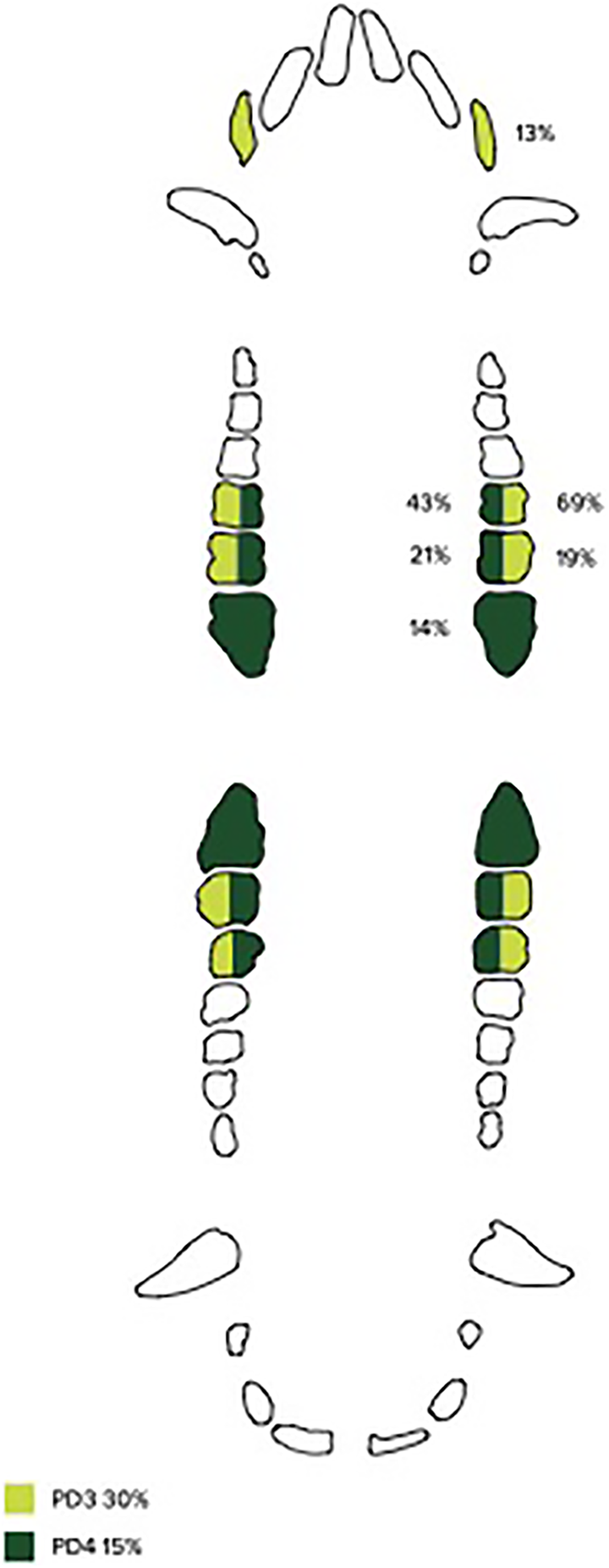
The frequency of periodontal disease (PD) Stage 3 and 4 in domestic pigs by tooth. Stage 3 PD affected 30% of pigs (light green color) with a total of 16 maxillary and mandibular teeth effected. Of these 16 teeth, 69% were maxillary and mandibular first molar teeth, 19% were maxillary and mandibular second molars, and 13% were maxillary third incisors. Stage 4 PD affected 15% of pigs (dark green color) with a total of 14 maxillary and mandibular teeth effected. Of these 14 teeth, 43% were maxillary and mandibular first molars, 21% were maxillary and mandibular second molars, and 14% were maxillary and mandibular third molars. The remaining 21% of teeth (not depicted here) were made up of 2 mandibular fourth premolars and 1 maxillary second premolar.
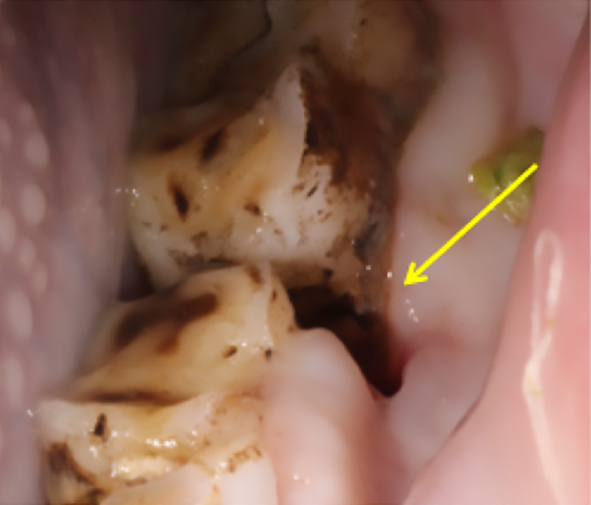
Mucogingival defect (yellow arrow) between left mandibular fourth premolar (308) and first molar (309) teeth.
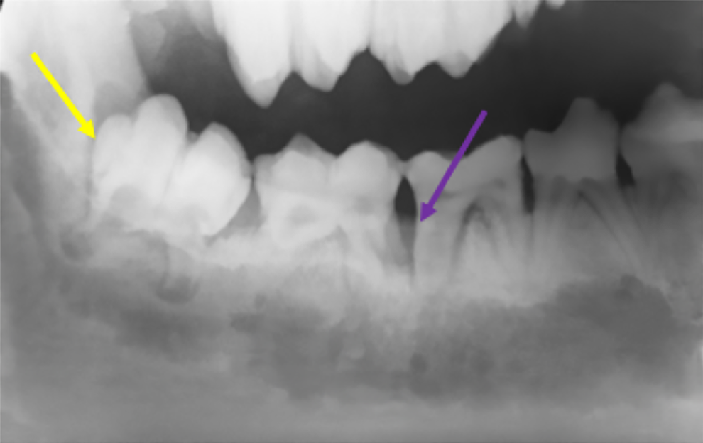
Intraoral radiograph of right mandible in a domestic pig. Stage 4 PD associated with the right mandibular first molar tooth (409). The purple arrow demonstrates the severe bone loss in the interdental space between tooth 409 and 410. A partially erupted right mandibular third molar tooth (411) is depicted by the yellow arrow.
Missing Teeth
Out of the 23 pigs, 21 had missing teeth (21/23; 91%) with a mean age of 4.4 years (± 3.0). A total of 106 missing teeth were categorized into tooth groups including incisor, first premolar, premolar (second, third, and fourth), and molar. Of these, 2 pigs (2/21; 10%) had a total of 3 (3/106; 3%) missing incisors, 20 pigs (20/21; 95%) had a total of 64 (64/106; 60%) missing first premolars, 7 pigs (7/21; 33%) had a total of 13 (13/106; 12%) missing premolars (other than first premolars), 8 pigs (8/21; 38%) had 24 (24/106; 23%) missing maxillary or mandibular third molars, and 1 pigs (1/21; 5%) had a missing maxillary second molar (1/106; 1%) and mandibular first molar (1/106; 1%).
Partially Erupted Teeth
Out of 23 pigs, 17 (17/23; 74%) had partially erupted teeth beyond the age of anticipated full eruption for the tooth identified. Mean age of pigs with partially erupted teeth was 3.7 years (± 1.9). Partially erupted teeth were separated into categories by tooth type including incisors, canines, and molars for a total of 55 teeth. There were no partially erupted premolar teeth. Of the 17 pigs, 1 pig (1/17; 6%) had 2 (2/55; 4%) partially erupted incisors, 1 pig (1/17; 6%) had 3 (3/55; 5%) partially erupted canines, 15 pigs (15/17; 88%) had a total of 48 (48/55; 87%) partially erupted maxillary or mandibular third molars, and 1 pig (1/16%) had a total of 2 (2/55; 4%) partially erupted maxillary first molars.
Unerupted Teeth
Unerupted teeth were noted in 12 pigs (12/23; 52%) beyond the age of anticipated permanent dental eruption for the tooth identified. Mean age of pigs with unerupted teeth was 4.3 years (± 3.1). Partially erupted teeth were categorized into incisors, canines, premolars, and molars for a total of 26 teeth. Of the 12 pigs with unerupted teeth, 11 pigs (11/12; 92%) had unerupted incisors for a total of 22 (22/26; 85%) teeth, 1 pig (1/12; 8%) had an unerupted canine for a total of 1 (1/26; 4%) tooth, 2 pigs (2/12; 17%) had unerupted premolars for a total of 2 (2/26; 8%) teeth, and 1 pig (1/12; 8%) had an unerupted molar for a total of 1 (1/26; 4%) tooth (Figure 4).
Supernumerary Roots
Supernumerary roots were detected in 10 pigs (10/23; 43%) with affected teeth separated into categories, canine and first premolar teeth, for a total of 25 teeth. Of the 10 pigs, 5 pigs (5/10; 50%) had bilateral supernumerary maxillary canine roots for a total of 10 (10/25; 40%) teeth. All 5 pigs with supernumerary canines were female (intact and spayed). (Figure 5) The remaining 15 teeth (15/25; 60%) had supernumerary first premolar roots in 8 pigs (8/10; 80%) of mixed gender.
Intraoral radiograph of right maxillary supernumerary canine root and persistent second deciduous incisor. The supernumerary root of the right maxillary canine tooth (104) is referenced by the yellow arrow; a persistent deciduous right maxillary second incisor tooth (502) is shown by the purple arrow.
Persistent Deciduous Teeth
Persistent deciduous teeth were noted in 10 pigs (10/23; 43%) with a mean age of 4.7 years (± 3.23) and were separated into categories by tooth type including maxillary second incisor, mandibular third incisor, and other (all other persistent deciduous teeth) for a total of 19 teeth. Notably, all pigs in the study population were >2 years in age and only 5 pigs were < 3 years in age. Of the 10 pigs, 9 pigs (9/10; 90%) had persistent deciduous maxillary second incisors for a total of 15 (15/19; 79%) teeth, 1 pig (1/10; 10%) had a persistent deciduous mandibular third incisor for a total of 1 (1/19; 5%) tooth, and 2 pigs (2/10; 20%) had other persistent deciduous teeth for a total of 3 (3/19; 16%) teeth (Figure 5).
Open Apices
Open apices, beyond the anticipated age for apical closure and excluding male canine teeth, were detected in 13 pigs (13/23; 57%) with a mean age of 2.9 years (± 0.64).Teeth with open apices were separated into categories by tooth type including canines, incisors (overall), maxillary first incisor, maxillary second incisor, maxillary third incisor, mandibular first incisor, and mandibular second incisor for a total of 96 teeth. Of the 13 pigs, 5 pigs (5/13; 38%) had open apices of canine teeth for a total of 14 teeth (14/96; 15%), 13 pigs (13/13; 100%) had open apices of incisors for a total of 82 teeth (82/96; 85%), 9 pigs (9/13; 69%) had open apices of maxillary first incisors for a total of 18 teeth (18/96; 19%), 5 pigs (5/13; 38%) had open apices of maxillary second incisors for a total of 10 teeth (10/96; 10%), 1 pig (1/13; 8%) had open apices of maxillary third incisors for a total of 2 teeth (2/96; 2%), 13 pigs (13/13; 100%) had open apices of mandibular first incisors for a total of 26 teeth (26/96; 27%) teeth, and 13 pigs (13/13; 100%) had open apices of mandibular second incisors for a total of 26 teeth (26/96; 27%, (Figure 6).
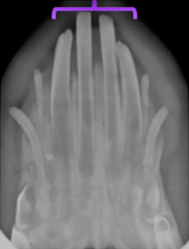
Intraoral radiograph of the mandibular incisors. All 4 first and second incisors have open apices (purple bracket), and the lateral mandibular incisors are still deciduous. This radiograph is not from a patient within the current study population, but from a juvenile pig unrelated to this study.
Tooth Fractures
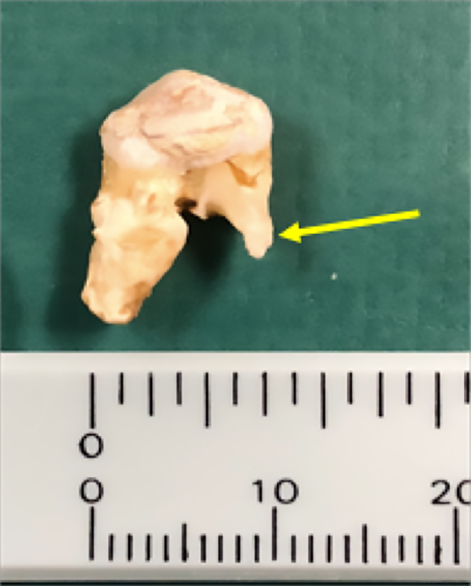
Tooth fractures were found in 5 pigs (5/23; 22%) and were separated into categories by fracture type including complicated crown fracture, complicated crown root fracture, and uncomplicated crown fracture (CCF, CCRF, and UCF) for a total of 7 teeth. Of the 5 pigs with tooth fractures, 1 pig (1/5; 20%) had a CCF involving 2 teeth (2/7; 29%), 2 pigs (2/5; 40%) had a CCRF involving 2 teeth (2/7; 29%), and 2 pigs (2/5; 40%) had an UCF involving 3 teeth (3/7; 43%, (Figure 7).
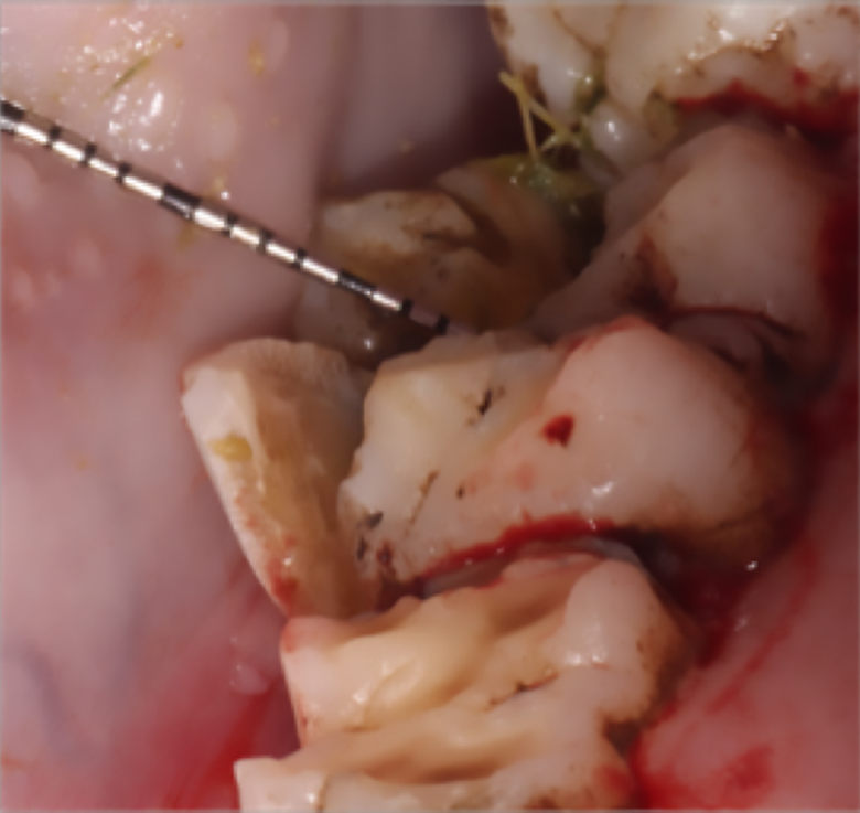
Photo of the left mandibular molar teeth. The periodontal probe is isolating a complicated crown root fracture of the left mandibular second molar tooth (310).
Malocclusion
Out of 23 pigs, 4 pigs (4/23; 17%) had a malocclusion. Two pigs (2/4; 50%) had a class III malocclusion, and 2 pigs (2/4; 50%) had a class I malocclusion (distoversion) of right and left maxillary second premolar (106, 206).
Tooth Resorption
Tooth resorption was noted in 7 pigs (7/23; 30%), and it was categorized as either external inflammatory tooth resorption or external replacement resorption in a total of 13 teeth. 19,20 Three pigs (3/7; 43%) had external inflammatory tooth resorption for a total of 7 teeth (7/13; 54%), and 4 pigs (4/7; 57%) had external replacement resorption for a total of 6 teeth (6/13; 46%, Figures 8 and 9).
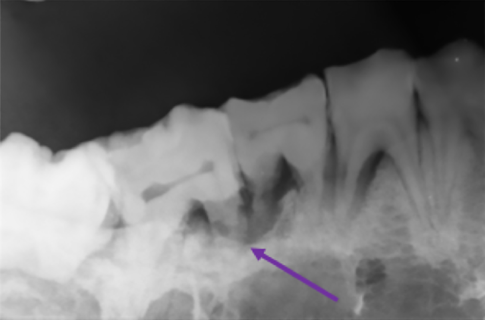
Intraoral radiograph of the right mandible of a domestic pig. External inflammatory tooth resorption is depicted by the purple arrow associated with the right mandibular second molar tooth (410).
Photograph of a mandibular premolar with tooth resorption in a domestic pig. The yellow arrow is referencing external inflammatory tooth resorption of the mesial root of the mandibular premolar.
Miscellaneous
Out of 23 pigs, 2 pigs (2/23; 9%) had periapical lucencies secondary to significant horizontal bone loss (perio-endo lesions) for a total of 2 teeth (1 premolar and 1 molar), 4 pigs (4/23; 17%) had endodontic disease detected by observing a periapical lucency, a wide bizarre pulp canal, and/or intrinsic staining for a total of 4 teeth (3 premolars and 1 molar), 2 pigs (2/23; 9%) had dental caries in 6 teeth (4 canines and 2 first premolars), and 1 pig (1/23; 4%) had evidence of generalized severe abrasion. Gingival enlargement was noted in 1 pig (1/23; 4%), and 1 pig (1/23; 4%) had a palatal foreign body (Figure 10).
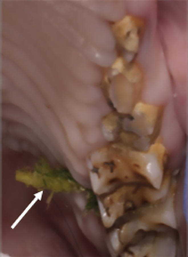
Intraoral photograph of a palatal foreign body in a domestic pig. Foreign material is visible on the palatal aspect of the left maxillary first and second molar tooth (209, 210) interdental space (white arrow).
Discussion
The study presented was the first study to the author’s knowledge to utilize in vivo detailed dental charting and intraoral radiography to document dental anomalies and pathology of the domestic pet pig population. Within the study population, PD had similar rates to dogs (87%) and mainly affected first molar teeth. 17 The most common anomaly was missing teeth (91%) with the first premolar being the most commonly affected. Supernumerary roots were seen in maxillary canines in female pigs only and the first premolars in males and females. The most common persistent deciduous tooth was the maxillary second incisor tooth (79%). The most common teeth with open apices were the mandibular first and second incisors followed by the maxillary first incisor teeth. External inflammatory and replacement tooth resorption were present in this population documenting not only the presence but also the application of the Andreasen classification system for tooth resorption in pigs. 19,20
Study sample size was a limitation. Significant statistical analysis with population inference for each dental pathology noted could not be performed with regard to age, gender, weight or breed given the small sample size. A larger prospective study with 75-100 pigs that included a wider variety of breeds and equal gender and age representation would be necessary to calculate population prevalence and determine possible impacts of age, gender, weight, and breed. In addition, the study population might also have been affected by bias due to client self-selection for dental treatment planning. This survey of 23 domestic client-owned pigs found 87% had evidence of at least 1 stage of PD. This percentage was consistent with previous findings in dogs in that >80% over the age of 3 years exhibited PD. 17 The representation of different stages of PD in affected pigs revealed that 95% of pigs had stage 1 PD, 75% had stage 2 PD, 30% had stage 3 PD, and 15% showed stage 4 PD. This data is in accordance with previously reported 15% of pigs having severe periodontitis in a population of 99 wild boars in Sweden but it varies from the 20% PD prevalence in 107 wild and 52 domestic post-mortem pigs in Australia. 10,13 The most commonly affected teeth in this study were maxillary and mandibular first molars. Previous reports of PD in pigs referenced “cheek teeth” (premolars and molars), specifically first molar teeth, as the most common sites for severe PD. 10,13 The higher overall percentage of PD in this study is likely secondary to the large number of pigs with gingivitis (stage 1 PD) which can only be evaluated antemortem with a dental probe (Figure 11). In addition, dental radiographs most likely revealed more subtle evidence of bone and attachment loss that would have been missed in the other studies without radiographs. Routine monitoring for PD including intraoral radiographs, dental charting and close attention to the maxillary and mandibular first molars may help decrease oral pain and progression of PD in porcine patients.
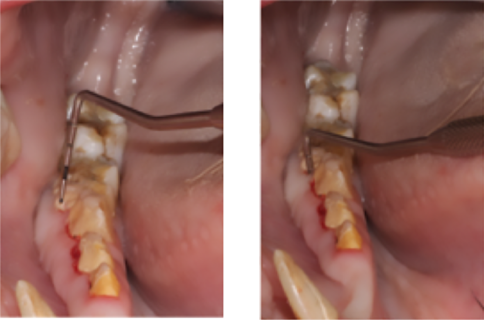
Intraoral photograph of a periodontal probe being inserted into a deep periodontal pocket in a domestic pig. The photo on the left depicts a periodontal pocket associated with the right mandibular first molar tooth prior to insertion of the probe, while the photo on the right shows the probe inserted into the periodontal pocket measuring 8.5mm.
In this study, 91% of pigs had missing teeth or hypodontia. The most common tooth absent from the dental arcade was the maxillary or mandibular first premolar followed by maxillary or mandibular third molars. Previous studies have reported similar findings stating the most common dental anomaly was hypodontia (62%) and 92% were missing first premolars. 9 Another study revealed that 69% of pigs had missing teeth of which 62% were first premolars. 13 This is the first data collection to mention third molars as the second most common tooth affected by hypodontia.
Seventy-three percent of pigs had partially erupted teeth, while 52% had unerupted teeth. The most common partially erupted teeth were maxillary and mandibular third molars. Incisors were the most common unerupted teeth. Previous eruption data states that all incisors (maxillary and mandibular) should be present by ∼24 months of age (2 years) and all molars by 26-30 months (2-2.5 years). 4,6 -8 Partially erupted teeth may relate back to the lack of published eruption dates in Vietnamese pot-bellied pigs and Vietnamese pot-bellied mini-pigs specifically.
Supernumerary roots were present in 43% of pigs, most commonly maxillary and mandibular first premolar and maxillary canines. Notably, the supernumerary canine roots were seen exclusively in the maxilla of female pigs. Supernumerary roots of first premolars were not noted in a study of cheek teeth development in mini-pigs in Japan. 7 To the author’s knowledge, no previous data has been recorded in regard to supernumerary roots in pigs. It is possible that this is a breed predisposition among Vietnamese pot-bellied pigs, however, there is not sufficient data to support this theory.
Persistent deciduous teeth were noted in 43% of pigs with the most commonly affected tooth being the maxillary second incisor. Unfortunately, the only deciduous exfoliation data in recent research did not involve incisors or canines, only premolars. 7 The current literature agrees that complete dentition of pigs should be present in the oral cavity by 2 to 2.5 years of age. 4,6 -8 For the persistent deciduous teeth noted in the study population (in which all pigs were >2 years) only 5 pigs were less than 3y. Therefore, based on the previously published eruption dates, the deciduous teeth noted in this population would be persistent. 4,6 -8
Open apices were noted in 57% of pigs in teeth other than male canine teeth. Incisors accounted for 85% of teeth with open apices; mandibular first and second incisors made up the majority of these teeth. Maxillary first incisors were the next most common teeth with open apices. Apical closure has been reported by 30 months for all teeth; however, the publication did not evaluate mini-pigs or Vietnamese pot-bellied pigs/Vietnamese pot-bellied mini-pigs. 4 Previous research in Japanese Clawn strain mini-pigs did not evaluate for closure of all apices in question but did report apical closure of all cheek teeth except third molars by 29 months. 7 It may be possible that the maxillary and mandibular first and mandibular second incisors erupt later in Vietnamese pot-bellied pigs and mini-pigs than previously reported for other pig breeds. Further research with Vietnamese pot-bellied pigs and mini-pigs <2 years is warranted to publish eruption dates for these specific breeds.
External inflammatory and replacement tooth resorption have not been defined in previous porcine literature, however, apical tooth resorption, a result of chronic inflammation, has been described in reference to tusk pathology. 11 While the percentages of external resorption in this population was low, it is important to acknowledge the presence and application of the Andreasen classification system to tooth resorption for porcine patients. 19,20 The pathogenesis is suspected to be similar to that of dogs and humans with external inflammatory resorption and chronic inflammation while replacement resorption may be a naturally non-painful occurring process affecting older animals. 19,20
Tooth fractures were only noted in 22% of the study population represented most commonly by CCRF (9%) and UCF (9%). These percentages are similar to the 8.1% of Swedish wild boars with tooth fractures; however, fractures were not defined as complicated crown fracture, complicated crown root fracture, uncomplicated crown fracture, etc. 13 A study of Croatian wild boars and tusk abnormalities revealed the most common anomaly to be tusk fractures. 12 It is possible given the domesticated indoor lifestyle of most pigs within the study population, traumatic injury to tusks is less likely, especially considering most pigs had received previous tusk trims.
Presence of other pathology including malocclusion, perio-endo lesions, endodontic disease, abrasion, foreign body, and gingival enlargement were noted in the population but not in significant enough numbers to draw any substantial conclusions or allow for population inference. Previously reported “tooth wear” in Swedish wild boars was 70% of the population, significantly less than in the current study population. 13 This may be due to the lack of chewing, rooting, grazing, and other normal porcine oral behaviors exhibited in the wild by domestic pigs. Dental caries were noted in 9% of pigs affecting only canine and first premolar teeth. This is similar to previous reports of 11% within a population of Swedish wild boars. 13 The most common inciting causes of dental caries are diet (highly fermentable carbohydrates) and genetics. 21 Given the similar rates between domestic and wild pigs, it is hard to draw any conclusions in regard to diet. The data collection also did not evaluate diet in relation to presence of dental caries.
With the recent surge in the popularity of pigs and mini-pigs as companion animals, there is a dire need for small animal practitioners and veterinary dentists to become better acquainted with porcine oral anatomy and pathology. The current study described in detail the presence and distribution of an array of porcine dental anomalies and pathology and presented the first in vivo population percentage, supported by detailed dental charting and intraoral radiographs, of domestic pet pigs affected by PD. Veterinary dental specialists and general practitioners now have the information necessary to institute and justify routine oral health care programs, improve pathology recognition, develop more successful treatment plans, and prevent oral pain in the domestic pet pig population. Further research with larger study numbers and randomized pig involvement is warranted to calculate true population prevalence of the described dental anomalies and pathology, but the study demonstrates that pet pigs are susceptible to similar dental conditions as other pet populations and require the same level of dental care.
Footnotes
Acknowledgments
The authors would like to acknowledge the Livestock Medicine and Surgery Service at Colorado State University Veterinary Teaching Hospital for collaborating on this project and stressing the importance of oral health in pigs, the veterinary dental technicians who supported the data collection (Megan Kirchner-Stephenson, Valerie Hamilton, Ashley Konig), and Maddi Funk from the CATS lab.
Declaration of Conflicting Interests
The author(s) declared no potential conflicts of interest with respect to the research, authorship, and/or publication of this article.
Funding
The author(s) received no financial support for the research, authorship, and/or publication of this article.