






Abstract
Parosteal osteosarcoma (OSA) is a subtype of surface OSA that is considered slow growing with a less aggressive biological behavior compared to central OSA and, therefore, carries a better prognosis with surgical excision. Parosteal OSA has a predilection for the skull in dogs with documented more frequent occurrences on the zygoma, maxilla, or coronoid process of the mandible, all of which present unique challenges to the oral surgeon. Ten cases of parosteal OSA of the canine maxillofacial bones are summarized and discussed in this report. The most common bone of origin was the caudal maxilla and/or zygoma in 50% of tumors. Exophthalmos was a prominent clinical sign noted in 40% of dogs. Median survival time was 14 months, with a one-year survival rate of 60%. Metastasis to the lungs was suspected in three cases. The histopathological diagnosis of parosteal OSA is crucially dependent on diagnostic imaging. When a primary bone tumor of the maxillofacial bones is suspected, it is essential for the oral surgeon and pathologist to collaborate to provide informed guidance on surgical planning, anticipated long-term outcomes, and clinical monitoring for these patients. The goals of this retrospective study are to identify key clinical and radiographic features, improve recognition for accurate diagnosis, and establish treatment recommendations for canine maxillofacial parosteal OSA.
Introduction
Osteosarcoma (OSA) is the most common malignant bone tumor diagnosed in veterinary species, accounting for up to 85% of all primary bone tumors in dogs.1–6 Oral OSA is the fourth most common non-odontogenic oral tumor in the dog, accounting for up to 10% of all canine oral masses and up to 12% of all canine OSAs.3,4,7
OSA is classified as a central or peripheral type based on the location of the tumor either within or on the surface of the bone from which it originates. Central OSA arises from medullary bone, whereas peripheral OSA arises from the periosteum on the bone surface.1,2,5,8–13 Peripheral OSA (also known as surface or juxtacortical OSA) can be further differentiated into parosteal OSA, periosteal OSA, and high-grade surface OSA. Parosteal OSA, believed to arise from the fibrous layer of the periosteum, typically exhibits limited to no invasion into cortical bone. 14 Parosteal OSA tends to grow slowly, exhibit a low risk of metastasis, and generally has a favorable prognosis after surgical removal.1,2,5,9,12,14 However, large and long-standing lesions may destroy underlying cortical bone. There is documented malignant transformation of parosteal OSA to high-grade surface OSA and distant metastasis in both veterinary and human literature.2,8,9,12,13,15–18 Periosteal OSA, originating from cambium cells of the periosteal layer, is more likely to cause bony lysis with extension into the medullary cavity and exhibit a biological behavior that is intermediate between parosteal and high-grade OSA.2,5,8,9,12,14 High-grade surface OSA is a rare, poorly differentiated, biologically aggressive bone tumor that carries a prognosis similar to central OSA.1,8,9,12
Parosteal OSA has been documented in dogs in the cervical vertebrae, 8 mandible, 19 zygomatic arch,2,17,20 long bones,8,10,13,15 and carpus. 10 In dogs, it appears to have a predisposition for the skull, particularly the zygoma.2,17 This contrasts with parosteal OSA in humans, which is most frequently diagnosed in the appendicular skeleton, particularly the femoral bone.2,9,11,16–18 There is limited literature available about the surgical treatment of parosteal OSA of the maxillofacial bones in dogs.17,20 Evaluation of these cases helps to refine understanding of the distribution, presentation, and demographics of canine patients with maxillofacial parosteal OSA. It also aids in understanding the biological behavior of this condition and the implications for clinical outcomes.
Materials and Methods
Records from a specialty veterinary oral pathology database (Specialty Oral Pathology for Animals) from 2019 to 2024 were queried for the diagnosis of parosteal or peripheral OSA. An additional case was included from a veterinary diagnostic lab (Texas Veterinary Medical Diagnostic Laboratory). Histology and imaging were reviewed for each case to confirm the diagnosis.
Histologic inclusion criteria were based on previously published features of parosteal OSA in dogs.1,2,14 These features included well-developed trabeculae of neoplastic bone, with or without small amounts of cartilage, that were separated by spindle cells with low mitotic count and mild to moderate nuclear atypia. 14 The histological features of parosteal OSA overlap with other benign and malignant tumors of bone. For case selection, clinical and radiologic inclusion criteria were as essential as tumor histomorphology. Tumors were excluded if histologic, radiographic, and clinical features could not rule out periosteal OSA, central OSA, high-grade OSA, multilobular osteochondrosarcoma (MLO), ossifying fibroma, osteoma, or other proliferative fibro-osseous lesions. Specifically, tumors that had more cartilage than bone were excluded since this feature favors periosteal OSA over parosteal OSA. Tumors with lobular organization were excluded if lobules regularly included cartilage and parosteal OSA could not be differentiated from MLO.
Cases were included if three-dimensional (3D) imaging supported a tumor location on the periphery of the bone of origin with an outward growth pattern. Since parosteal OSA arises from the periosteal surface of the bone, cases were excluded if the diagnostic imaging showed a tumor originating centrally within bone or if central osteolysis was present. An exception to this was made for one case where the tumor originated from, and breached through, the thin palatine bone. Remaining inclusion criteria supported a diagnosis of parosteal OSA for this case.
For cases that met the inclusion criteria, medical records and pathology submission forms were reviewed. Data collected for analysis included sex, breed, weight, age at time of diagnosis, clinical signs, and tumor anatomic location and descriptive features. Tumor duration was documented and defined as the time from the date the owner or veterinarian first noted physical evidence of, or clinical signs associated with the tumor, to the date of histopathological diagnosis.
All diagnostic head imaging and, when available, radiology reports were reviewed by a board-certified veterinary dentist (JJ), a veterinary dentistry resident (GK), and a board-certified veterinary radiologist. Cortical bone invasion of the parent tumor bone was visually classified as absent (tumor was confined to the periosteum) or present (tumor invasion into cortical bone). The presence or absence of the following radiographic features was recorded: a lobular growth pattern, as defined by a rounded, undulating, or scalloped border, and the presence of a string sign, defined as a lucent line or cleft separating the tumor from the cortex.
Tumor dimensions were measured, and tumor volume was approximated using the formula for the volume of an ellipsoid (Volume = [length × width × height]×[π/6]). The type of surgical treatment performed was documented. Histologic margins were reported for cases where excisional biopsies were performed. Margins were classified as complete (narrowest tumor margin >5 mm), narrow (narrowest tumor margin <5 mm), or incomplete (tumor cells extended to the margins of the sample).
Imaging modalities for oncologic staging or monitoring for local recurrence were summarized, which included radiographs, ultrasound, computed tomography (CT) and/or cone beam computed tomography (CBCT) of the skull, thorax and/or abdomen. When available, cytologic analyses of lymph node fine-needle aspirates were documented. The outcome information included dates of local recurrence, metastasis, and, when applicable, the date and reason for euthanasia. If the patient was still alive at the time of record review, the date of last follow-up was recorded. Outcomes calculated were time to metastasis, time to local recurrence, and overall survival time. Time to metastasis and overall survival time were defined as the time from the date the owner or veterinarian first noted the oral mass to the date of metastasis and death or last follow-up, respectively. Time to local recurrence was defined as the time from the date of surgical removal or debulking to the date of local recurrence noted by the owner or veterinarian. The patients’ medical records were reviewed, with follow-up occurring no less than 4 months after the initial diagnosis.
Results
The initial search of electronic records identified 15 cases, five of which were excluded. One tumor had abundant cartilage and could not be differentiated from either periosteal OSA or chondrosarcoma. Three tumors were excluded because they could not be differentiated from MLO. One tumor was predominantly fibrous tissue with non-mineralized chondro-osseous matrix. The ten remaining tumors had clinical, radiologic, and histologic features that supported the diagnosis of parosteal OSA.
Signalment and Clinical Presentation
Patient signalment is presented in Table 1. Table 2 provides a summary of tumor location, size, duration, treatment and outcome. The median age at the time of diagnosis was 8.5 years (range 7–13 years; mean 9.1 years). There were 6 males (all neutered) and 4 females (3 spayed and 1 intact). Breeds represented included Labrador Retrievers (3), mixed-breed dogs (2), and one of each of the following breeds: Boston Terrier, Rat Terrier, Australian Shepherd, Staffordshire Bull Terrier and Yorkshire Terrier. The median weight at time of diagnosis was 17.1 kg (range 5.4–37.7 kg; mean 21.1 kg). Follow-up times ranged from 4 to 54 months.
Patient Signalment.
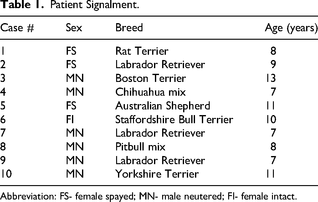
Abbreviation: FS- female spayed; MN- male neutered; FI- female intact.
Tumor Location, Surgical Treatment, and Follow Up.
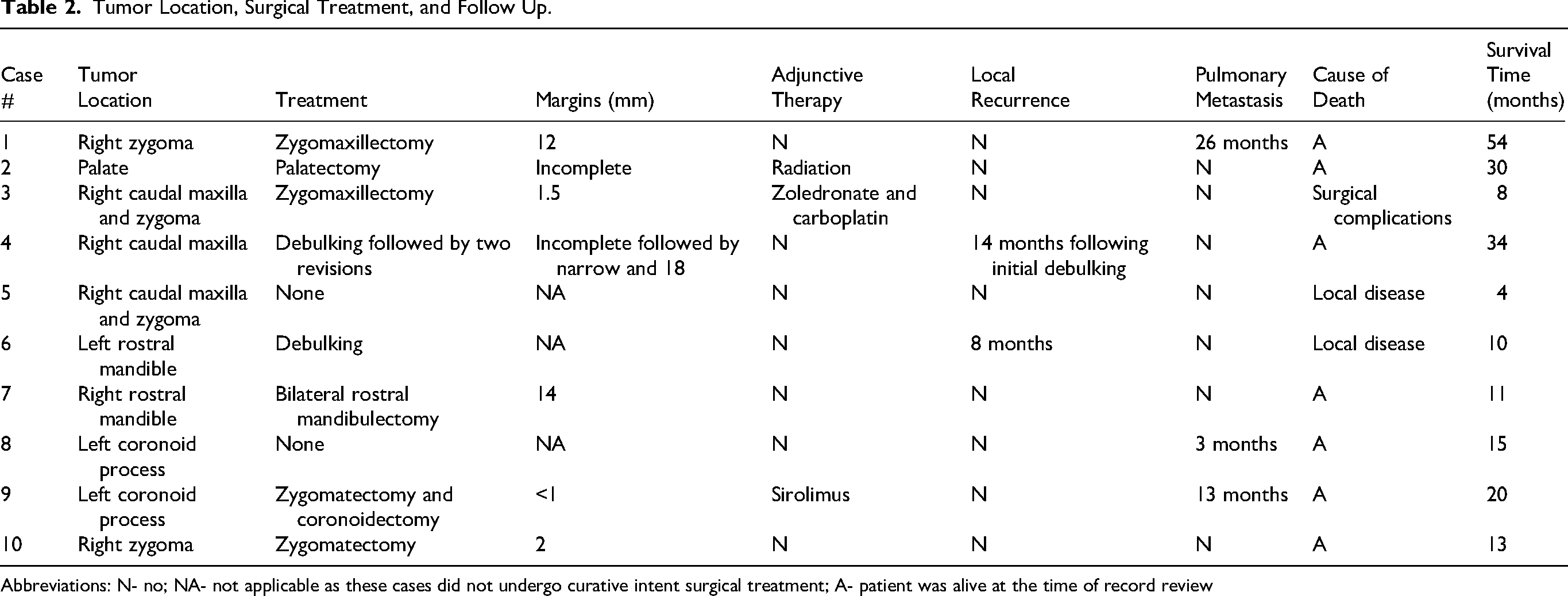
Abbreviations: N- no; NA- not applicable as these cases did not undergo curative intent surgical treatment; A- patient was alive at the time of record review
One tumor (1/10) involved the caudal maxilla alone, 2/10 involved only the zygoma, 2/10 involved both the caudal maxilla and zygoma, 2/10 involved the rostral mandible, 2/10 involved the coronoid process of the mandible, and 1/10 involved the palatine bone. Tumor volume ranged from 0.47 to 58.9 cm3. In all ten cases, the gross appearance of the tumor was clinically described as firm or bony, with 3/10 cases also being described as smooth.
Associated clinical signs at presentation included exophthalmos and pain or difficulty when opening the mouth. Exophthalmos was reported in 4/10 cases, of which 2 tumors involved the coronoid process, 1 tumor was associated with the caudal maxilla and zygoma, and 1 tumor was confined to the zygoma. Both cases involving the coronoid process also had decreased temporomandibular joint (TMJ) range of motion. Additionally, 1/3 cases involving the caudal maxilla showed pain when opening the mouth.
In 4 cases, physical examination or advanced imaging showed mandibular or retropharyngeal lymph node enlargement at initial presentation. In 3 of these cases, cytology was performed and they were reported as reactive lymphoid tissue.
Maxillofacial Imaging
Diagnostic imaging modalities and characteristics are summarized in Table 3. All ten cases had advanced 3D imaging of the skull, with CT and/or CBCT images available for review. CT was performed in 6/10 cases, and CBCT was performed in 5/10 cases. Intraoral dental radiographs were additionally performed in 8/10 cases. Cortical invasion was present in 8/10 cases and absent in 2/10 cases. The presence of a string sign was identified in 4/10 cases (Figure 1A). A lobular pattern was described in all ten cases (Figure 2A).

Case #10. (A) CBCT – transverse view showing an osseous mass arising from the periosteum of the right zygoma (asterisk) and causing mild disruption of the subjacent cortical bone. There is presence of a string sign separating the tumor from the underlying cortex (arrow). (B) Low magnification histomicrograph of the mass arising from the zygoma surface. Pre-existing zygomatic bone is on the right of the image and the bony mass abuts the lateral periosteal surface (arrows). At the base of the mass, neoplastic tissue invades zygomatic bone in a localized area (bracket). HE stain, bar = 3 mm. Abbreviations: CBCT – Cone beam Computed Tomography, HE – Hematoxylin and Eosin.

Case #5. (A) CBCT – transverse view showing an osseous mass arising from the right caudal maxilla and zygoma (asterisk). There is invasion of cortical bone present. (B) Microhistograph of the parosteal OSA with irregular trabeculae of neoplastic bone (arrows) that are generally parallel and have basophilic staining. Neoplastic spindle cells are distributed throughout the intertrabecular spaces. HE stain, bar = 200 um. Abbreviations: CBCT – Cone beam Computed Tomography, OSA – osteosarcoma, HE – Hematoxylin and Eosin.
Diagnostic Imaging Modalities and Characteristics.
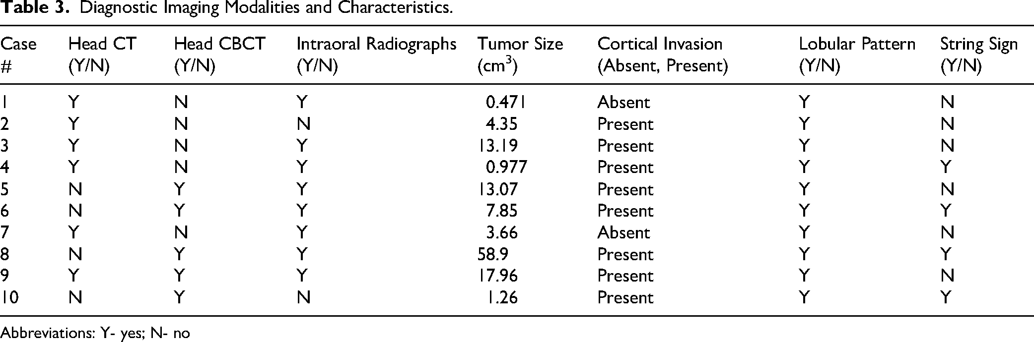
Abbreviations: Y- yes; N- no
Histological Findings
Histologically, each parosteal OSA bulged from the periosteal surface of bone with little to no destruction of the subjacent cortex (Figure 1B). The histologic organization of neoplastic tissue included thin trabeculae of bone that generally had a radiating pattern with parallel organization of bony plates. The osteoid often had basophilic staining even in decalcified sections (Figure 2B). The neoplastic spindle cells both lined bony trabeculae and contributed to fibrovascular stroma between trabeculae. A small amount of cartilage was identified in three tumors. Lobules of neoplastic tissue were often at the periphery of the mass (Figure 3B). The mitotic count ranged from less than 1 figure to 4 figures per 10 high-power fields (2.37 mm2). Five tumors had less than 1 mitotic figure in (2.37 mm2). Osteoclasts were noted in six tumors. None of the examined histologic sections had evidence of tumor necrosis. Inflammation was seen in two tumors (cases #6 and #7), each of which was a rostral mandibular tumor with surface ulceration attributed to occlusal trauma. Further histopathology findings are shown in Figure 4.

Case #4. (A) CT image with contrast enhancement – transverse view showing local recurrence of parosteal OSA within the soft tissue of the right commissure (asterisk). This CT was performed 14 months following the initial debulking procedure of the parosteal OSA associated with the right caudal maxilla. (B) Microhistograph of the recurrent parosteal OSA with small lobules (arrows) of neoplastic tissue originating within fibrous tissue at the edge of the main mass. The histologic features were identical to the original tumor. HE stain, bar = 300 um. Abbreviations: CT – Computed Tomography, OSA – osteosarcoma, HE – Hematoxylin and Eosin.

Case #9. (A) Low magnification microhistograph of parosteal OSA originating from the periosteal surface of the coronoid process of the mandible. HE stain, bar = 5 mm. (B and C) High magnification microhistograph shows spindle to polygonal cells lacking in atypia surround bony trabeculae. The osteoid of neoplastic bone has smudgy basophilic staining with occasional foci of cartilage (arrows). HE stain, bar = 100 um. (D) High magnification microhistograph shows forming adjacent to the tumor, reactive periosteal bone (bottom of image) must be differentiated from neoplastic bone (not shown in this image). HE stain, bar = 1 mm. Abbreviations: OSA – osteosarcoma, HE – Hematoxylin and Eosin.
Treatment
Two cases were diagnosed through incisional biopsy without further surgical attempts for complete excision. One case had an incisional biopsy with debulking and no additional surgical treatment. Seven cases underwent one or more surgeries with the goal of complete resection. Of these cases, 3 had complete margins, 1 had incomplete margins, and 3 had narrow margins. Noteworthy complications following any type of maxillofacial surgery were reported in only one dog. In case #3, dehiscence occurred following the zygomaticomaxillectomy procedure, resulting in an oronasal fistula. This required two additional procedures to attempt to close the defect. However, due to lack of patient compliance for an awake visual exam, records are unclear whether primary closure was achieved. Additionally, persistent nasal discharge was reported by the owner 3 months following the final repair attempt.
Three dogs received adjunctive oncologic treatments during the study period. Case #3 received zoledronate and carboplatin prior to and following zygomaticomaxillectomy with narrow margins. Case #2 received multiple sessions of intensity modulated radiation therapy following palatectomy with incomplete margins. Case #9 began a mammalian target of rapamycin (MTOR) inhibitor 13 months following coronoidectomy (Figure 5) with narrow margins for treatment of pulmonary metastatic disease.

Case #9: Parosteal OSA of the left coronoid process (arrows). (A and B) 3-D skull reconstructions. (C and D) CBCT images of transverse (C) and sagittal (D) views. (E) 3-D skull reconstruction following left zygomatectomy and coronoidectomy. Abbreviations: CBCT – Cone beam Computed Tomography, OSA – osteosarcoma, 3-D – 3 dimensional, HE – Hematoxylin and Eosin.
Follow Up
Local tumor recurrence was documented in two cases; both of these patients had gross evidence of tumor regrowth. In case #4, recurrence was evident at 14 months following surgical excision with incomplete margins. In case #6, recurrence was evident at 8 months following incisional biopsy/debulking with no further attempts for complete surgical excision. Noteworthy, in case #4, which involved the caudal maxilla, the local recurrence appeared as mineralization of the adjacent mucosa at the commissure rather than the associated maxillary bone (Figure 3A). Two revision surgeries were performed (one with narrow margins, one with complete margins), and no evidence of additional local recurrence was noted during the study period.
Screening for metastasis was performed in 8/10 cases and included thoracic radiographs (7/8), thoracic CT (4/8), abdominal radiographs (3/8), abdominal CT (4/8) and abdominal ultrasound (1/8). During the study period, 3/10 cases developed evidence of pulmonary metastasis. In cases #1 and #9, pulmonary nodules that were suspected as metastases were seen at 26 and 13 months, respectively. Case #1 had complete margins reported following surgical excision of the tumor, and case #9 had narrow margins reported following surgical excision of the tumor. The remaining case of documented metastasis (case #8) had pulmonary nodules consistent with metastases on thoracic radiographs 3 months following initial diagnosis via incisional biopsy, and the family declined further treatment. Twelve months following diagnosis of pulmonary metastasis this patient was reported to be alive with no respiratory symptoms.
Two cases had documented regional lymphadenopathy following excisional biopsy. In one case where margins were defined as narrow (case #9), the mandibular lymph nodes were sampled twice during the follow-up period, and cytology indicated reactive lymphoid tissue in the first aspirate and normal lymphoid tissue in the second. In the other case, where margins were defined as incomplete prior to further excision (case #4), cytology indicated reactive lymphoid tissue of the prescapular lymph nodes at the time of tumor recurrence and normal lymphoid tissue of the mandibular lymph nodes at the time of revision surgery.
Overall survival times ranged from 4 to 54 months with a median survival time of 14 months. In cases where complete and narrow margins were obtained, mean survival times (MST) were 33 and 13.6 months, respectively. The case with incomplete margins had a survival time of 30 months. Dogs that underwent surgery for curative intent, regardless of margins, had a MST of 24.3 months compared to 9.6 months for surgery limited to incisional biopsy or debulking. For the three dogs that received adjunctive oncologic treatments, the median survival time was 20 months compared to a median survival time of 13 months for dogs that did not. Of these, cases #2 and #9 were still alive at the end of the study period, 30 and 20 months following diagnosis, respectively.
Three of the 10 dogs were euthanized during the study period; in all of these cases the reason for euthanasia was attributed to the oral mass. For case #3, surgical excision was narrow without recurrence, although euthanasia was performed 8 months after diagnosis due to complications secondary to severe, persistent post-operative oronasal fistula. For case #5, surgical excision was not elected following incisional biopsy, and euthanasia was performed 4 months after diagnosis due to reported oral pain. For case #6, euthanasia was performed 10 months after diagnosis due to tumor growth following incisional biopsy and debulking.
Discussion
There are several case reports detailing parosteal OSA in dogs. To the authors’ knowledge, this is the first report that evaluates and documents multiple cases of canine parosteal OSA exclusively associated with the maxillofacial bones. In agreement with previous publications, the data from this study reported maxillofacial parosteal OSA to be a tumor of middle-aged to older dogs.2,4 While Labrador Retrievers accounted for approximately one-third of the cases in this small case series, a variety of dog breeds and sizes were represented. In cases of appendicular OSA, large-breed dogs appear to be significantly predisposed. 21 There was no sex predisposition noted in this case series. All patients, except for one intact female, had been sterilized. While there is literature that postulates an association between sterilization status and appendicular OSA in dogs,22–25 the small sample size of this study limits interpretation of this association.
The most common clinical signs associated with maxillofacial parosteal OSA were exophthalmos (4/10 cases) and difficulty or pain when opening the mouth (2/10 cases). When these clinical signs were present, the associated tumors were involving the caudal maxilla, zygoma, and/or coronoid process. These clinical signs likely developed secondary to the proximity and mass effect of the tumors on the orbit and its associated structures. It is worth noting that of the four dogs that showed exophthalmos and/or difficulty or pain opening the mouth, three had the largest tumors in the study (>13 cm3). These three cases also had long-standing clinical signs prior to obtaining a diagnosis of parosteal OSA, ranging from 2 to 7 months. Noting these common presentations of parosteal OSA is essential to aid quick diagnosis and treatment, as conditions of these maxillofacial bones may easily go unnoticed by both the dog owner and veterinarian. Space-occupying lesions of the caudal maxillofacial bones should be considered as a differential diagnosis in cases of exophthalmos and pain upon reduced TMJ range of motion.
A predilection for the caudal maxilla and/or zygoma was observed with half of the cases involving bones in these regions. Complex maxillofacial anatomy, potential intraoperative complications, and proximity to the orbit and cranial vault are among the reasons that oncologic surgery in these locations is challenging. The two cases involving the coronoid process of the mandible also presented challenges regarding surgical approach due to the proximity of the zygomatic arch. While the majority of dogs included in this study underwent curative intent surgery, there were three cases where treatment was limited to incisional biopsy with or without debulking due to owner preference. Two out of these three cases involved the caudal maxilla, coronoid process, and/or zygoma. The decision to not move forward with curative intent surgery may be related to potential complications, cost, and/or aesthetic considerations involved with these locations. It is worth noting that for the three dogs euthanized during the study period, the reasons for euthanasia were attributed to the owner's decision to not pursue curative intent surgery (cases #5 and #6) and to oronasal fistula complications following bisphosphonate administration, chemotherapy, and zygomaticomaxillectomy (case #3). While oronasal fistula formation following maxillectomy procedures is documented to be as low as 11%, 26 this potential complication should be discussed with owners prior to any surgery involving the palate and/or maxilla.
Advanced diagnostic imaging was a requirement for inclusion in this study as CT and/or CBCT not only enable 3D evaluation of the maxillofacial bones, but also provide an increased sensitivity to tumor location and extent of surrounding bone involvement27–29 which is crucial for the diagnosis of parosteal OSA. While intraoral dental radiographs were available for evaluation in several cases, 3D imaging was superior for identifying the bone layer of tumor origin. When available, 3D imaging such as CT or CBCT should be utilized for diagnosis and treatment planning of maxillofacial tumors.
The most consistent radiographic feature in this case series was the presence of a lobular growth pattern in all ten cases. In dogs, lobulation of craniofacial bone tumors is strongly associated with the diagnosis of MLO; therefore, this should be recognized as a source of potential confusion. 30 In humans, parosteal OSA appears as a lobular, ossified, exophytic mass 9 and this study confirms the same is true for canine parosteal OSA. String sign, which is the presence of a radiolucent line separating the tumor from the underlying cortex has been observed in 65% of human cases in cross-sectional imaging. 9 In this case study, a string sign was seen in 4/10 cases. While the majority of the cases in the study did not exhibit this radiographic presentation, the string sign is still a useful characteristic that may aid in the diagnosis of parosteal OSA. Another distinguishing characteristic of parosteal OSA on radiographic imaging is the origin from the periosteal surface of the bone and absence of central bone invasion. In this study, no tumors involved central bone; however, 80% of parosteal OSA lesions had some degree of cortical bone destruction. A previous veterinary study of zygomatic arch parosteal OSA did not provide quantitative data but states, “Many of the cases presented here had cortical invasion”. 2 Potential factors to explain cortical destruction/invasion include large tumor size, long duration of the tumor, and anatomic differences in bone. Pertinent to this last point, it is important to consider that radiographic features of parosteal OSA have been extrapolated from the human medical literature where the femur is the most common location for parosteal OSA.9,11 In dogs, the zygoma and caudal maxilla are common locations for parosteal OSA, both of which have a thin cortex compared to the human femur.
The diagnosis of parosteal OSA relied heavily on both histopathologic and radiographic characteristics. Five cases were excluded because the histomorphology was equivocal. Histologically, parosteal OSA is characterized by thin, radiating and often parallel trabeculae of neoplastic bone that often has basophilic staining of osteoid. However, these features are not pathognomonic and differentials include periosteal OSA, high grade surface OSA, central OSA (both low and high grade), MLO, ossifying fibroma and osteoma. Tumor location and growth pattern are most important in differentiating parosteal OSA from central OSA, and ossifying fibroma. Limited nuclear atypia and mitotic count differentiate parosteal OSA from surface high-grade OSA. Osteoma is often an exophytic periosteal tumor; however, compared to parosteal OSA, osteoma is less cellular and broad bony trabeculae mature to form dense lamellar bone. 31 Differentiating parosteal OSA from periosteal OSA and MLO is most challenging since each of these tumors proliferates from the surface of the parent bone and histologic criteria are based on subjective interpretation. In periosteal OSA, cartilaginous matrix predominates yet tumors generally also have radiating trabeculae of bone similar to parosteal OSA. 14 The argument that periosteal OSA is more likely than parosteal OSA to destroy subjacent cortical bone may apply to tumors of long bones that have a thick cortex but may be less pertinent to canine maxillofacial tumors.
Differentiating between parosteal OSA and MLO is complicated by morphologic similarities since both tumors may have bone and cartilage separated into lobules by bands of fibrous tissue. Like parosteal OSA, MLO often involves bones of the maxilla, palate, zygomatic process, and orbit.14,30 In MLO, multiple lobules are invariably present and consist of cartilage or cartilaginous bone surrounded by plump spindle cells and fibrous septae.14,30 This contrasts with the trabecular organization of parosteal OSA. As shown in Figure 2A, parosteal OSA may have lobules demarcated by fibrous tissue that resembles periosteum; however, the neoplastic bone is trabecular, with or without cartilage, and spindle cells are present between trabeculae.
Interpretation of incisional biopsies is particularly challenging since biopsy size and biopsy selection site may produce a sample that lacks sufficient tumor architecture or is not representative. Oral biopsies are often superficial and are obtained from the visible portion of the mass. The gingival and/or mucosal surface may be traumatized with inflammation and granulation tissue contributing to distortion of tumor architecture. Punch biopsies from a bony mass often fragment, resulting in loss of tumor architecture. For incisional biopsies, a provisional diagnosis with differentials is often most prudent. These limitations also highlight the importance of providing pathologists with a detailed history, photographs, and advanced diagnostic imaging.
A third of the cases received some form of oncologic treatment throughout the study period. While low case number prohibits statistical confirmation of treatment benefit, patients who received oncologic treatments had a median survival time of 7 months longer than those who did not. A case report of metastatic parosteal OSA in the humerus of a dog documents treatment with metastasectomy, amputation, stereotactic body radiation therapy, six doses of carboplatin, metronomic chlorambucil, and toceranib with a survival time of 623 days following histopathological diagnosis and 849 days following pulmonary metastatic disease. 13 Central OSA of the appendicular and non-maxillofacial axial skeleton is well documented to be associated with a poor prognosis due to survival times ranging from 4 to 6 months in cases managed by surgery alone and an early time to metastasis.3,6,32,33 Central OSA within the maxillofacial bones is considered to be relatively less aggressive, with increased survival times ranging from 5 to 17 months, and rare cases reporting survival over 4 years.4,7,32,34–38 The data from this study shows that dogs with maxillofacial parosteal OSA had an overall median survival time of 14 months, with a MST of over 2 years for dogs that underwent curative intent surgery. However, as the majority of dogs in this study were still alive at the time of writing this manuscript, these values likely underestimate true potential survival times.
Few patients in this study had complications related to treatment. It is important to note that in case #3, single doses of zoledronate and carboplatin were administered less than one month prior to surgical treatment of zygomaticomaxillectomy, which may have contributed to a persistent oronasal fistula. Zoledronate is a bisphosphonate medication that alters physiologic bone remodeling and, as a result, increases the risk of necrotic bone accumulation and risk of a condition called medication-related osteonecrosis of the jaw (MRONJ). However, as only a single dose was administered less than a month prior to surgery, it is unknown what true role it had in development of dehiscence. Caution is still recommended regarding this class of medications as dentoalveolar surgery is considered a risk factor for MRONJ. 39 Additionally, the recent administration of concurrent chemotherapy may have also played a role in delayed wound healing due to immunosuppression. While case #2 received multiple sessions of intensity modulated radiation therapy, this commenced one month following excisional surgery in order to minimize the risk of osteoradionecrosis. No complications were reported in this patient. These are factors that should be considered when planning multistage treatment in cases of maxillofacial oncologic surgery.
Regarding canine mandibular OSA it has been shown that histologic margins may not consistently predict repercussions such as local regrowth or metastasis. 32 However, achieving tumor free histologic margins has been found to be the primary prognostic indicator in dogs with maxillofacial OSA.34,35,37,38 In this case series, histological margins varied greatly, yet local recurrence following surgical excision occurred in only two cases where surgical margins were incomplete or following incisional biopsy with debulking. Of note, in one of these cases (case #4), recurrence appeared as mineralization within the adjacent soft tissue. A possible explanation for this, beyond progression of remaining tumor cells following initial procedure, is iatrogenic wound implantation of neoplastic cells through seeding of local soft tissue during removal or closure of the primary tumor site. Regardless, during excision of maxillofacial OSA, one tissue plane beyond the periosteum should be removed with the tumor to ensure complete removal of neoplastic cells. Based on the information from this case series, the authors recommend complete surgical excision with histological margins of 5 mm or greater to provide the longest disease-free survival time. However, this study shows that when aggressive surgical therapy is not feasible, narrow to incomplete excision may still provide significant patient benefit and improved longevity.
Pulmonary metastasis of axial OSA in dogs at initial diagnosis and at follow up has been documented as 11–13% and 35–46%, respectively.3,4,32,35 When observed in this case series, metastasis was slow and was diagnosed in the lungs. This occurred at 3, 13, and 26 months following diagnosis in three dogs. It is unknown if case #8 truly had pulmonary metastasis at 3 months following tumor diagnosis as this dog is still alive over a year later with no repeat thoracic imaging and no clinical signs consistent with progression of pulmonary metastatic disease. Since this dog was 8 years of age, the radiographic lung lesions may have represented benign osseous metaplasia. Notably, this dog had thoracic radiography alone. While not always an available option, CT is the preferred method for oncologic screening as it provides a higher diagnostic yield for metastasis than radiographs. 40 As only four dogs in this study had thoracic CT imaging, the incidence of pulmonary metastasis cannot be accurately established.
Mandibular and/or retropharyngeal lymph node enlargement was reported in 4/10 cases. In all samples with cytological analysis, reactive lymphoid tissue was diagnosed. Lymph node metastasis in central OSA is an uncommon finding, reported in 6.1% of cases evaluated at necropsy. 4 The results of this study suggest that lymphadenopathy should be investigated through cytology or biopsy for canine patients with maxillofacial parosteal OSA; however, it should not be a limiting factor for pursuing surgical treatment without confirmation of metastasis.
There were several limitations of this paper, as there inherently are with retrospective studies. Treatment and clinical staging were not standardized. Challenges also existed in the effort to determine prognostic data accurately. A third of the included cases never pursued curative intent surgery, which minimized interpretation of the data in these cases and may have affected accurate histopathological diagnosis. Additionally, some cases had follow-up times as short as 4 months, which may have underestimated true survival times. Regarding staging and follow-up, only a third of cases had repeat head and thorax CT to evaluate for tumor regrowth and metastasis. Additional studies with longer follow-up times and a strict protocol regarding treatment, follow-up and imaging are recommended.
In conclusion, parosteal OSA of the maxillofacial bones in dogs is an uncommon neoplasm that relies significantly on both histological and diagnostic imaging features for diagnosis. Parosteal OSA can occur in middle-aged to older dogs with no apparent breed or sex predilection. Anatomic locations include caudal maxilla, zygoma, coronoid process of the mandible, rostral mandible, and palate. Dogs with caudal tumors may show exophthalmos and/or pain or difficulty opening the mouth. Radiologic and histologic features overlap with other primary bone tumors; in particular, features of parosteal OSA may be misinterpreted as MLO. Destruction of cortical bone does not preclude the diagnosis of parosteal OSA, particularly for large tumors and tumors that occur on the surface of bone that normally has a thin cortex (eg, maxilla, palate, zygoma). Parosteal OSA carries a favorable prognosis compared to central OSA, particularly for patients that receive curative intent surgery. Pulmonary metastasis is a risk, believed to occur late in the course of disease.
Footnotes
Acknowledgements
The authors would like to thank Robert Cole, DVM, DACVR for his contribution towards reading and interpreting all diagnostic imaging. The authors would also like to thank all veterinarians and owners who allowed the participating dogs to be included in this study.

Ethical Considerations
Ethical approval was not required
Consent to Participate
Not applicable
Consent for Publication
Not applicable
Funding
The authors received no financial support for the research, authorship, and/or publication of this article.
Declaration of Conflicting Interest
The authors declare no conflict of interest regarding the research, authorship, and/or publication of this study.
Data Availability
The data that support the findings of this study are available on request from the author.