






Abstract
Desmoplastic small round cell tumor (DSRCT) is an aggressive small round cell sarcoma that typically occurs intra-abdominally in adolescents and young adults, and is characterized by a recurrent t(11;22)(p13;q12) translocation leading to generation of the EWSR1-WT1 fusion gene, which codes for a chimeric protein with transcriptional regulatory activity. DSRCT has a characteristic histologic appearance of nests of uniform small cells within prominent fibroblastic stroma and immunohistochemically it shows multidirectional differentiation, with expression of epithelial, neural, and muscle markers. We illustrate a case of DSRCT that presented as a large intra-abdominal mass, which harbored EWSR1 rearrangement by fluorescence in situ hybridization and EWSR1-WT1 fusion transcripts by reverse transcription–polymerase chain reaction (RT-PCR), and which histologically had an entirely solid morphology, lacking evidence of desmoplastic stroma. This purely solid variant emphasizes that even when occurring at a typical location, DSRCT may be difficult to recognize when lacking nonclassical morphology. This is of clinical relevance, as DSRCT with this pattern could be misdiagnosed as Ewing sarcoma if RT-PCR is not performed, with resulting prognostic and therapeutic implications.
Desmoplastic small round cell tumor (DSRCT) is an aggressive small round cell sarcoma that typically occurs as disseminated intra-abdominal disease in adolescents and young adults, 1 and is characterized by a recurrent t(11;22)(p13;q12) translocation leading to generation of the EWSR1-WT1 fusion gene, which codes for a chimeric protein with transcriptional regulatory activity.2-4 DSRCT has a characteristic histologic appearance, of nests of uniform small cells within prominent fibroblastic stroma, and immunohistochemically it shows multidirectional differentiation, with expression of epithelial, neural, and muscle markers. 5 The prognosis of DSRCT is almost uniformly dismal, as most patients present with metastatic disease and are poorly responsive to chemotherapy, 1 with overall 3- and 5-year survival rates of approximately 45% and 15%, respectively. 6 While DSRCTs were initially treated with similar multi-agent intensive chemotherapy regimens to those used for Ewing sarcoma, 7 complete pathologic response was infrequent. For patients without extra-abdominal metastatic disease, multimodal therapy including multi-agent intensive chemotherapy, aggressive debulking surgery and radiation therapy is currently considered to represent the standard of care, 8 with prolonged progression-free survival noted. 7
We illustrate a case of DSRCT that presented as a large intra-abdominal mass, which harbored EWSR1-WT1 fusion transcripts by reverse transcription–polymerase chain reaction (RT-PCR), and which histologically had an entirely solid morphology, lacking evidence of desmoplastic stroma. This case arose within the abdominal cavity of a 16-year-old female who presented with abdominal pain and an enlarging abdominal mass. Computed tomography scan showed a bulky maximally 18 cm solid abdominal mass which appeared to arise within the gastrosplenic ligament and was seen to compress and involve the spleen, and which extended inferiorly into the left iliac fossa. Several enlarged lymph nodes were identified around the splenic hilum as well as in the left anterior diaphragmatic area. The patient received 12 weeks of neoadjuvant chemotherapy with vincristine, cyclophosphamide, doxorubicin, ifosfamide, and etoposide without significant response before undergoing resection of the main tumor mass with splenectomy.
Macroscopically, the resected lesion comprised a predominant firm, solid maximally 20 cm white mass attached to the surface of the large bowel, with 3 white nodules up to 1.2 cm in diameter on the splenic surface. Several other lymph nodes were identified around the splenic artery. Histologically, attached to but not infiltrating the large bowel wall was a lobulated, cellular tumor composed of solid sheets of small round cells with small amounts of amphophilic cytoplasm and round to ovoid monomorphic hyperchromatic or vesicular nuclei (Figure 1A-D). There were prominent mitotic figures, extensive geographic necrosis, and foci of lymphovascular invasion. Prominent surrounding fibroblastic stroma was absent despite sampling of multiple tumor areas (Figure 1A-D). The nodules attached to the spleen showed similar morphology, and the spleen was focally infiltrated by tumor. Twelve tumor nodules were present within the surrounding fibroadipose tissue, some of them with surrounding nodal architecture and consistent with effaced lymph nodes. The tumor showed diffuse immunoreactivity for desmin and diffuse nuclear expression of WT1 (carboxy terminal) with focal expression of neuron-specific enolase (NSE), while smooth muscle actin (SMA), myogenin, myoD1, cytokeratin AE1/AE3, CD99, and neurofilament were negative. Fluorescence in situ hybridization (FISH) showed rearrangement of EWSR1 at 22q12 and RT-PCR showed EWSR1-WT1 fusion transcripts, as well as absence of EWSR1-FLI1 or EWSR1-ERG fusions, confirming the diagnosis of desmoplastic small round cell tumor.
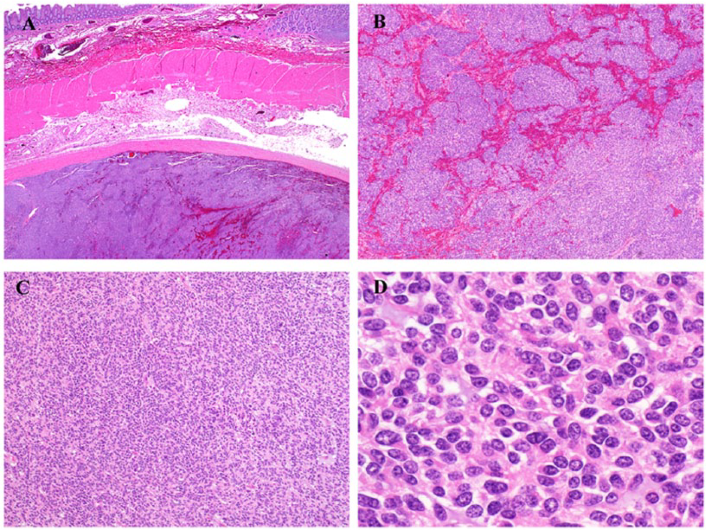
(A, B) Desmoplastic small round cell tumor (DSRCT). A large circumscribed and lobulated mass is attached to the outer aspect of the bowel wall (A). This comprises markedly cellular distributions of uniform small round cells present as sheets or nests. There is intervening hemorrhage in areas, but prominent desmoplastic stroma is not discernible. (C, D) The tumor has a sheet-like architecture in most places (C), and at high magnification the uniformity of the cells is appreciable. These have small ovoid or rounded nuclei with even chromatin and small amounts of amphophilic cytoplasm. The lack of surrounding desmoplastic stroma is noticeable, and without this feature this could be mistaken for Ewing sarcoma. The use of fluorescence in situ hybridization alone for detection of EWSR1 gene rearrangement is therefore not sufficient to confirm the diagnosis of DSRCT, and reverse transcription–polymerase chain reaction should be performed if possible, to confirm the presence of EWSR1-WT1 fusion transcripts, which are diagnostic in the correct clinical and pathologic context.
This purely solid pattern of desmoplastic small round cell tumor illustrates that its diagnosis can be difficult when there is variant morphology, even when occurring at a typical location. There is morphologic and (because of the nonspecific immunoprofile of DSRCT) immunophenotypic overlap with other small round cell neoplasms that may occur intra-abdominally, including Ewing sarcoma and alveolar rhabdomyosarcoma. Making the correct diagnosis is of clinical relevance, as DSRCT with this pattern could be misdiagnosed as Ewing sarcoma if RT-PCR is not performed, with resulting prognostic and therapeutic implications.
A range of morphologic appearances has been described in DSRCT, 9 including cases showing cytologic atypia with larger, spindled or sometimes giant and bizarre nuclei,9,10 or large epithelioid cells with anaplasia resembling carcinoma.11,12 Some tumors contain cytoplasmic clearing or vacuolation, or can show rhabdoid morphology, with intracytoplasmic eosinophilic inclusions representing bundles of intermediate filaments, 13 or there can be rosette or tubule formation. 14 Immunohistochemically, DSRCT shows variable expression of markers of several cell lineages. Tumors may not express all antigens, and can lack those that are typically expressed such as cytokeratin and desmin. 13 The majority of DSRCTs express desmin and epithelial membrane antigen, 15 most express cytokeratins and variably express a range of neural markers (including chromogranin, synaptophysin, CD56, NSE, neurofilament protein, and S100 protein),9,15 muscle-specific actin, or alpha-SMA. 15 Up to almost 90% of DSRCT are positive with antibodies toward the carboxy terminus of the Wilms tumor (WT1) protein.15,16 CD99 is typically negative, although variable positivity can be seen in up to half of cases.15,16 Genetically, DSRCT is associated with a characteristic EWSR1-WT1 gene fusion, arising from a reciprocal t(11;22)(p13;q12) translocation juxtaposing the 5′ region of the Ewing sarcoma (EWSR1) gene on chromosome 22 with the 3′ DNA-binding segment of WT1 (the Wilms’ tumor suppressor gene) on chromosome 11.2-4,17 EWSR1-WT1 fusion appears no longer specific for DSRCT, as it has been recently described in a small round cell tumor of the cauda equina that was clinically low grade, 18 and which comprised cells in cords and nests as well as some rosette-like structures, rare mitoses and a low Ki-67 proliferation index and features of smooth muscle differentiation and focal CD99 and Neu-N expression by immunohistochemistry. 18
This case illustrates a rare, wholly solid pattern of DSRCT presenting as a predominant intra-abdominal mass, which leads to the potential for misinterpretation as another small round cell neoplasm. This may be especially challenging when assessing limited material, such as in core biopsy. A diagnosis of DSRCT should be considered for any intra-abdominal small round cell neoplasm, particularly if occurring in adolescent/young adult patients. Accurately distinguishing DSRCT from other intraabdominal neoplasms (including Ewing sarcoma) is important to guide the most appropriate treatment protocol and to facilitate informed discussion of prognosis with the patient so the surgical pathologist needs to be aware of the morphologic and immunohistochemical spectrum. Additionally, as the diverse range of neoplasms harboring EWSR1 gene rearrangements is now well recognized,19-22 the use of FISH alone for detection of EWSR1 gene rearrangement is not sufficient to confirm a diagnosis of DSRCT, and RT-PCR should be performed if possible, to confirm the presence of EWSR1-WT1 fusion transcripts, which are diagnostic in the correct clinical and pathologic context.
Footnotes
Declaration of Conflicting Interests
The author(s) declared no potential conflicts of interest with respect to the research, authorship, and/or publication of this article.
Funding
The author(s) disclosed receipt of the following financial support for the research, authorship, and/or publication of this article: The authors acknowledge support from the NIHR Royal Marsden/ICR Biomedical Research Centre.