






Abstract
Desmoplastic small round cell tumor (DSRCT) is a rare malignant tumor that occurs mainly in the retroperitoneum of children and young adults. In its prototypical form, DSCRT displays characteristic morphology with nested primitive small round cells in a desmoplastic stroma and a distinctive immunophenotype with polyphenotypic differentiation. However, DSCRT can also exhibit a broader clinical, histologic and immunohistochemical spectrum and, therefore, cause diagnostic difficulties. Given that DSCRT is an aggressive and nearly universally fatal disease, making the correct diagnosis is critically important. Herein, we report three patients with DSRCT and unusual clinical, morphologic or immunohistochemical characteristics, in order to highlight its remarkable diversity and increase awareness of this unusual, distinctive neoplasm.
Introduction
Desmoplastic small round cell tumor (DSRCT) is a rare and highly aggressive mesenchymal neoplasm that typically originates in the abdominal cavity in young male patients.1,2 Characteristic radiologic findings of abdominopelvic DSRCT include the presence of multiple heterogeneously dense masses whereas diffuse serosal involvement is common. 3 However, occasional cases of DSRCT have been associated with atypical clinical features including, arising in elderly patients, females4,5 and uncommon anatomic sites, such as subcutaneous tissue, head and neck, brain and cranial fossa, upper and lower extremity, visceral organs, and in bone.6–16 Atypical presentations of DSRCT are described in about 24% of tumors overall, 44% in the consultation setting. 17
Histologically, DSRCT is characterized by round or angulated nests of primitive small round cells enmeshed in a prominent reactive desmoplastic stroma. The tumor cells are usually relatively monomorphic and have round-to-oval, hyperchromatic nuclei, inconspicuous nucleoli, and scant eosinophilic cytoplasm. Mitoses are numerous and necrosis is typically present. DSRCT has a distinctive immunoprofile showing polyphenotypic differentiation, with co-expression of epithelial, muscle and neural markers.1–14,18 Most tumors are positive for keratins/epithelial membrane antigen (EMA), express desmin in a paranuclear dot-like pattern, exhibit neural markers (neuron-specific enolase (NSE) and CD56) and show nuclear positivity for the carboxy (C) terminus of the Wilms tumor protein 1 (WT1). Keratin may also show a dot-like pattern, with CAM5.2 being the most sensitive marker. Nuclear expression of WT1 (C-terminus) is highly specific for DSRCT, observed in more than 90% of tumors. Other immunohistochemical (IHC) markers expressed with less frequency include: neuroendocrine markers (synaptophysin, chromogranin), smooth muscle actin (SMA), CD99, and S100-protein. 19 The immunohistochemical pattern suggests the neoplastic cells have features that overlap with cell types of multiple different lineages.19,20 Cytogenetic analysis shows a characteristic translocation, t(11;22)(p13;q12), resulting in generation of the EWSR1::WT1 fusion gene. 21 This chimeric protein product acts as a transcriptional activator factor, upregulating expression of PDGFRα, FGFR4, VEGFR2, and IGF2, activating MYC, PAX2, TALLA1 and NTRK3, and blocking p53. 22
Although DSRCT has characteristic clinicopathologic features, a minority of tumors exhibit unusual findings that can cause diagnostic challenges. In the current case series, we report three DSRCTs from the files of two institutions (Dartmouth Hitchcock Medical Center, Lebanon, NH and University of Miami, Miami, FL) with unconventional morphologic or clinical features to highlight and increase awareness of the remarkable diversity of this rare neoplasm.
Case Series (Table 1 and supplementary table)
Patient 1: A 51-year-old woman presented with a progressively enlarging abdominal mass. CT scan showed a left upper quadrant intra-abdominal mass measuring at least 16.5 × 12.5 cm, with involvement of the abdominal wall. A subsequent CT-guided biopsy was interpreted as high grade epithelioid leiomyosarcoma, and although the patient was deemed operable the resection was delayed due to insurance issues and lack of follow-up. A repeated CT scan at six months showed metastatic disease to the lungs and pleura, whereas the primary site also showed progression with an increase in size and peritoneal implants throughout the abdomen and pelvis. The patient was no longer a surgical candidate and started on chemotherapy.
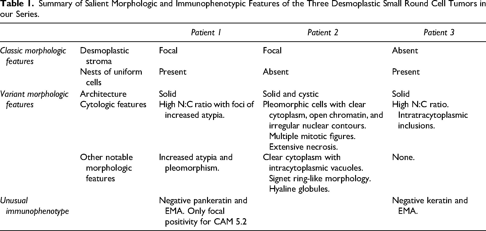
Summary of Salient Morphologic and Immunophenotypic Features of the Three Desmoplastic Small Round Cell Tumors in our Series.
The biopsy was reviewed as a consultation at the University of Miami. Histologic sections showed sheets of monomorphic round cells with a small amount of eosinophilic to clear cytoplasm and high nuclear-to-cytoplasmic (N:C) ratios with round to ovoid, hyperchromatic nuclei. The tumor cells were arranged in nests surrounded by reactive fibrous tissue. Patchy necrosis and high mitotic activity were noted (Figure 1). By immunohistochemistry, the tumor cells showed expression of SMA, CAM 5.2 (focal), CD56 (focal), and WT1 (C-terminus). They were negative for pankeratin, EMA, and WT1 (amine (N)-terminus). The estimated Ki67 proliferation index was approximately 65%. Next-generation sequencing (NGS) performed by a commercial platform (Foundation One, Cambridge, MA) was positive for EWSR1::WT1 fusion. This result in conjunction with the morphologic and immunohistochemical profile confirmed the diagnosis of DSRCT.

Desmoplastic small round cell tumor presenting as an intraabdominal/abdominal wall mass in a 51-year-old woman. (A) The tumor is composed of solid sheets of malignant small cells (H&E, 23X). (B) Patchy areas of necrosis are evident (H&E, 200×). (C) Malignant cells infiltrate fibrous tissue and show foci of crush artifact (H&E, 200×) (D) The tumor cells exhibit round to ovoid, hyperchromatic nuclei and scant, lightly eosinophilic cytoplasm (H&E, 600×).
Patient 2: A 27-year-old man presented with two peritoneal and omental masses measuring 13.6 cm and 8.9 cm in greatest dimension respectively. The tumors deemed resectable but eventually the patient developed widespread metastatic disease. Histologic examination of the initial biopsy showed a population of infiltrating and nested epithelioid malignant cells that were associated with small cystic spaces. At the periphery, the cells had significantly smaller nuclei with less cytoplasm. More centrally, the tumor cells had variable size. Many of the neoplastic cells exhibited clear cytoplasm with multiple intracytoplasmic vacuoles surrounded by eosinophilic cytoplasm. Occasional cells with signet ring-like morphology were also present. The nuclei showed pleomorphism with irregular nuclear contours and open chromatin. Some cells contained rare hyaline globules. Multiple mitotic figures and necrosis were identified (Figure 2). The neoplastic cells were positive for pankeratin, EMA, desmin, WT1 (C-terminus) and did not show expression for CD99, S100-protein, or WT1 (N-terminus). Fluorescence in situ hybridization (FISH) detected EWSR1 rearrangement. The overall clinical, morphological, immunohistochemical and FISH results were compatible with DSRCT.

Peritoneal desmoplastic small round cell tumor. (A) At low power, solid sheets of tumor cells are separated by fibrous bands, with hyperchromatic areas peripherally and pale, clearer areas centrally (H&E, 40×). (B) At intermediate magnification, distinct morphologic patterns are evident. Neoplastic cells with high N:C ratio and dense eosinophilic cytoplasm are seen on the left, whereas those on the right demonstrate more voluminous clear cell cytoplasm (H&E 100×). (C) The cells are epithelioid with moderate pleomorphism, vesicular chromatin, clear to eosinophilic cytoplasm and occasional intra-cytoplasmic eosinophilic material (H&E, 100×). (D) A subset of cells exhibits abundant clear, granular to finely vacuolated cytoplasm somewhat resembling lipoblastic differentiation (H&E, 600×).
Patient 3: A 22-year-old man presented with right flank pain, and computed tomography (CT) scan demonstrated a right renal mass without adenopathy or metastatic disease. Subsequent radical nephrectomy revealed a 7.8 cm mass involving the renal parenchyma, with invasion of the renal vein and sinus. Histologic examination showed sheets of round cells with multiple areas of necrosis lacking a desmoplastic stroma. The tumor cells were small with high N:C ratio, slightly coarse chromatin and small or inconspicuous nucleoli. Intracytoplasmic inclusions were occasionally seen (Figure 3). By immunohistochemistry, the tumor cells were positive for WT1 (C-terminus), CD99, desmin (perinuclear dot-like pattern), CD56, TLE1, and MDM2. They were negative for EMA, keratin AE1/3, keratin 7, WT1 (N-terminus), myogenin, MyoD1, and carbonic anhydrase 9

An unusual renal primary desmoplastic small round cell tumor. (A) The tumor invades the renal parenchyma in a pushing fashion. It is unencapsulated and composed of sheets of rounds cells with high N:C ratio (H&E, 40×). (B) Solid sheets of predominantly round cells are seen in a background with inconspicuous capillaries (H&E, 200×). (C) Higher magnification shows round cells with irregular nuclear contours, coarse chromatin, inconspicuous nucleoli and increased mitotic activity (H&E, 600×). (D) WT1 C-terminus immunohistochemical stain shows multifocal nuclear positivity (Immunohistochemistry, 600×).
Discussion
The latest (fifth edition) of the WHO Classification of Soft Tissue and Bone Tumors published in 2020 describes the essential diagnostic criteria for DSRCT which include: classic morphologic features of small round cells in solid nests with a desmoplastic stroma and positive immunohistochemistry for keratin, desmin in a dot-like pattern and C-terminus of WT1. Desirable, although not required for the diagnosis, is detection of an EWSR1 gene rearrangement or EWSR1::WT1 fusion. 23 However, it is recognized that DSRCT can exhibit variation from its usual morphology, 21 showing a pure solid pattern, tubules, glands, rhabdoid features, spindle cell or signet ring cell morphology, and, very rarely, an absence of significant stromal desmoplasia.1,6,17,20,21,23,24 Also, a minority of patients can be associated with unusual clinical features that can lead to a differential diagnosis that does not include DSRCT.
We identified three such patients of unusual DSRCT. One tumor (Patient 3) exhibited a lack of desmoplasia, which is one of the most characteristic features of this DSCRT. Patient's 2 tumor was uniquely composed of pleomorphic cells with intracytoplasmic vacuoles mimicking lipoblastic differentiation, inclusions and hyaline globules, with occasional signet ring-like cells showing peripherally placed nuclei. Key morphologic and immunophenotypic features of the tumors in this study cohort are summarized in Table 1. The two patients with unusual clinical presentations (Patient 1 involving an older woman and Patient 3 presenting as a localized kidney mass without dissemination) also showed uncommon immunophenotypic features. In both, the neoplastic cells did not stain for EMA, which is expressed in 96% of DSRCT tumors, and they were negative for keratins. 25
To establish the diagnosis of DSRCT in a young or adolescent male with typical presentation, histologic assessment, and immunohistochemistry are most likely sufficient. However, in tumors with an unusual clinical presentation, morphologic features, immunohistochemical profile or a combination thereof, molecular testing may be necessary. 23 The differential diagnosis of DSRCT depends on location, age, and morphology of the tumor and may include malignancies from different lineages such as carcinomas, melanoma, hematolymphoid tumors and other defined subtypes of small round cell sarcomas. 18
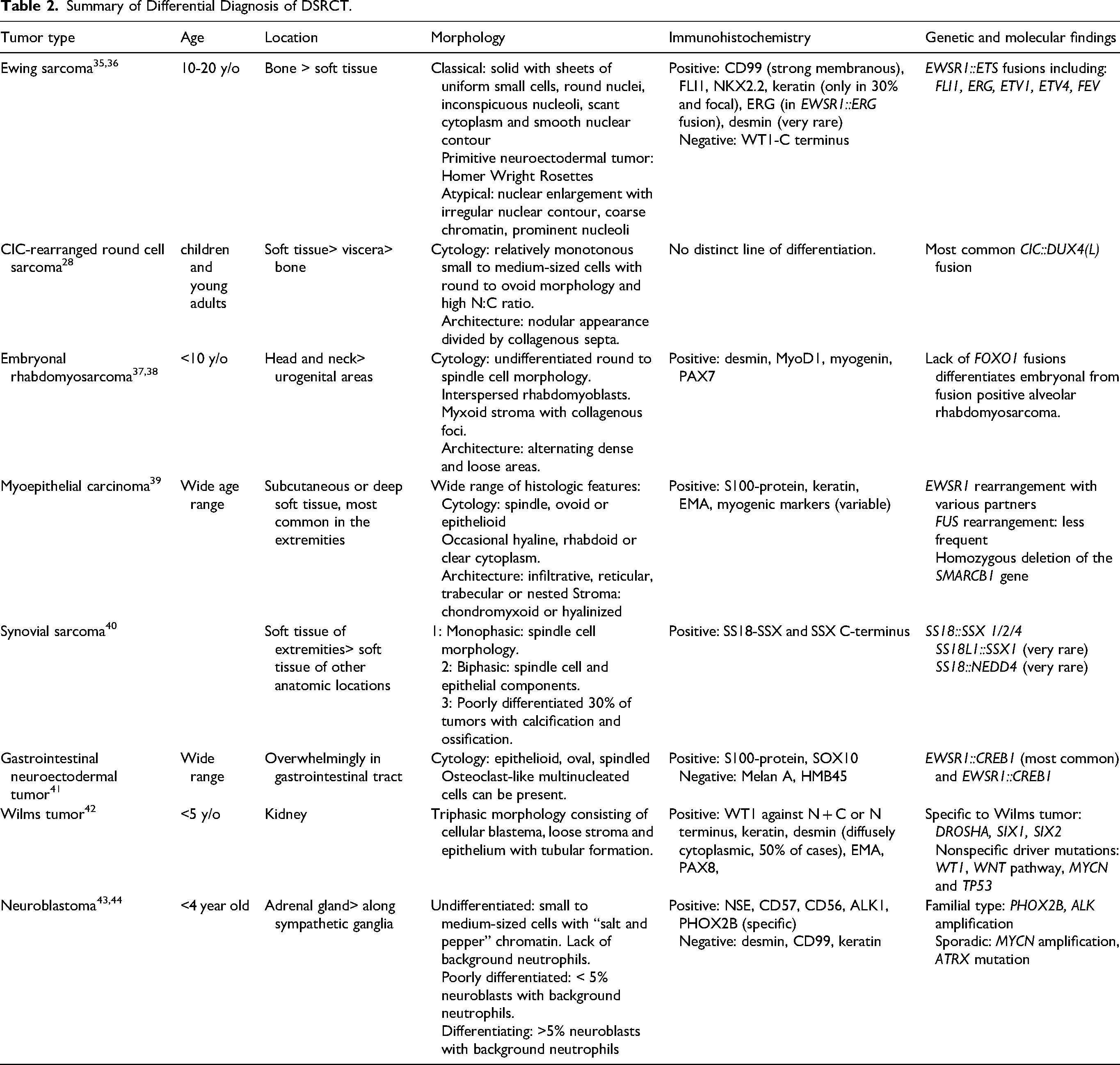
In younger patients, Ewing sarcoma, CIC-rearranged round cell sarcoma, embryonal rhabdomyosarcoma, myoepithelial carcinoma, poorly differentiated synovial sarcoma, gastrointestinal neuroectodermal tumor, germ cell tumors and hematolymphoid neoplasms should all be considered in the differential diagnosis (Table 2). Although Ewing sarcoma can express keratin in approximately 25% of tumors and desmin in ∼3%, the detection of EWSR1::ETS partner (FLI1 in ∼85% of tumors) distinguishes the two neoplasms.26,27 Round cell sarcomas with CIC or BCOR rearrangements may also exhibit a wide morphologic spectrum, including small to medium-sized neoplastic cells with scant, lightly eosinophilic to clear cytoplasm. In addition, in one-third of tumors, a nodular appearance with thick collagenous septa may be noted.3,28 In children, embryonal rhabdomyosarcoma may have an alternating loose and dense pattern of cellularity but exhibits a greater degree of cytologic pleomorphism and is positive for markers of rhabdomyoblastic differentiation such as MyoD1, myogenin or PAX7. 29 Myoepithelial carcinoma of soft tissue is an uncommon tumor, which typically arises in the extremities and is characterized by spindle, ovoid or epithelioid cells arranged in multiple nodules, lobules, or nests. The tumor cells can have vacuolated cytoplasm and show variable growth patterns. Although myoepithelial carcinoma can be positive for keratin and desmin and exhibits EWSR1 rearrangements in ∼50% of tumors, WT1 has never been reported as a partner, and it is expected to be negative for WT1 C-terminus by immunohistochemistry. 30 Synovial sarcoma has a wide anatomic and demographic distribution and may be monophasic, biphasic or poorly differentiated. The last could conceivably enter the differential diagnosis with DSRCT, but synovial sarcoma can be distinguished by detecting, in the great majority of tumors, the characteristic SS18::SSX1/2/4 fusion by molecular methods or immunohistochemistry using gene fusion surrogates.23,31 Gastrointestinal neuroectodermal tumor usually arises in the gastrointestinal tract and can histologically exhibit a mixture of spindle and epithelioid patterns. Diffuse positivity for S100-protein and SOX10 and detection of characteristic EWSR1::CREB1 (most common) or EWSR1::ATF1 help secure the diagnosis.32,33 Germ cell tumors include a wide variety of tumors, which can show a homogenous cellular proliferation with pale cytoplasm. 17 EWSR1 rearrangements have not been reported in these tumors. Tumors such as non-Hodgkin lymphoma/leukemia can be seen in all ages and at any location, and they may be distinguished easily from DSRCT with hematolymphoid markers. 17 In older patients, primary/metastatic poorly differentiated or sarcomatoid carcinomas lead the differential diagnosis.20,23,34
Summary of Differential Diagnosis of DSRCT.
In addition to the diagnostic considerations above, the differential diagnosis of renal DSRCT additionally includes blastemal-predominant Wilms tumor, neuroblastoma and malignant rhabdoid tumor of the kidney.29,45 Blastema-predominant Wilms tumor usually occurs in children younger than six years old. It is principally composed of small round blue cells and can be extremely challenging to differentiate from DSRCT. Glomeruloid bodies or focal epithelial components, which can be histologic clues for Wilms tumor, may be absent in a small biopsy. While these tumors share immunohistochemical features, the perinuclear dot-like pattern of desmin positivity is characteristic of DSRCT, in contrast to the diffuse cytoplasmic reactivity in Wilms tumor. Ultimately, molecular studies may be necessary for the diagnosis.30,31 Neuroblastoma also occurs in patients younger than five years old. 32 Cytologically the cells are medium-sized and exhibit more atypia than typical DSRCT. Neuroblastoma consistently expresses neuroendocrine markers but is negative for keratin, which is frequently positive in DSRCT.20,33 PHOX2B is also a sensitive and specific immunohistochemical marker for neuroblastoma and can aid in the differential with other histological mimickers. 46 Rhabdoid tumor of the kidney mostly affects children younger than two years of age. The neoplastic cells show a large nucleolus and cytoplasmic inclusions, which are not common features of DSRCT. Desmin expression can be seen in both DSRCT and rhabdoid tumor of the kidney, but loss of INI1 expression is only seen in the latter.47,48 It is also worth mentioning that renal DSRCT is an extremely rare tumor, with only 16 patients among 1600 in a recent study. 45
As the application of more sophisticated molecular techniques becomes widely available, driver or stochastic molecular events are being discovered at an accelerated pace. It is abundantly clear now that many fusions that were considered pathognomonic in the recent past are in fact “promiscuous”; identical driver fusions have been detected in various neoplasms of mesenchymal or other lineage with completely different morphology and natural history. EWSR1::WT1 is one of the most specific, limited to DSCRT in the recent 2020 WHO Classification. Nevertheless, very recently Schoolmeester et al 49 reported three unusual tumors involving the female genital tract with EWSR1::WT1 fusions where the morphologic and immunohistochemical features were incompatible with the diagnosis of DSRCT. Two tumors were low-grade spindle cell sarcomas, whereas the third tumor was a high-grade sarcoma with spindle and epithelioid features. Gene expression analysis comparing the low-grade tumors and two classic tumors of DSRCT revealed 1726 genes that were differentially expressed which reached statistical significance. The tumors in this study also expressed markers of smooth muscle differentiation which is unusual in DSRCT. It is interesting that our Patient 1 was a 51-year-old female whose tumor expressed SMA. To the best of our knowledge, six patients of non-desmoplastic small round cell tumors with EWRS1::WT1 have been described in the literature.49,50 It seems likely that these tumors represent different entities as they appear to be a relatively heterogenous group. The tumor of Patient 1 had high mitotic activity that helps distinguish it from the so-called low-grade small round cell sarcoma with EWSR1::WT1 fusion. 50
DSRCT is an aggressive tumor with a 5-year survival rate of 10-15%. Consequently, a misdiagnosis may have significant prognostic and management repercussions.51,52 There is no standard treatment protocol, and conventional multimodal therapy is not very effective. DSRCT treatment has mainly derived from the protocols of Ewing sarcoma, and many trials combine DSRCT with Ewing sarcoma due to the rarity of the former. However, due to DSRCT poor prognosis and response to treatment, targeted therapy toward EWSR1::WT1 downstream targets has been used with no significant advancements so far. 53 Effective therapeutic agents targeting downstream genes of the EWSR1::WT1 fusion are urgently needed to potentially improve outcomes for this highly aggressive tumor. 54 Therefore, a better understanding of this neoplasm is crucial to develop more effective treatments options. Correct identification of patients with DSRCT is critical to facilitate clinical trial enrollment and the study of new therapeutic protocols. 22
While DSRCT is largely characterized by its unique combination of morphologic, immunophenotypic and molecular features, deviation from its customary characteristics may lead to diagnostic challenges. In this case series, we highlight uncommon phenotypes of DSRCT with the hope to increase awareness about the diversity of this rare neoplasm.
Supplemental Material
sj-docx-1-ijs-10.1177_10668969221095178 - Supplemental material for Highlighting the Diversity of Desmoplastic Small Round Cell Tumor: A Case Series
Supplemental material, sj-docx-1-ijs-10.1177_10668969221095178 for Highlighting the Diversity of Desmoplastic Small Round Cell Tumor: A Case Series by Sepideh Nikki Asadbeigi, Julio A. Diaz-Perez, Andrew E. Rosenberg, Jason R. Pettus, Darcy A. Kerr and Konstantinos Linos in International Journal of Surgical Pathology
Footnotes
Declaration of Conflicting Interests
The author(s) declared no potential conflicts of interest with respect to the research, authorship, and/or publication of this article.
Funding
The author(s) received no financial support for the research, authorship, and/or publication of this article.
Ethical Approval
Not applicable, because this article does not contain any studies with human or animal subjects.
Informed Consent
Not applicable, because this article does not contain any studies with human or animal subjects.
Trial Registration
Not applicable, because this article does not contain any clinical trials.

Supplemental Material
Supplemental material for this article is available online.
References
Supplementary Material
Please find the following supplemental material available below.
For Open Access articles published under a Creative Commons License, all supplemental material carries the same license as the article it is associated with.
For non-Open Access articles published, all supplemental material carries a non-exclusive license, and permission requests for re-use of supplemental material or any part of supplemental material shall be sent directly to the copyright owner as specified in the copyright notice associated with the article.
