






Abstract
Desmoplastic small round cell tumor (DSRCT) is an aggressive small round cell neoplasm which predominantly occurs intra-abdominally in adolescents and young adults with a male predominance, and which is characterized by a recurrent t(11;22)(p13;q12) translocation leading to formation of the EWSR1-WT1 fusion gene, which generates a chimeric protein with transcriptional regulatory activity. Histologically, DSRCT has a characteristic morphology, of islands of monotonous small cells within prominent sparsely cellular fibroblastic stroma, and immunohistochemically it shows polyphenotypic multidirectional differentiation, with expression of epithelial, muscle, and neural markers. However, DSRCT can arise more rarely in other sites and exhibit a spectrum of both histologic features and immunoprofile, which may confuse diagnosis with other small round cell neoplasms. Correct diagnosis is important to ensure correct treatment and prognostication; DSRCT are almost universally fatal neoplasms with patients usually succumbing to disease within the first 2 years of diagnosis. While combination treatment strategies can confer a survival benefit, the overall prognosis remains poor. Further insight into the tumorigenic molecular changes generated by the fusion oncogene may lead to the generation of specific targeted therapies. We review DSRCT, discussing morphology and immunohistochemistry, molecular genetic findings, potential targeted treatments, and the differential diagnosis.
Introduction
Desmoplastic small round cell tumor (DSRCT) is an aggressive small round cell neoplasm that predominantly occurs intra-abdominally in adolescents and young adults with a male predominance. It was first proposed as a distinct neoplastic entity by Gerald and Rosai in 1991, 1 who described a distinctive type of highly aggressive small round cell tumor with a predilection for adolescent males, with predominant or exclusive intra-abdominal location, only inconstant and secondary organ involvement, and morphologically a nested growth pattern with marked desmoplastic reaction, as well as immunoreactivity for epithelial, neural, and muscle (desmin) markers. 1 The histopathologic features of DSRCT are well delineated: of islands and nests of uniform small round cells dispersed within a prominent fibroblastic stroma, as well as a polyimmunophenotypic immunoprofile, with its cells expressing epithelial, muscle and neural markers to varying degrees. DSRCT remains of unknown etiology; as previous ultrastructural and immunohistochemical studies showed variable incomplete epithelial, muscle or neural differentiation, it was believed to originate from a primitive progenitor cell with potential for multiphenotypic differentiation. 2 However, DSRCT is characterized by a recurrent t(11;22)(p13;q12) translocation, which leads to formation of the EWSR1-WT1 fusion oncogene, and as a translocation-associated neoplasm DSRCT does not recapitulate a normal “cell of origin” or line of differentiation. Additionally, there is high transient fetal expression of the WT1 gene within body cavity serosal linings. 3
Clinical Features and Management
DSRCT has a peak incidence in young adults, described as between 20 and 24 years, 4 with mean age of 28.3 years 5 (age range 10-41 years), 6 but rarely occurs in older patients, including those in middle age and elderly patients up to the eighth decade. 7 There is a striking male predominance of at least 4:1,5,6 although there does not appear to be a marked racial predilection, with age-adjusted incidence rates for patients of Afro-Caribbean and European descent of 0.5 cases/million and 0.2 cases/million, respectively. 4 The majority of DSRCTs present within the abdominal cavity, involving the pelvis, retroperitoneum, omentum and mesentery,3,6 often with widespread serosal involvement. Neoplasms can also occur as primary intrascrotal (paratesticular) masses arising in continuity with the peritoneum via the processus vaginalis or in isolation, or as primary neoplasms within the thoracic cavity including the pleurae. 8 Much more rarely, DSRCT can arise as primary disease at other anatomic sites, including the head and neck (including the sinonasal cavity, salivary glands, and brain),8-10 the hand, 8 and visceral organs such as small and large bowel,11,12 ovary, 13 and kidney.14,15 Tumors can present with widespread disseminated metastases including to the lungs and brain. DSRCT can also present with nodal and visceral metastases without evidence of a primary site. 16 However, DSRCT typically tend to present as painful abdominal masses with abdominal distension, acute abdomen or ascites, or may cause symptoms due to mass or pressure effects on adjacent structures. An unusual presentation includes of paraneoplastic phenomena, with severe hypertension with hypokalemia and metabolic alkalosis due to renin secretion in disseminated tumor. 17
Radiologically, these are typically multifocal peritoneal/omental masses, usually present in combination with a dominant soft tissue deposit (at least double the size of the next largest soft tissue deposit) with its epicenter in the abdomen or pelvis. 5 The most common computed tomography (CT) finding at presentation is of peritoneal/omental disease (95%), usually manifesting as multiple peritoneal or omental soft tissue lesions, with a solitary peritoneal mass present in a smaller number of cases. Dominant lesions tend to be large (≥5 cm in 16/16 cases in a large radiologic series and ≥10 cm in 11/16 cases). Nodal enlargement has been reported to be as high as 50%, most commonly in the retroperitoneum but with mediastinal and mesenteric nodes also recognized. Patients may also have synchronous liver and bony metastases. 5 Forty percent of patients in a large pediatric imaging study of DSRCT had metastatic disease to the liver, lungs, spleen, or bones at diagnosis. 18 Functional imaging with positron emission tomography (PET)/CT can be advantageous over anatomical imaging for the accurate staging of disease, and 18F-fluorodeoxyglucose (FDG) PET/CT has been shown to accurately detect 97.4% of DSRCT in this study. 18
DSRCT is an aggressive neoplasm with an almost uniformly poor prognosis; most patients present with metastases and respond poorly to chemotherapy 1 and die of disease within the first 2 years of diagnosis. Overall, 3- and 5-year survival rates have been reported as 44% and 15%, respectively. 19 Twenty-five of 35 patients with follow-up information in a large series died of widespread metastases, ranging from 8 to 50 months (mean, 25.2 months) from the time of diagnosis, 6 with median survival from diagnosis of approximately 22.3 months. 5 A recent large analysis of pediatric patients (age range: 0-21 years) showed similarly poor overall survival rates at 2 and 5 years of 52% and 18%, respectively, with overall median survival time of 2.1 years. 20 No statistically significant survival differences have been associated with gender or ethnicity. 4
DSRCTs were initially approached with similar multiagent intensive chemotherapy regimens used in the treatment of Ewing sarcoma. 21 Despite both being small round cell tumors, complete pathological response following chemotherapy was seldom observed in DSRCT, especially in patients with bulky disease presentations. The addition of aggressive debulking surgery and whole abdominal and pelvic radiation therapy significantly improved survival.22,23 Achieving a complete remission following aggressive multimodal therapy was initially thought to correlate with a good prognosis. 24 However, with longer follow-up, relapses were documented in a majority of patients. Despite these shortcomings, multimodal therapy including multiagent intensive chemotherapy, aggressive debulking surgery, and radiation therapy is now considered the standard of care for patients presenting without extra-abdominal metastases, 25 and prolonged progression-free survival has been documented in patients who had aggressive multimodality therapy. 26 A retrospective cohort analysis of 192 patients with DSRCT showed a statistically significant survival advantage for patients who received radiation after surgery compared with those who did not. 4 However, one of the challenges in delivering radiotherapy to the abdomen and pelvis is the accompanying toxicity; abdominopelvic radiotherapy may cause significant acute hematological and gastrointestinal toxicity requiring supportive measures. Late radiation-induced toxicity has included high rates of small bowel obstruction, and more advanced radiation techniques such as intensity-modulated radiation therapy (IMRT) may reduce the rate of acute and late side effects. 27 In the past 2 decades, efforts were directed at improving the poor prognosis of patients. Treatment intensification with myeloablative chemotherapy followed by autologous stem cell transplant failed to show survival improvement.28-30 Following cytoreductive surgery, the role of hyperthermic peritoneal perfusion with chemotherapy (HIPEC) using cisplatin remains unclear.31,32 Novel therapies are currently lacking. The discovery of downstream upregulation of PDGFA and IGF-R1 by the EWSR1-WT1 fusion transcript sparked interest in the study of PDGFR and IGF-1R pathway inhibitors.33-35 Imatinib, a KIT and PDGFRα inhibitor has limited activity,34,35 and while mulitkinase inhibitors sunitinib and pazopanib have been found to be active, evidence is limited to small case series.36,37 Ganitumab, an anti-IGF-1R monoclonal antibody has shown demonstrable activity in DSRCT, showing disease stabilization or partial response. 38 Other novel therapies with limited activity include trabectedin39,40 temsirolimus, an mTOR inhibitor, 41 low dose cyclophosphamide and vinorelbine 42 and irinotecan. 43 Androgen receptor expression in DSRCT is found in a minority of patients, but clinical responses following androgen deprivation have only been in a range of 3 to 4 months. 44 New therapies under investigation include targeting angiogenesis blockade by using bevacizumab, in combination with irinotecan and temozolomide 45 Finally, the ongoing discovery of tumor-associated surface antigens will prompt the development of targeted immunotherapies in the near future.46,47
Ultrastructure
Electron microscopic appearances of DSRCT have largely shown cells to have an undifferentiated, primitive appearance, with scanty numbers of organelles. 3 Ultrastructural features have ranged from undifferentiated small cells, often with prominent desmosome-like junctions, to larger epithelial elements,48,49 sometimes cytoplasmic lipid droplets 50 and myofibroblastic stroma around tumor islands. 49 Cells may also be joined by small junctions and can contain paranuclear aggregates of intermediate filaments 51 ; the latter have been frequently described and appear to be characteristic,2,48 but the absence of microfilaments with densities or Z-band-like material (suggestive of either smooth or skeletal muscle differentiation) is notable. 2 Some tumors can show dendritic-like processes containing microtubules and dense core granules2,52 (and these were shown to express at least one neural marker immunohistochemically). 2
Genetics
While previous ultrastructural and immunohistochemical studies showed DSRCTs to display epithelial, muscle, and neural phenotypes, the heterogeneity of electron microscopic and immunohistochemical findings was notable. It is now established that DSRCT is a translocation-associated neoplasm that does not recapitulate a normal line of differentiation. DSRCTs are consistently associated with EWSR1-WT1 gene fusions, arising from a characteristic reciprocal chromosomal translocation t(11;22)(p13;q12) which juxtaposes the 5′ region of the Ewing sarcoma gene EWSR1 gene on chromosome 22 with the 3′ DNA-binding segment of WT1 (the Wilms’ tumor suppressor gene) on chromosome 11,53,54 with genomic breakpoints in WT1 intron 7, and EWSR1 introns 7, 8, and 9.8,55 There is heterogeneity of the EWSR1-WT1 fusion transcripts generated,54,56 including differences in the combinations of EWSR1 exons that fuse with WT1 (including use of EWSR1 exons 7, 8, and 9, as well as variant transcripts due to aberrant splicing resulting in loss of EWSR1 exon 6 or WT1 exon 9). 54 Analysis of the fusion transcripts has shown in-frame fusions between the amino-terminal domain of EWSR1 and the last three zinc fingers of the WT1 DNA-binding domain (of which there are 2 alternatively spliced variants). 55 EWSR1-WT1 codes for a chimeric protein that acts as a novel transcription factor, which modulates transcription at WT1 target sites and deregulates several target genes.55,57 The zinc finger domain of WT1 has been shown to have affinity with certain promoter domains to influence or activate the expression of downstream proteins, 58 including PDGFA, PAX2, insulin-like growth factor 1 receptor, epidermal growth factor receptor and IL2 receptor β.58-61 It has been hypothesized that the induction of PDGFA (a potent fibroblast growth factor) by EWSR1-WT1 may contribute to the prominent desmoplasia of these neoplasms. 61
EWSR1 gene rearrangement can be routinely detected in paraffin-embedded material by fluorescence in situ hybridization (FISH), using a specific break-apart probe that flanks the common EWSR1 translocations. The EWSR1-WT1 fusion transcript can be identified by reverse transcription–polymerase chain reaction (RT-PCR). As the EWSR1 gene can fuse with a large number of partner genes to generate several different neoplasms, some of which (including Ewing sarcoma) are morphologic mimics of DSRCT,62-64 RT-PCR should ideally be performed in conjunction with FISH 65 in order to detect the characteristic EWSR1-WT1 fusion transcripts.
Multiple copies of EWSR1-WT1 and WT1-EWSR1 have also been demonstrated in 2 cases of DSRCT by FISH and metaphase comparative genomic hybridization, 66 comprising multiple subclones harboring 1 to 3 copies of der(11)t(11;22)(p13;q12) and/or der(22)t(11;22)(p13;q12); this is possibly secondary to duplication of the derivative chromosome due to non-disjunction and/or mitotic recombination between the normal and derivative chromosomes 11 and 22. 66 Other genomic imbalances described are of gain at chromosome 3 and polysomy of chromosome 5. 66 Secondary mutations (including in MET and PIK3CA) have been described in small numbers of advanced, heavily pretreated DSRCTs, which might have direct implications for specific targeted molecular therapies. 67
While EWSR1-WT1 fusion was thought to be specific for DSRCT, it has also been described in a clinically indolent low-grade small round cell tumor of the cauda equina, 68 which was composed of nests and cords of small round cells with some rosette-like structures, infrequent mitotic figures and a low Ki-67 proliferation index, and immunophenotypic features of smooth muscle differentiation, as well as focal CD99 and Neu-N expression. 68 It is therefore important to note that as the EWSR1-WT1 fusion is no longer specific to DSRCT, the documentation of these fusion transcripts by RT-PCR needs to be correlated with the clinical and histopathologic findings for each case.
Pathologic and Immunohistochemical Findings
Macroscopically, DSRCT comprises multiple firm, white nodules on the peritoneal surface that can be admixed with necrosis or hemorrhage. These can form larger, pale or white masses (or sometimes a dominant mass) with a homogeneous fleshy firm cut surface and sometimes cystic degeneration. Histologically, DSRCTs are composed of variably sized, sharply demarcated hypercellular nests and islands of typically uniform small cells with round or ovoid hyperchromatic or vesicular nuclei, inconspicuous nucleoli, minimal amounts of cytoplasm and indistinct cell borders (Figures 1A-F, and 2A-B). There are numerous mitoses and frequent necrosis, including in tumor islands and individual cells. Typically, there is a striking desmoplastic stromal reaction that divides the tumor into sharply delineated islands (Figure 1A-E). This stroma is relatively cellular and contains spindle-shaped fibroblasts and smooth muscle actin (SMA)–positive myofibroblasts in collagenous or looser extracellular matrix (Figure 2C). 3 Stromal vascularity can be prominent, and can include large vessels with eccentrically thickened walls or capillary tufts. 3 However, the amount of stroma is variable and it may not be a prominent component. 15 It is important to note that there can be marked variation from tumor to tumor and within different areas of an individual neoplasm, including variation in cellularity, architecture, amount and appearance of the stromal components, cell cytology and immunoreactivity, 8 so that typical features may not be represented when sampled material is limited such as in core biopsy specimens.
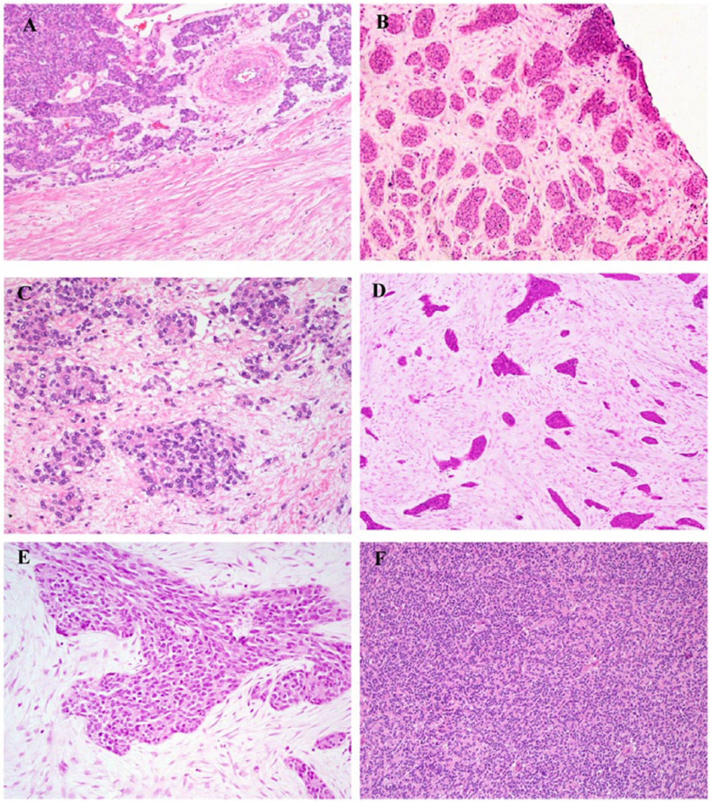
Desmoplastic small round cell tumor (DSRCT). These neoplasms are associated with a characteristic histologic appearance, typically comprising variably sized and sharply demarcated hypercellular nests and islands of uniform small cells with round or ovoid hyperchromatic or vesicular nuclei, inconspicuous nucleoli, minimal amounts of cytoplasm and indistinct cell borders. There can be marked variation in the distribution of the small cell component and the stroma (A), which can lead to sampling error in biopsy specimens. The sharp interface between cell nests and the surrounding stroma is often striking (1D). DSRCT has a spectrum of morphologic appearances, and can show rosette formation (C), or sometimes a more uniformly solid appearance (F), mimicking other small round cell neoplasms such as Ewing sarcoma.
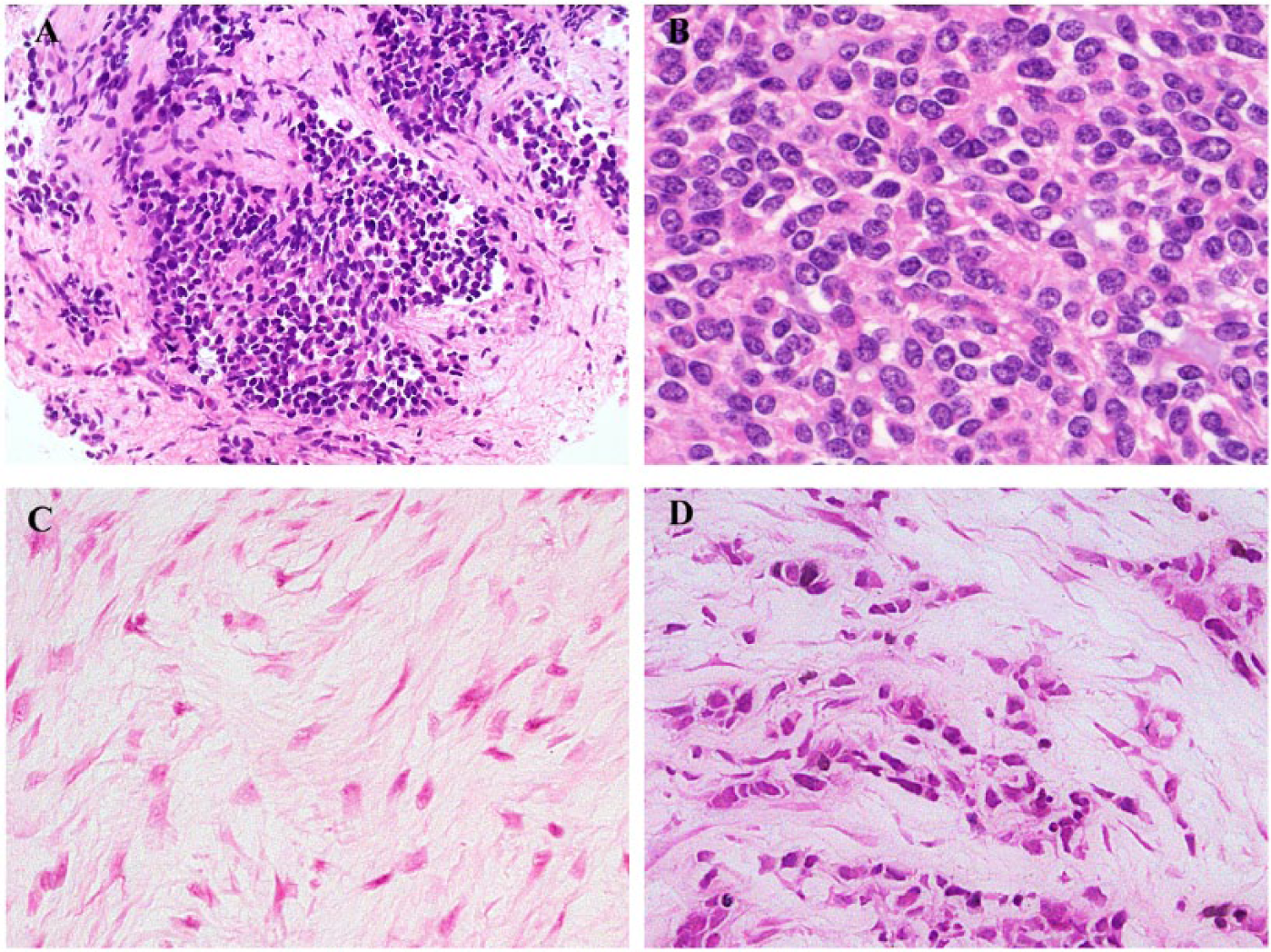
(A, B) The cells of desmoplastic small round cell tumor (DSRCT) are typically uniform and round to ovoid, with small hyperchromatic or vesicular nuclei with evenly dispersed chromatin and scanty cytoplasm. (C) The stroma of DSRCT is relatively cellular and contains spindle-shaped fibroblasts and smooth muscle actin-positive myofibroblasts in loosely collagenous extracellular matrix. (D) Other rarer variant morphology seen in DSRCT includes cells dispersed in single rows within the stroma in a pattern reminiscent of lobular breast carcinoma.
Up to a third of DSRCTs have been described to show a wide range of morphologic appearances. 6 Occasional examples show greater amounts of cytologic atypia with larger nuclei or even giant bizarre nuclei, 69 and rarely, tumors can be predominantly composed of cells with atypical nuclei or spindled morphology. 6 The large cell variant predominantly comprises large epithelioid cells with areas of anaplasia mimicking metastatic carcinoma, 52 and some neoplasms contain greater amounts of cytoplasm, which can show clearing or vacuolation. There can also be rosette (Figure 1C) or tubule formation, 70 and focally, tumor cells may have intracytoplasmic eosinophilic inclusions composed of bundles of intermediate filaments 71 giving a rhabdoid appearance. Some tumors show focal epithelial differentiation, with the presence of glands or pseudoglands, and single rows of cells may also be present within the stroma in a pattern reminiscent of lobular breast carcinoma (Figure 2D).
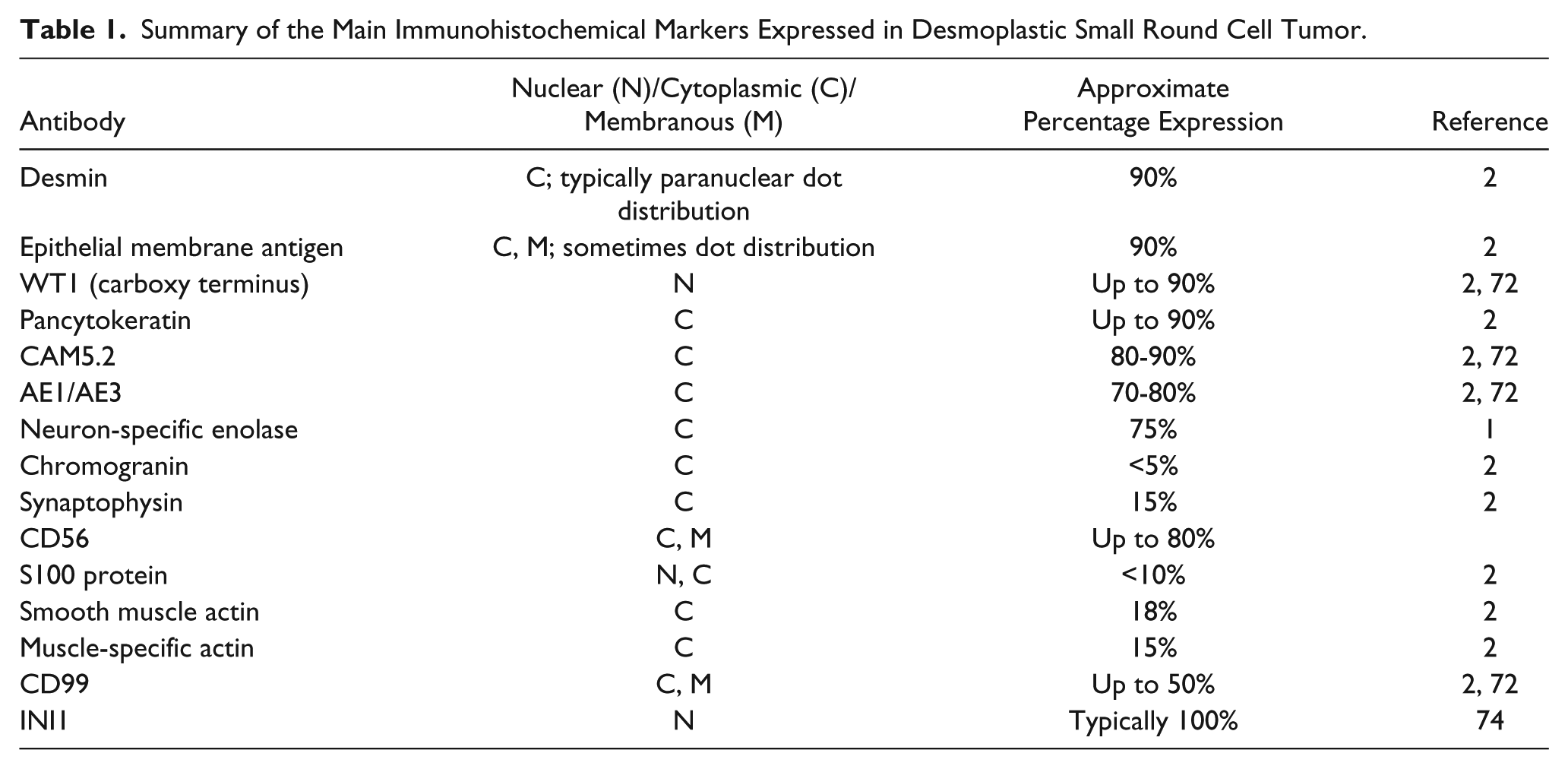
DSRCT classically has a polyphenotypic immunoprofile, variably expressing markers toward several cell lineages (Table 1), although not all antigens are expressed in every neoplasm, and it is important to note that commonly expressed markers such as desmin and cytokeratin may be negative 71 (or negative in the tissue available on diagnostic core biopsy specimens). There is expression of desmin in the large majority of cases (Figure 3A-B), 2 epithelial membrane antigen (EMA) in the majority, 2 cytokeratins (including AE1/AE3 and CAM5.2 (Figure 3C-D), although not CK5/6 or CK20) and neural markers, including neuron-specific enolase (NSE) and CD57 in most cases, 2 and smaller numbers expressing chromogranin, synaptophysin, CD56 (Fig. 3E), neurofilament protein and S100 protein.2,6 Small numbers of cases may also express variable muscle-specific actin or α-SMA. 2 Desmin expression is typically in a paranuclear dot distribution. Dot expression can also be seen with keratin, and positivity for both desmin and cytokeratins can be diffuse, with the most sensitive myogenic and epithelial markers shown to be desmin and CAM5.2. 72
Summary of the Main Immunohistochemical Markers Expressed in Desmoplastic Small Round Cell Tumor.
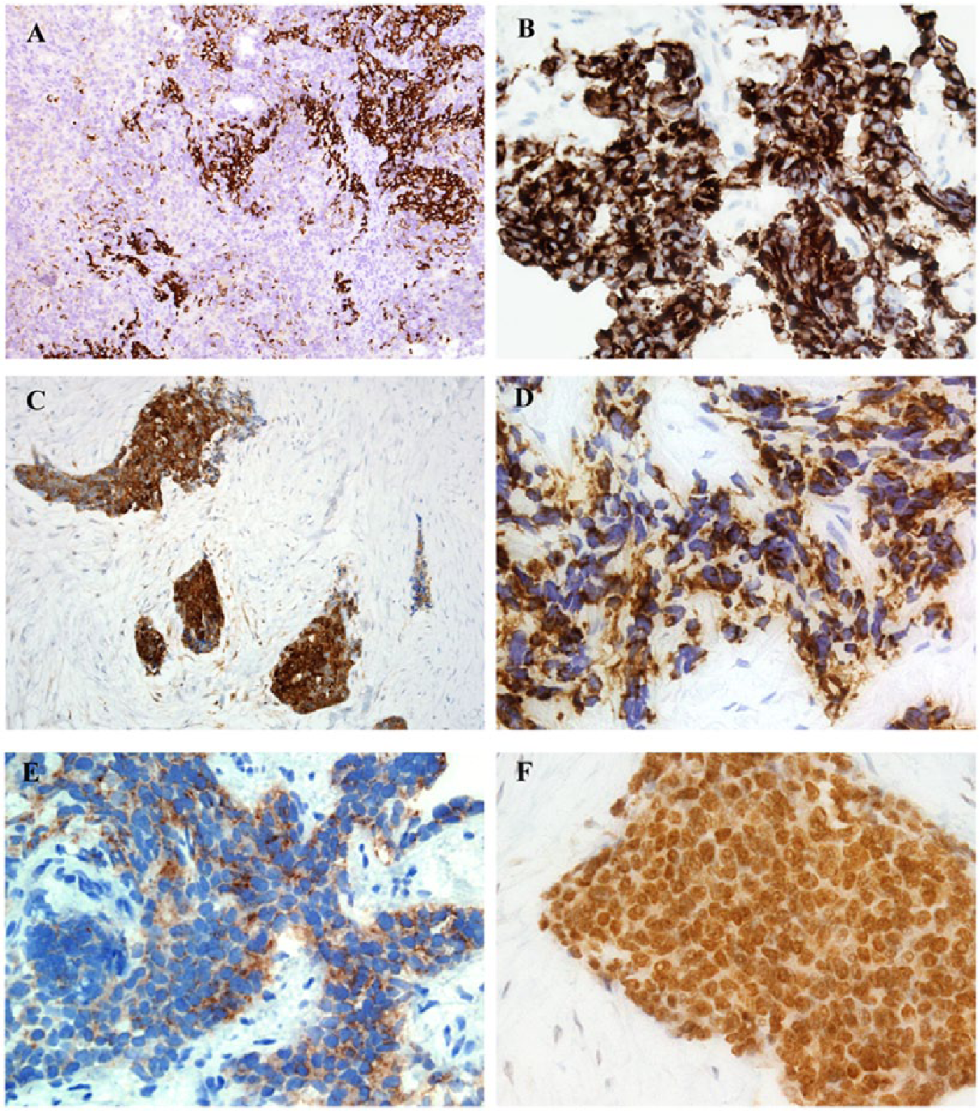
The large majority of desmoplastic small round cell tumors (DSRCTs) express desmin, although this is to variable degrees, from focal to diffuse, and often in a dot-like distribution. (A) Areas of prominent strong desmin expression (right of field), but this is patchier in other areas (left), and desmin positivity may be scanty or absent in small samples or biopsy material. (C, D) Most DSRCTs are positive for cytokeratins, including AE1/AE3 (C) and CAM5.2 (D). The AE1/AE3 expression in this example is diffuse, while the CAM5.2 expression is more focal and also dot-like in areas. CAM5.2 has been shown to be the most sensitive epithelial marker for DSRCT. (E) Most DSRCTs also show variable expression of neural or neuroendocrine markers, including CD56. (F) DSRCT is immunoreactive for antibodies selectively directed toward the carboxy terminus of the Wilms tumor (WT1) protein in two-thirds to up to almost 90% of cases.
DSRCT is immunoreactive for antibodies selectively directed toward the carboxy terminus of the Wilms tumor (WT1) protein (Figure 3F) in two-thirds to up to almost 90% of cases.2,72 CD99 is typically negative, but its variable expression can occur in approximately one-third to just over half of cases.2,72 Rarely, however, DSRCT can be negative for C-terminal WT1 and instead show nuclear expression with the amino (N)-terminal antibody, with atypical expression patterns due to these neoplasms harboring variant EWS-WT1 fusion transcripts with some cases expressing full length WT1.2,73 Tumors may also be positive for CD15, MOC-31, and Ber-EP4 and small numbers may express CD117, calretinin, 72 and NB84. 6 While INI1 expression is typically retained in nuclei of DSRCT, 74 widespread loss of nuclear expression (ie, in >90% of neoplastic cells) has been described in a tumor with classical histologic features and lacking rhabdoid morphology. 75 DSRCT may very occasionally express NKX2-2, a homeodomain transcription factor which is involved in neuroendocrine/glial differentiation and is a downstream target of the EWSR1-FLI1 fusion oncogene, and which is a sensitive (but not wholly specific) immunohistochemical marker of Ewing sarcoma.76,77 This is important to note as most EWSR1-associated tumors are negative for NKX2-2. 76 While desmin expression is often diffuse, the early myogenic regulatory nuclear transcription factors myogenin and MyoD1 are consistently not expressed. DSRCT is typically negative for CK5/6, CK20, glial fibrillary acidic protein, peripherin, CA19-9, thrombomodulin, α-fetoprotein, carcinoembryonic antigen, TAG-72 (B72.3), placental alkaline phosphatase, S100 protein, HMB45, and myoglobin. 2
Differential Diagnosis
Because of the characteristic divergent phenotype of DSRCT that overlaps with a spectrum of other round cell neoplasms, the differential diagnosis is wide and includes Ewing sarcoma, rhabdomyosarcoma, carcinoma and small cell mesothelioma. Diagnosis can be more challenging in core biopsy specimens, as some of the distinctive features such as the prominent stromal pattern may not be easily appreciable, and the full immunophenotypic spectrum may not be represented in limited material. Multifocal cytokeratin expression in the context of an abdominal or pleural neoplasm can lead to diagnostic confusion with sarcomatoid carcinomas and mesotheliomas. As up to one-third of DSRCT have been described to display a wide range of morphologic features, 6 it is important to have an index of suspicion for less common histologic features. 6 While EWSR1-WT1 fusions are no longer entirely specific for DSRCT, these are absent in the neoplasms in the principal differential diagnosis described below.
DSRCT can often be mistaken for Ewing sarcoma, which can arise within the retroperitoneum. The cells have similar cytologic features, a proportion of DSRCTs express CD99, Ewing sarcomas can express cytokeratins and rarely desmin, and the characteristic prominent stroma may not be represented in biopsy specimens. Additionally, Ewing sarcoma can sometimes show surrounding desmoplastic, fibrotic, or sclerosing hyalinized stroma, and its adamantinoma-like variant displays nests of cells with peripheral palisading and a host desmoplastic response. However, DSRCTs typically show more variable expression of CD99, rather than the diffuse membranous positivity typical of Ewing sarcoma. Ewing sarcoma lacks WT1 positivity, 78 and most tumors contain characteristic translocations involving EWSR1 and the ETS family of transcription factors, particularly those generating EWSR1-FLI1 and EWSR1-ERG fusions.
An emerging group of aggressive, primitive round cell neoplasms resemble Ewing sarcoma but lack evidence of EWSR1 gene rearrangements or other gene rearrangements associated with small round cell neoplasms such as FOXO1, DDIT3, or SS18. CIC-DUX4 gene fusion appears to be the most common genetic abnormality in these undifferentiated round cell neoplasms 79 ; it has been demonstrated in up to two-thirds of these round cell tumors lacking EWSR1 rearrangements in children and young adults, particularly in males,79-81 but the most frequent site of these is within the limbs. More rarely, CIC-FOXO4 fusions are harbored by similar undifferentiated Ewing-like sarcomas,82,83 including those with desmoplastic stroma. 82 CIC-DUX4 sarcomas can have similar morphology to DSRCT and may cause diagnostic confusion as they are usually strongly positive for WT1, 78 and may occasionally be focally positive for desmin, cytokeratin, EMA, and S100 protein, 80 but these may be associated with a slightly greater degree of morphologic heterogeneity and pleomorphism, greater prominence of nucleoli, spindle cell elements and myxoid changes and generally have minimal or absent intervening collagen.79,84 Another group of aggressive undifferentiated round cell sarcomas arising in adolescents or young adults with an apparent male preponderance is associated with BCOR-CCNB3 gene fusions, and while these occur most frequently within bones, some arise in the deep soft tissues.85,86 These tumors express nuclear CCNB3 immunohistochemically and this is thought to be a relatively specific marker, 85 with about 90% also expressing bcl-2 and approximately two-thirds showing positivity for CD117 and CD99.85,86
Poorly differentiated synovial sarcomas (SS) are composed of monotonous sheets or fascicles of relatively uniform ovoid to rounded cells with focal expression of cytokeratin and EMA, and have been documented in abdominal or retroperitoneal sites with bulky disease, which can cause confusion with DSRCT.23,87,88 Diffuse, moderate to strong nuclear expression of TLE1 is seen in the majority of SS,89,90 but is not associated with DSRCT. In addition, SS has a specific t(X;18)(p11.2;q11.2) translocation, in which the SS18 gene on chromosome 18 fuses with one of the SSX genes located on the X chromosome (usually SSX1 or SSX2), which remains specific for synovial sarcoma.
Clear cell sarcoma (CCS) and clear cell sarcoma–like tumor of the gastrointestinal tract (CCSLGT) are both associated with EWSR1 rearrangements, and can mimic DSRCT as these are both aggressive, infiltrative neoplasms that can present with disseminated intra-abdominal disease and often arise in young adults.64,91 These are characteristically centered in the muscularis propria of the stomach or bowel, with secondary extension into the submucosa and subserosa. CCS typically comprises nests of bland, uniform rounded cells often with prominent regular nuclei and moderate amounts of clear to eosinophilic cytoplasm (rather than the scanty cytoplasm of DSRCT), separated by thin fibrous septa, and sometimes there are tumoral giant cells and melanin pigment. CCSLGT are usually composed of relatively monomorphic medium to large epithelioid or ovoid cells with variable amounts of pale eosinophilic or sometimes clear cytoplasm and centrally sited round vesicular nuclei, and may contain macronucleoli, or show cellular spindling and pleomorphism. An additional distinctive feature is CD68-positive, multinucleated osteoclast-like giant cells.92-94 CCS typically diffusely express S100 protein, with most also positive for HMB45, MelanA, and MiTF, while CCSLGT express S100 protein but are negative for HMB45 and MelanA. 64 Most CCS are associated with EWSR1-ATF1 fusion transcripts and EWSR1-CREB1 in smaller numbers, whereas CCSLGT typically harbor EWSR1-CREB1 or sometimes EWSR1-ATF1 fusions; neither of these gene fusions are associated with DSRCT.
Both alveolar and embryonal rhabdomyosarcomas occur in a young patient population. Alveolar rhabdomyosarcoma (ARMS) is a round cell neoplasm and embryonal rhabdomyosarcoma (ERMS) is composed of spindled to ovoid cells so when the latter predominates can simulate a small round cell tumor. Both express desmin and can mimic DSRCT, particularly ARMS in which cellular nests are separated by prominent fibrous septa that can resemble the stroma of DSRCT. ARMS occur predominantly within the extremities, as well as the trunk and head and neck. While ARMS may occur in the abdominal cavity with widespread metastatic disease, primary occurrence in the abdominal cavity is unusual. Desmin expression is typically diffuse and strong, and nuclear positivity for myogenin and MyoD1 is usually also widespread, the latter in contrast to DSRCT in which these are always absent. The majority of ARMS also harbor PAX3/7-FOXO1 gene fusions, which are specific for these neoplasms. Malignant peripheral nerve sheath tumor (MPNST) has a large range of morphologies, including rarely of small cells, but MPNSTs occur more frequently in patients with neurofibromatosis type-1 and may be seen to originate from a preexisting benign nerve sheath neoplasm (most often neurofibroma) or from a nerve. Histologically, MPNSTs show at least focal atypia, and tend to display at least focal areas of cells with “nerve sheath” morphology, with elongated, buckled, or tapered hyperchromatic nuclei. 95
The presence of dot keratin expression in DSRCT may lead to confusion with metastatic small cell carcinoma or Merkel cell carcinoma, particularly as DSRCT can rarely be composed predominantly of large epithelioid cells. 52 Both of these neoplasms characteristically occur in an older patient population, and in addition there may be clinical or radiologic evidence of a primary site of carcinoma. Small cell carcinoma often shows nuclear molding and sometimes a stippled chromatin pattern. Carcinoma may also express lineage-specific markers such as TTF-1, but is negative for desmin and WT1. With DSRCT occuring in the peritoneum, tunica vaginalis or less frequently within pleura, 96 mesothelioma is part of the differential diagnosis, but the morphology is usually markedly different, and the absence of CK5/6 and thrombomodulin expression in DSRCT, along with expression of CD15, MOC-31, and Ber-EP4 argue against a mesothelial neoplasm. 2
Intra-abdominal disease can affect the ovaries including bilaterally, so that ovarian tumors can be thought to represent the primary neoplasm. 97 Ovarian small cell carcinoma of hypercalcemic type (SCCOHT) can closely resemble DSRCT. Recent whole-exome sequencing has shown that virtually all SCCOHTs lack functional SMARCA4/BRG1 98 ; this gene and the related SMARCB1 gene are mutated in almost all atypical teratoid/rhabdoid tumors and malignant rhabdoid tumors, such that it has been proposed that SCCOHT should be renamed “malignant rhabdoid tumor of the ovary.” 98 All ovarian SCCOHTs in a recent study have been shown to lack immunohistochemical BRG1 expression and expressed INI1. Morphologic mimics of SCCOHTs, including DSRCT, showed nuclear positivity for BRG1, with variable staining proportions (with heterogeneous positivity in DSRCT). 99 DSRCT and blastemal-predominant Wilms’ tumor (WT) can share similar histologic features, 100 with coexpression of desmin and cytokeratin, and paranuclear dot-like desmin expression can be seen with equal frequency in DSRCT and blastemal-predominant WT, causing diagnostic confusion. 100 The best methods of distinguishing the two are the detection of EWSR1-WT1 fusion transcripts and selective immunoreactivity for carboxy-terminus WT1 for DSRCT, and dual immunoreactivity for both amino and carboxy termini of WT1 for WT. 100
Neuroblastoma also typically arises within the retroperitoneum and small numbers of DSRCT can express NB84. Neuroblastoma occurs in a younger patient population; while it can rarely arise in adults, it predominantly presents between the ages of 18 and 21 months, with most diagnosed by 5 years. Clinically, there may be a recognizable site of origin within the sympathetic ganglia, the adrenal medulla or the organ of Zuckerkandl, and patients may have elevated urinary catecholamines and associated metabolites. The cells of neuroblastoma tend to be medium-sized and generally show more atypia than the uniform cells of DSRCT, and often there is a prominent lobular architecture. In addition, neuroblastoma often shows at least focal palely eosinophilic neurofibrillary matrix resembling neuropil as well as variable ganglionic differentiation. Most neuroblastomas consistently express NB84, neurofilament protein, chromogranin, synaptophysin and CD56. Epithelioid GISTs occur in intra-abdominal, retroperitoneal or pelvic sites, and are usually composed of sheets of rounded cells that may be sometimes relatively small and uniform, resembling DSRCT. Epithelioid GIST may show larger cells with clear cytoplasm, or more spindled cells in other areas, without intervening collagenous stroma. While this is patchier in epithelioid GIST, these still typically express CD117 and DOG1 at least focally, along with CD34 and sometimes h-caldesmon, none of which are typically positive in DSRCT. Most GISTs also contain KIT or less frequently PDGFRA mutations that are not described in DSRCT. Endometrial stromal sarcomas (ESS) occur in older female patients, and comprise cellular distributions of closely packed cells in sheets and nests, which can resemble DSRCT. Low-grade ESS can also show variable expression of cytokeratin and desmin, leading to further diagnostic confusion. Low-grade ESS typically has an architectural pattern of large nests, sheets and nodules rather than irregular islands of cells in desmoplastic stroma, and is associated with small thick-walled vessels. It typically shows diffuse positivity for CD10 as well as estrogen and progestogen receptors (ER and PgR), and harbors characteristic fusions including JAZF1-SUZ12, JAZF1-PHF1, and EPC1-PHF1. High grade ESS also typically lacks the intense desmoplastic stroma of DSRCT, may show areas of larger, mildly pleomorphic round or ovoid cells 101 and is associated with YWHAE-NUTM2 gene fusions.
Conclusions
DSRCT is a highly aggressive round cell neoplasm predominating in adolescent and young adult patients, associated with a characteristic EWSR1-WT1 gene fusion, which still has an almost universally fatal clinical outcome. While it has a characteristic histologic appearance, it can show a spectrum of morphologic variation. Correct recognition is crucial, not only because of the difference in prognosis between DSRCT and other round cell neoplasms but also because of the potential for designing therapies toward the molecular targets of the EWSR1-WT1 fusion in the near future, which is particularly critical as conventional chemotherapeutic agents have only shown severely limited efficacy in treating either local or metastatic disease. Specifically pertinent to the surgical pathologist is the fact that the morphology overlaps with a variety of neoplasms of different lineages, which is compounded by the polyimmunophenotypic but variable immunoprofile, the potential for occurrence at unusual sites and the demonstration that the EWSR1-WT1 fusion is no longer specific for DSRCT. An index of suspicion is therefore required in diagnosing DSRCT, as well as knowledge of the widespread variation in histologic and immunohistochemical features. Confirmatory molecular diagnosis with RT-PCR in the context of appropriate morphologic and clinical findings is optimal, as the finding of EWSR1 rearrangement alone is not sufficient for confirmation of the diagnosis due to the ability of the EWSR1 gene to combine with other genes to generate other neoplasms in the differential diagnosis.
Footnotes
Declaration of Conflicting Interests
The author(s) declared no potential conflicts of interest with respect to the research, authorship, and/or publication of this article.
Funding
The author(s) disclosed receipt of the following financial support for the research, authorship, and/or publication of this article: We acknowledge support from the NIHR Royal Marsden/ICR Biomedical Research Centre.