






Abstract
Background:
Standard open calcaneal osteotomies to correct hindfoot malalignment have been associated with high complication rates, including nerve damage and wound infection. This has resulted in the development of minimally invasive techniques such as the percutaneous endoscopically assisted calcaneal osteotomy (PECO), which in cadaver studies has been shown to be potentially safer than open surgeries. The aim of this study was to demonstrate the safety and effectiveness of PECO in a clinical setting, with regard to neurovascular injury rates, infection, and short-term radiographic and functional outcomes.
Methods:
Forty-one (41) patients with planovalgus or cavovarus foot deformities underwent treatment using PECO. Outcomes were analyzed at 6 months and primarily included neurovascular outcomes and wound infection rates. Secondarily, radiographic and functional (visual analog scale for pain [VAS], 36-Item Short Form Medical Outcomes Survey [SF-36], and Foot Function Index [FFI] scores) outcomes were also assessed.
Results:
There were no reported cases of superficial wound infections, lateral calcaneal or sural nerve dysesthesia. Minor complications occurred in 6/41 feet. The mean postoperative hindfoot correction was 8.3 ± 2.2 mm (range: 6-15mm) compared to preoperative status. Compared to preoperative status, significant improvements (P = .001 for all) were seen in the VAS, SF-36, and FFI at 6 months postoperatively.
Conclusions:
PECO resulted in minimal complications with no lateral calcaneal or sural nerve dysesthesias and no wound complications. It also resulted in significant improvements in postoperative radiographic and functional outcomes from baseline to 6-months postoperatively, demonstrating its use as a safe and effective means of treating hindfoot malalignment.
Level of Evidence:
Level IV, retrospective case series.
Keywords
Calcaneal displacement osteotomies are commonly used by surgeons to correct hindfoot deformity and restore the weight-bearing axis of the foot in isolation or in conjunction with other procedures. Depending on the pathology being addressed, medializing or lateralizing calcaneal osteotomies are the most frequently performed to correct valgus or varus deformities respectively. By restoring foot alignment and the weight bearing axis it has been shown that not only are peak pressures diminished in the hindfoot, but functional outcomes can be significantly improved in a variety of pathologies.2,10,11
When performing calcaneal osteotomies, open techniques using a lateral oblique incision have traditionally been considered the mainstay of operative treatment.6,9,12,13 Not only are open approaches associated with rates of neurovascular injury and wound complications that range from 5% to 28%, but they can often adversely affect concomitant incisions required for other elements of hindfoot reconstructions.1,7,22 In an attempt to reduce these operative complications, promote early recovery, and improve function, minimally invasive techniques (MIS) to perform calcaneal osteotomies have recently been developed with promising results.7,8,19 One of these is the percutaneous endoscopically assisted calcaneal osteotomy (PECO), which utilizes 3 portals: 2 placed on the lateral calcaneal tuberosity (distal and proximal) and 1 superomedial to the calcaneal osteotomy. In a cadaveric study on PECO, all important neurovascular structures in the hindfoot were a mean distance of approximately 12 mm from the nearest operative portal or osteotomy cut, potentially decreasing injury to these structures. 22 More specifically, this study also showed that less than 10% of lateral calcaneal nerve branches were injured using PECO, compared to a potential 80% injury rate using an open technique; however, this has not yet been tested in a clinical setting. 22
As such, the principal aim of this study was to determine the clinical rate of neurovascular injury, specifically the lateral calcaneal and sural nerves, and wound infection rates with the use of the PECO technique, as a follow-up to our initial anatomic cadaveric study. 22 The secondary aim was to determine whether PECO was associated with improved short-term postoperative radiographic and functional outcomes.
Methods
This study is a retrospective review of all patients who underwent calcaneal osteotomies using the PECO technique between March 2011 and May 2013. The surgeries were performed by a single surgeon at a tertiary care academic center for correction of either planovalgus or cavovarus foot deformities that had failed conservative treatment for at least 6 months. All patients were followed up for a minimum of 6 months, and patients who had undergone previous hindfoot surgeries were excluded. This study was approved by the Institutional Review Board.
Forty-one (41) patients were included in the data analysis. Patients included 20 males and 21 females. The average age of the patients was 41.1 ± 16.4 years (range: 18-68 years), and all patients where followed for 6 months.
PECO Surgical Technique
The full technical details of the PECO technique have previously been described. In short, it uses 3 surgical portals, with 2 placed laterally and 1 medially (1 superior to the lateral calcaneal tuberosity and anterior to the Achilles, 1 inferior to the lateral calcaneal tuberosity and anterior to the plantar fascia insertion, and 1 superior to the medial calcaneal tuberosity and anterior to the Achilles insertion but posterior to the neurovascular bundle) (Figure 1). A 4-mm dry scope cannula with blunt trochar and a suture threaded through its distal eyelet is then used with the surgical portals established to guide sutures that shuttle a Gigli saw from the superolateral portal safely around the medial calcaneal tuberosity and out the inferior lateral portal, touching the periosteum of the dorsal, medial, and plantar calcaneus (Figure 2). Generally, a straight oblique cut is performed, but if the operator is facile with the technique, a dome osteotomy can also be created by changing the Gigli saw angle through the arc of the cut. Completion of the osteotomy is confirmed by an easily mobile posterior calcaneus. A ¼-inch osteotome is then used to gently rotate and distract the osteotomized calcaneus in order to translate the tuberosity either medially or laterally as required to correct the hindfoot to an anatomic axis in line with the tibia. Two percutaneous cannulated 6.5-mm screws are inserted across the osteotomy site from posterior inferior to anterior superior in the calcaneal osteotomy with start points proximal to the weight bearing portion of the calcaneus and distal to the Achilles tendon insertion.
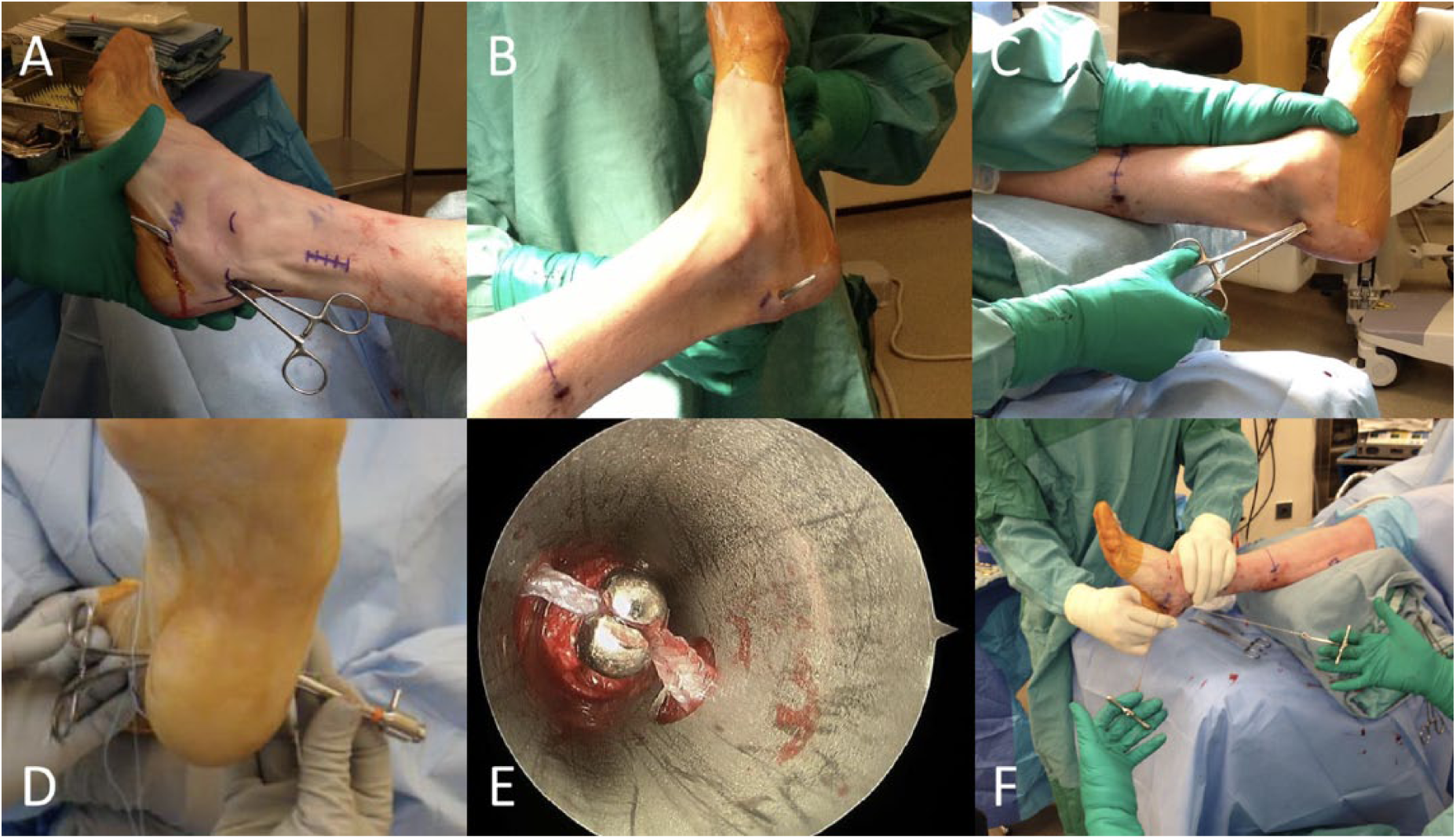
Percutaneous endoscopically assisted calcaneal osteotomy (PECO) Technique. (A) Two lateral portals with a blunt hemostat creating a tunnel directly over the lateral calcaneal cortex. (B) Creation of the medial portal. (C) Dissection on bone and placement of right-angle clamp. (D) Placement of endoscopic cannula. (E) Endoscopic retrieval of the shuttling suture loop via a right-angle clamp. (F) The Gigli saw is then passed using this suture loop, and the osteotomy is completed.21,22
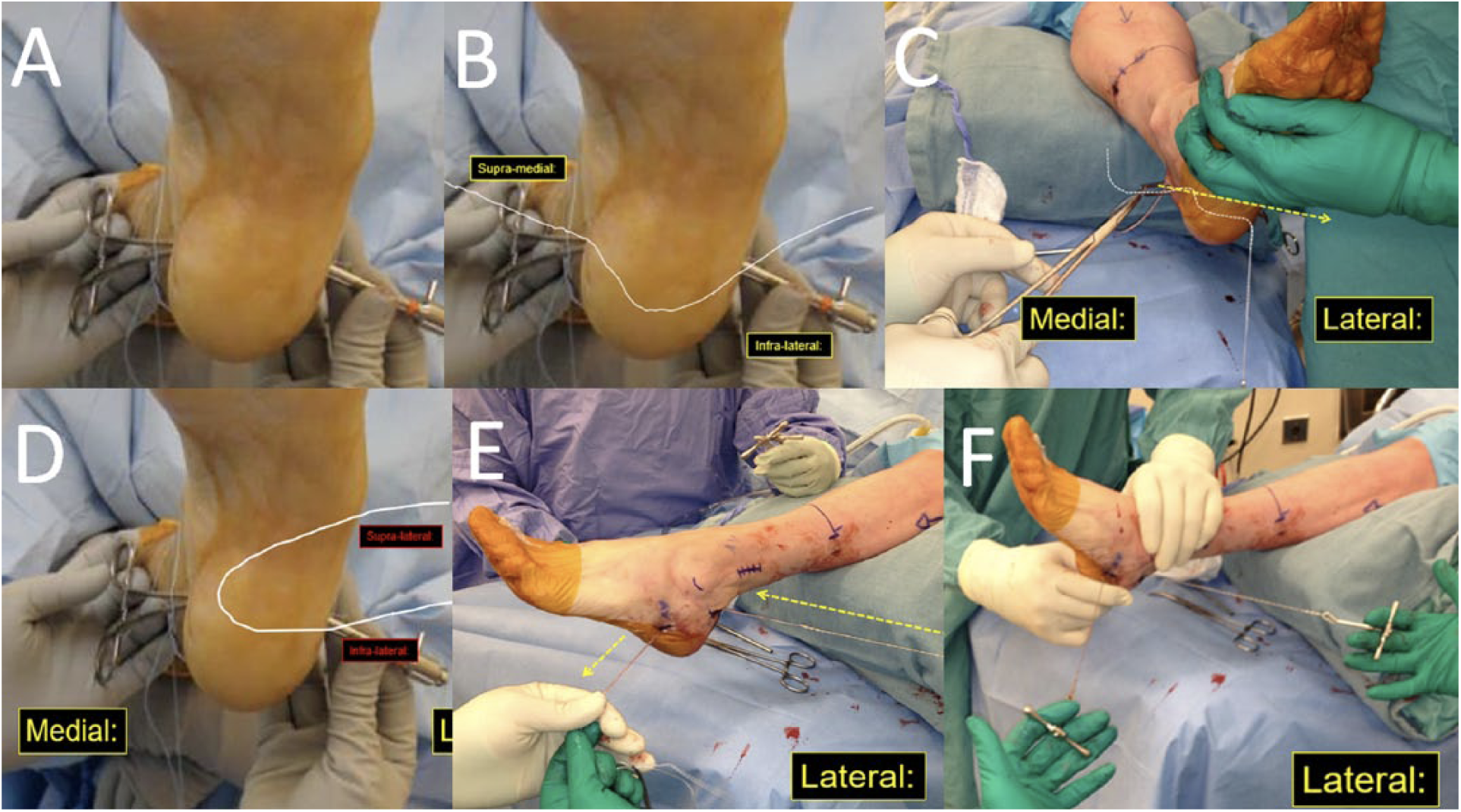
Technical details of how the Gigli saw is passed around the calcaneus. White lines represent the suture shuttles and the dotted lines with arrows the direction of pull. (A) Initial endoscopic suture retrieval (Figure 1C). (B) The suture shuttle is then taken out the medial portal. (C) The suture shuttle is then retrieved from the medial side and taken out the superolateral portal. (D) Location of suture shuttle once passed around the calcaneus. (E) Suture shuttling Gigli saw around calcaneus. (F) Gigli saw in situ ready for osteotomy.
Outcomes
Operative complications of PECO were collected, with primary outcomes measures being neurovascular injury and wound infection rates at 6 months. Secondarily, functional outcomes were assessed preoperatively and 6 months postoperatively and included the visual analog scale for pain (VAS), 36-Item Short Form Medical Outcomes Survey (SF-36), 24 and Foot Function Index (FFI) 3 scores.
Radiographic outcomes were also assessed for union across the osteotomy site and the magnitude of calcaneal shift. Translation was measured by using the difference between preoperative (Figure 3A) and 6-month postoperative hindfoot alignment views (Figure 3B) 17 (measuring the perpendicular distance between the longitudinal mid axis of the tibia and the lowest point on the calcaneus) and using intraoperative axial calcaneal views (Figure 3C).
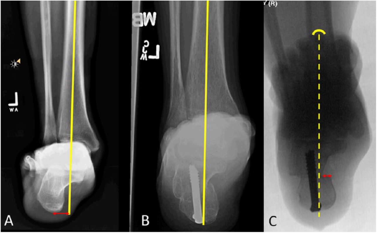
Radiological measurements of calcaneal translation. (A) Preoperative radiograph. Arrow represents the distance from the midaxial line to the lowest weight-bearing portion of the calcaneus. (B) Postoperative radiograph at 6 months. Central weight-bearing portion matches the midaxial line. (C) Intraoperative fluoroscopy. Midaxial line (dotted) positioned from second ray to central portion of the calcaneus. Translation measured as distance from medial proximal wall to medial distal wall of calcaneus (arrow).
Statistical Methods
Data was summarized using descriptive statistics and presented as mean ± standard deviation (SD) unless otherwise noted. All of the data was tested for normality. The data from the SF-36 and FFI were not normally distributed, but the VAS data was normally distributed. The Wilcoxon sign rank test was used to compare between pre- and postoperative outcome results for non-normally distributed data. The paired t test was used to compare pre- and postoperative outcome results for normally distributed data. Comparisons were based on the means of the pre- and postoperative SF-36, VAS, and FFI (pain, disability, activity limitations, and total scores) data. Significance was established at P <.05 for both the Wilcoxon sign rank and paired t test.
Results
Surgical Complications
There were no reported cases of superficial wound infections nor sural or lateral calcaneal nerve dysesthesia (Table 1). Other complications from the PECO technique occurred in 6 of 41 feet (14.6%) The complications were as follows: (1) painful scar at the medial portal in 1/41 feet (2.4%); (2) painful hardware that required hardware removal in 4/41 feet (9.8%); and (3) plantar nerve symptoms in 1/41 feet (2.4%). All the plantar nerve symptoms resolved with conservative treatment at 3 months postoperatively. There was no distinct difference in sural nerve injuries between lateralizing and medializing calcaneal osteotomies and no reported cases of persistent tarsal tunnel syndrome.
Complications From the PECO Surgical Technique.
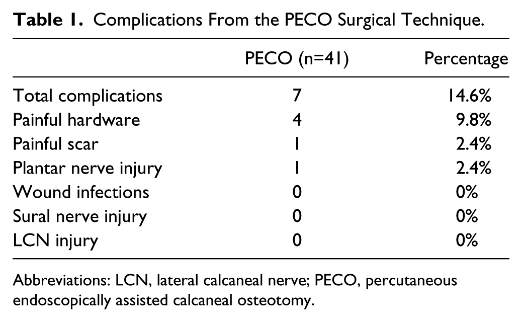
Abbreviations: LCN, lateral calcaneal nerve; PECO, percutaneous endoscopically assisted calcaneal osteotomy.
Radiographic Outcomes
The mean intraoperative translation was measured at baseline in 38/41 patients (92.7%) and was 8.5 ± 1.7 mm (range: 6-15 mm). Postoperative hindfoot alignment techniques were used to assess the mean improvement in hindfoot alignment 6 months after surgery in 38/41 patients (92.7%). The mean postoperative hindfoot correction was 8.3 ± 2.2 mm (range: 6-15 mm) with the resultant anatomic hindfoot alignment in the majority within 1 standard deviation of the norm compared with the malaligned preoperative status. There were 3 cases that required corrections that were beyond the normal range (1 lateral side [15.2 mm], and 2 medial side [15.4 mm and 15.2 mm, respectively]). There was no case of undercorrection. There was no significant difference in outcomes between patients who had corrections beyond normal range and patients with overcorrection (P > .05 for all variables). There were no cases of radiographic nonunion.
Functional Outcomes
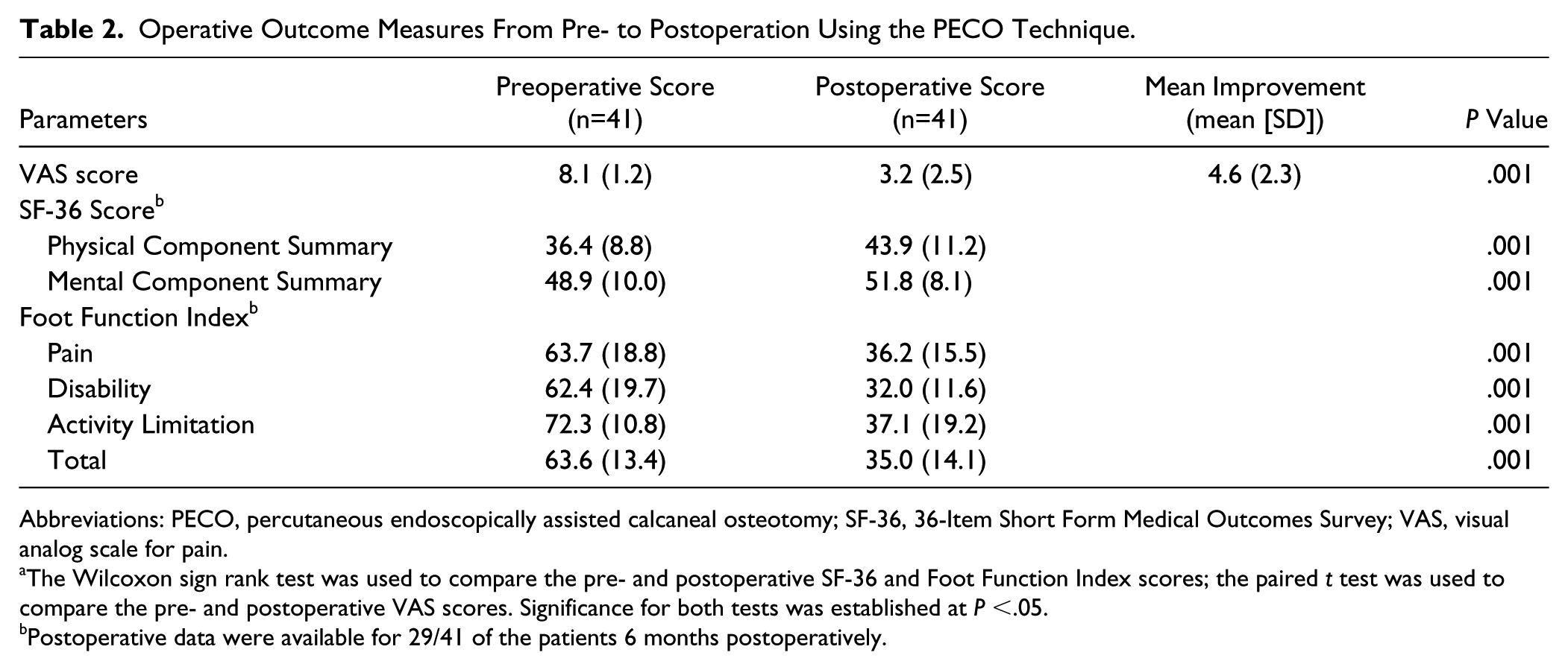
To assess the functional outcomes of PECO the VAS (n=41/41; 100%), SF-36 (n=29/41; 70.7%), and FFI (n=29/41; 70.7%) scores from 6 months postoperatively were compared to preoperative scores (Table 2). The highest possible score (indicating most severe pain) on the VAS scale was 10. The mean preoperative VAS score (8.1 ± 1.2) was significantly improved postoperatively (3.2 ± 2.5; P = .001).
Operative Outcome Measures From Pre- to Postoperation Using the PECO Technique.
Abbreviations: PECO, percutaneous endoscopically assisted calcaneal osteotomy; SF-36, 36-Item Short Form Medical Outcomes Survey; VAS, visual analog scale for pain.
The Wilcoxon sign rank test was used to compare the pre- and postoperative SF-36 and Foot Function Index scores; the paired t test was used to compare the pre- and postoperative VAS scores. Significance for both tests was established at P <.05.
Postoperative data were available for 29/41 of the patients 6 months postoperatively.
Two components of the SF-36 questionnaire were scored reflecting physical (PCS) and mental (MCS) component health scores (Table 2). The mean preoperative SF-36 PCS score (36.4 ± 8.8) was significantly improved postoperatively (48.9 ± 10.0; P = .001) as was the mean MCS score (43.9 ± 11.2 vs 51.8 ± 8.1; P = .001). This is indicative of improved physical and mental health in patients after undergoing PECO surgery.
Foot function was assessed pre- and postoperatively using 4 measures: (1) pain, (2) disability, (3) activity limitation, and (4) total FFI score (Table 2). The mean preoperative pain, disability, activity limitation, and total scores were 63.7 ± 28.8, 62.4 ± 19.7, 72.3 ± 10.8, and 63.6 ± 13.4, respectively, which decreased to 36.2 ± 15.5, 32.0 ± 11.6, 37.1 ± 19.2, and 35.0 ± 14.1 postoperatively (P = .001 for all). The reduction in FFI scores provided evidence of significant improvement in all 4 measures from baseline to 6 months postoperation (Figure 4).
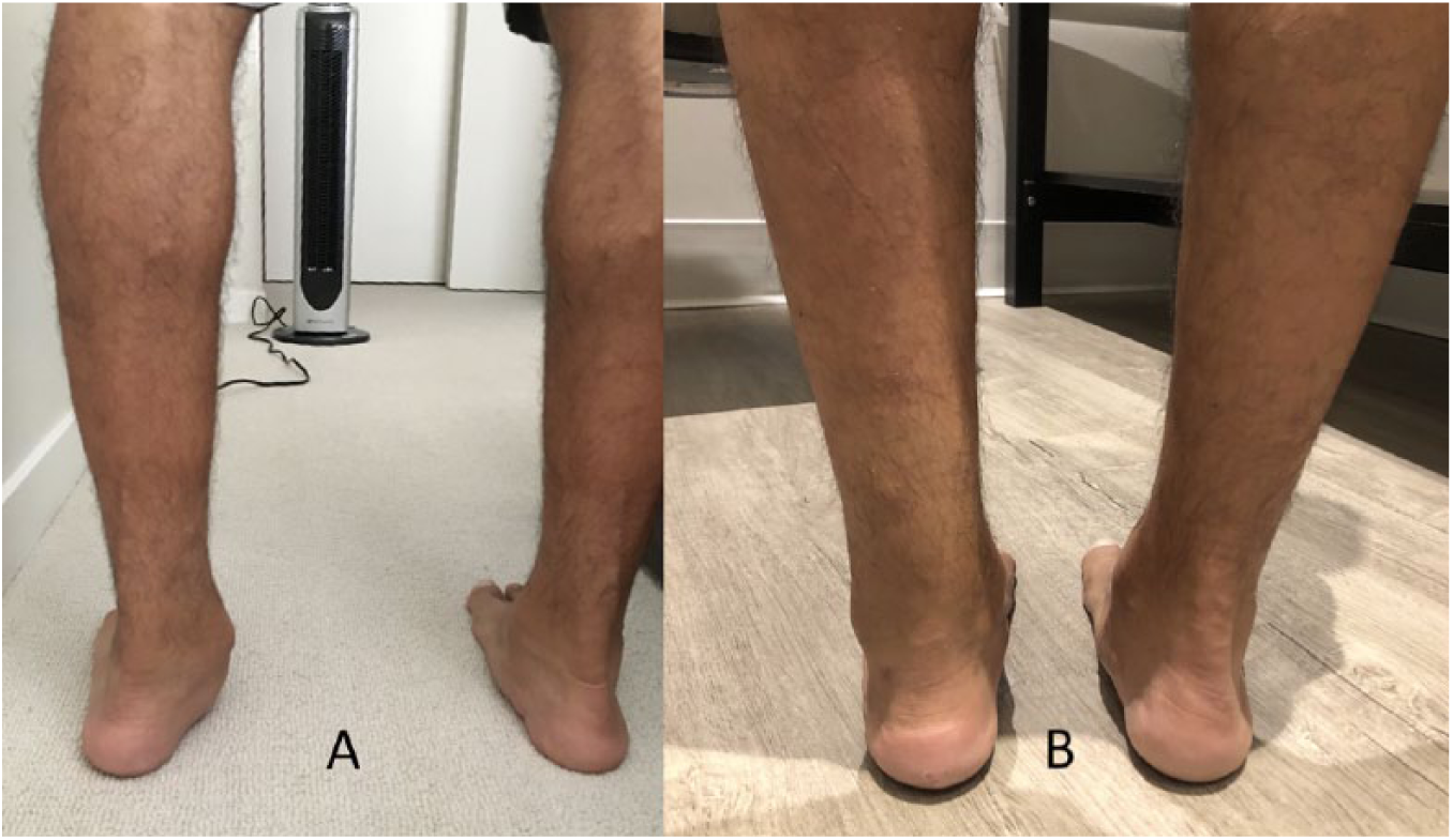
Clinical photograph of a patient with left hindfoot valgus malalignment (A) preoperatively and (B) 6 months after a medializing percutaneous endoscopically assisted calcaneal osteotomy.
Discussion
The PECO operative technique was developed as an alternative to more traditional open osteotomy techniques that while achieving good results, are known to have a high incidence of complications including neurovascular damage and wound infections.1,7 The results of this study support the findings of our previous cadaveric study 22 and show that the PECO technique results in minimal complications, including no reported cases of sural or lateral calcaneal nerve dysesthesia nor superficial wound infections. In addition, patients undergoing PECO obtained improved short-term functional and radiographic outcomes at 6 months postoperatively, including less pain, improved mental and physical health, and improvement in 4 parameters of the FFI (pain, disability, activity limitation, and total score).
This is comparable with other recent studies that have looked at the outcomes of MIS calcaneal osteotomies.7,8 Kendal et al in 2015 compared MIS to open calcaneal osteotomies in a heterogenous group of clinical conditions and looked at short-term outcomes at 30 days postoperatively. In their series, the MIS technique had fewer wound complications (6.45% vs 28%), less infection (3% vs 20%), and less sural neuropathy (0 vs 3). 7 They also demonstrated a displacement of 9.4 mm and union rate of 97% with their MIS technique, which was comparable to our findings on the PECO technique. Although they did not examine functional outcomes, all their reported outcomes are comparable to what we showed in our study. Similarly, Kheir et al in 2015 retrospectively reviewed 29 patients who had MIS calcaneal osteotomies performed primarily as part of tibialis posterior reconstruction for flexible planovalgus. They reported no wound infections or neurovascular injuries with union rates of 100% and restoration of hindfoot alignment in all cases. 8
The mean postoperative correction achieved with PECO in this study was comparable to previous reports for both minimally invasive and open osteotomies. 7 Although we observed a wide range of corrections in terms of hindfoot realignment, ranging from 6 to 15 mm, there was no significant difference in outcomes between patients who had correction to normal range and those with correction beyond the normal range (P > .05 for all variables). However, we cannot rule out that correction beyond the normal range may have affected the operative outcomes for patients as the number of patients in this group was small. In addition, most of the patients underwent concomitant surgeries, such as planovalgus or cavovarus foot reconstruction, that may have affected operative outcome.
Calcaneal osteotomies have a high rate of patient satisfaction following surgery, 20 with up to 92% of patients being satisfied with the operation 3-5 years after surgery. 23 Short-term follow-up of calcaneal osteotomies has shown clear gains in foot function measured with the AOFAS scale. 2 In a long-term follow-up study (mean 51 months; range 38-62 months), open calcaneal osteotomies were reported to improve the AOFAS score from 48.8 preoperatively to 85.5 postoperatively, and to have a reduction in the VAS scores from 7.3 to 1.7. 23 The reported improvements in the pre- and postoperative (3-5 year) VAS scores are comparable to the short-term improvements in our study. Likewise, we showed substantial gains in foot function using the FFI. Given the similar short-term benefits of calcaneal osteotomies performed using PECO and open operative techniques, this suggests that the clinical benefits of PECO will likely also be long-term.
The lack of sural or lateral calcaneal nerve injury in this study indicates that PECO is a safe technique clinically, with a lower nerve injury rate than in our cadaveric studies. 22 It also brings to question the historical notion that lateral calcaneal pain and dysesthesia after a calcaneal osteotomy is due to lateral calcaneal wall prominence produced by displacement of the tuberosity, which is addressed in open surgery by punching the prominent bone down. 18 In the case of the PECO, the prominent wall is left in situ regardless of the amount of displacement or wall prominence, yet in this study there were no reported cases of sural or lateral calcaneal dysesthesia. The disparity in dysesthesia between the open and PECO technique may be due to the possibility that the open technique results in elevated rates of cutaneous nerve injury,5,16,22 and thus results in subsequent scarring or tethering of the injured nerve to the overlying open incision scar 20 against a prominent calcaneal wall.
Strengths and Limitations
The patient-reported instruments used in this study are widely accepted measures for assessing foot and ankle operative outcomes. The SF-36 survey is routinely used to assess the outcomes of foot and ankle surgery4,14 as is the FFI. 14 The FFI has been shown to be similar to other patient-reported measures of foot function (including but not limited to the AOFAS, Lower extremity functional scale [LEFS], and Western Ontario and McMaster Universities Osteoarthritis Index [WOMAC]) in terms of scale, reliability, and validity. 15 Therefore, the instruments were appropriate and the measured short-term operative outcome data reflects real clinical benefits to patients.
One of the limitations of this study is that the follow-up (6 months) is relatively short and it remains to be determined in a clinical setting whether the benefits of PECO are sustained over a longer period of time. This time period of 6 months is however adequate to assess the primary outcome of the presence or absence of neurovascular injury and is over a longer duration than other similar outcome studies assessing MIS calcaneal osteotomies. 7 The surgeries were also performed by a single surgeon. As such, to better understand whether the clinical gains demonstrated in this study are consistent across surgeons, a larger multisurgeon study is required.
Conclusions
The PECO technique had a low complication rate and resulted in no wound complications or injuries to the sural or lateral calcaneal nerves, which are concerns related to open procedures. In addition, PECO demonstrated significant improvement in short-term postoperative radiographic and functional (VAS, SF-36, and FFI scores) outcomes from baseline to 6 months, supporting its use as a safe and effective means of treating hindfoot malalignment in the clinical setting.
Supplemental Material
FAI800983-ICMJE – Supplemental material for Neurovascular and Clinical Outcomes of the Percutaneous Endoscopically Assisted Calcaneal Osteotomy (PECO) Technique to Correct Hindfoot Malalignment
Supplemental material, FAI800983-ICMJE for Neurovascular and Clinical Outcomes of the Percutaneous Endoscopically Assisted Calcaneal Osteotomy (PECO) Technique to Correct Hindfoot Malalignment by Andrea Veljkovic, Michael Symes, Alastair Younger, Chamnanni Rungprai, Kaniza Zahra Abbas, Peter Salat, Joshua Tennant and Phinit Phisitkul in Foot & Ankle International
Footnotes
Acknowledgements
We would like to thank Jane Kondejewski and Dr Natalie Purcell for helping us with the preparation of this manuscript.
Declaration of Conflicting Interests
The author(s) declared no potential conflicts of interest with respect to the research, authorship, and/or publication of this article. ICMJE forms for all authors are available online.
Funding
The author(s) received no financial support for the research, authorship, and/or publication of this article.
Ethics
This study obtained ethical approval from the International Review Board (IRB) at the University of Iowa Hospital and Clinics.

References
Supplementary Material
Please find the following supplemental material available below.
For Open Access articles published under a Creative Commons License, all supplemental material carries the same license as the article it is associated with.
For non-Open Access articles published, all supplemental material carries a non-exclusive license, and permission requests for re-use of supplemental material or any part of supplemental material shall be sent directly to the copyright owner as specified in the copyright notice associated with the article.
