






Abstract
Background:
A calcaneal osteotomy is a common component of surgical management for various foot and ankle deformities. Open and minimally invasive surgical (MIS) calcaneal osteotomies can be performed. The purpose of this study was to compare complication rates including infection, wound dehiscence, nerve damage, hardware failure, reoperation, and nonunion between these cohorts.
Methods:
All patients who underwent an open or MIS calcaneal osteotomy between March 2021 and July 2024 at a single institution were identified and retrospectively reviewed. All procedures were performed by one of 2 fellowship-trained orthopaedic foot and ankle surgeons on patients >18 years of age at the time of surgery. These procedures were performed in combination with other procedures to correct planus or cavus feet. Demographic information and postoperative complications were recorded for each patient with a minimum of 12 months follow-up.
Results:
Forty-four patients who underwent open calcaneal osteotomy and 26 patients who underwent MIS calcaneal osteotomy met the inclusion criteria for the current study. Patients were followed for a mean 25.3 ± 11.1 months. There was no significant difference in the rate of nonunion (P = .703) or nerve complications (P = .410) between cohorts. There were fewer postoperative minor wound complications in the MIS cohort (1/26, 3.8%) compared with the open cohort (9/44, 20.5%) (P = .036) observed in the MIS cohort; however, the P = .036 difference in minor wound complications was found to be not significant after false discovery rate (FDR) adjustment.
Conclusion:
Data from the current study indicate that open and MIS calcaneal osteotomy are associated with no difference in major complication rates or amount of calcaneal correction. With the numbers available, no significant difference in total or minor complication rates could be detected after FDR adjustment, though fewer minor wound complications were observed in the MIS cohort (3.8% vs 20.5%). Both approaches were associated with significant improvements in patient-reported function and mobility; significant improvement in pain scores was observed in the MIS cohort only after FDR adjustment.
Level of Evidence:
Level IV, retrospective cohort study.
Introduction
In patients with various foot and ankle deformities, a calcaneal osteotomy is often a necessary component of surgical correction. As the architecture of the foot functions as a tripod, the position of the calcaneus is an incredibly important factor in whole foot alignment. Modern orthopaedic foot and ankle surgeons can select from a variety of calcaneal osteotomy techniques based on patient-specific deformities.
F.C. Dwyer first popularized this surgery for deformity correction in 1959.2,6 Historically, calcaneal osteotomies are performed as an open surgery, often on the lateral aspect of the calcaneus. Like most open hindfoot procedures, these operations have been associated with a significant rate of wound complications, infection, and damage to surrounding neurovascular structures.2,7,11,12,15,19
As minimally invasive surgery techniques are increasing in popularity across orthopaedics, many have applied these techniques to operations about the hindfoot. For the correction of planovalgus and cavovarus deformities, Toepfer et al 20 found few instances of soft tissue injury (4.2%) with a percutaneous minimally invasive (MIS) calcaneal osteotomy. Similarly, Kheir et al 13 reported an excellent union rate and no soft tissue or neurovascular complications following MIS calcaneal osteotomies. Waizy et al 21 reported shorter operating times (22 vs 29 minutes) and no difference in radiation exposure (7 vs 10 cGycm2) or fluoroscopy time (10 vs 23 seconds) in 60 consecutive patients undergoing MIS vs open calcaneal osteotomies.
Although favorable outcomes have been reported following the open and MIS calcaneal osteotomy for correction of various hindfoot deformities, current literature lacks robust direct comparisons of complications between the 2 techniques. Accordingly, the current study analyzed the complication rates between 2 cohorts of patents at a single institution. We hypothesized that MIS calcaneal osteotomy would have significantly fewer complications in comparison to the open cohort.
Methods
Following institutional review board approval, consecutive patients who underwent a medializing or lateralizing calcaneal osteotomy procedure between March 2021 and July 2024 were included in the current retrospective review. Patients who previously underwent an ipsilateral partial calcaneal resection were excluded from our current review, as were revision cases. All procedures were performed by one of 2 fellowship-trained orthopaedic foot and ankle surgeons at a single institution. All patients were at least 18 years old at the time of surgery and were followed for a minimum of 12 months following their procedure.
A total of 70 consecutive patients met the inclusion criteria for the current study and were included in our retrospective review. Patient medical records were reviewed for demographic data, including sex, age, follow-up duration, and BMI. For treatment of various hindfoot deformities, 44 patients underwent an open calcaneal osteotomy and 26 patients underwent an MIS calcaneal osteotomy. As our study surgeons began to use MIS techniques more regularly, they began to shift their practice to do all MIS calcaneal osteotomies in patients who previously would have been offered an open osteotomy. There is no clinical difference or indication that dictated which technique was used during a particular patient’s case.
Although all medializing osteotomies (open, n = 18, and MIS, n = 16) were performed as a sliding medial displacement osteotomy for correction of planovalgus deformities, all lateralizing osteotomies (open, n = 4, and MIS, n = 10) were performed as a lateralizing Dwyer-type, closing wedge osteotomy with lateral shift as well as a closing lateral wedge for correction of cavovarus deformities (Figures 1-4).16,17 The majority of procedures in both cohorts were performed for planovalgus deformities, which reflects the more common occurrence rate reported in the literature in comparison to cavovarus deformities.16,17 Patients in both cohorts did undergo additional procedures at the time of their calcaneal osteotomy. Additional procedures included a combination of an Evans osteotomy, Cotton osteotomy, first metatarsal joint dorsiflexion osteotomy, FDL transfer to the navicular, Achilles lengthening, posterior tibial tenotomy, posterior tibial tendon transfer to midfoot, extensor hallucis longus tendon transfer to the medial cuneiform, gastrocnemius recession, spring ligament repair, hallux interphalangeal fusion, midfoot capsulotomy, Kidner procedure, plantar fascia release, and/or transfer of peroneus longus to peroneus brevis (Supplemental Table 1).
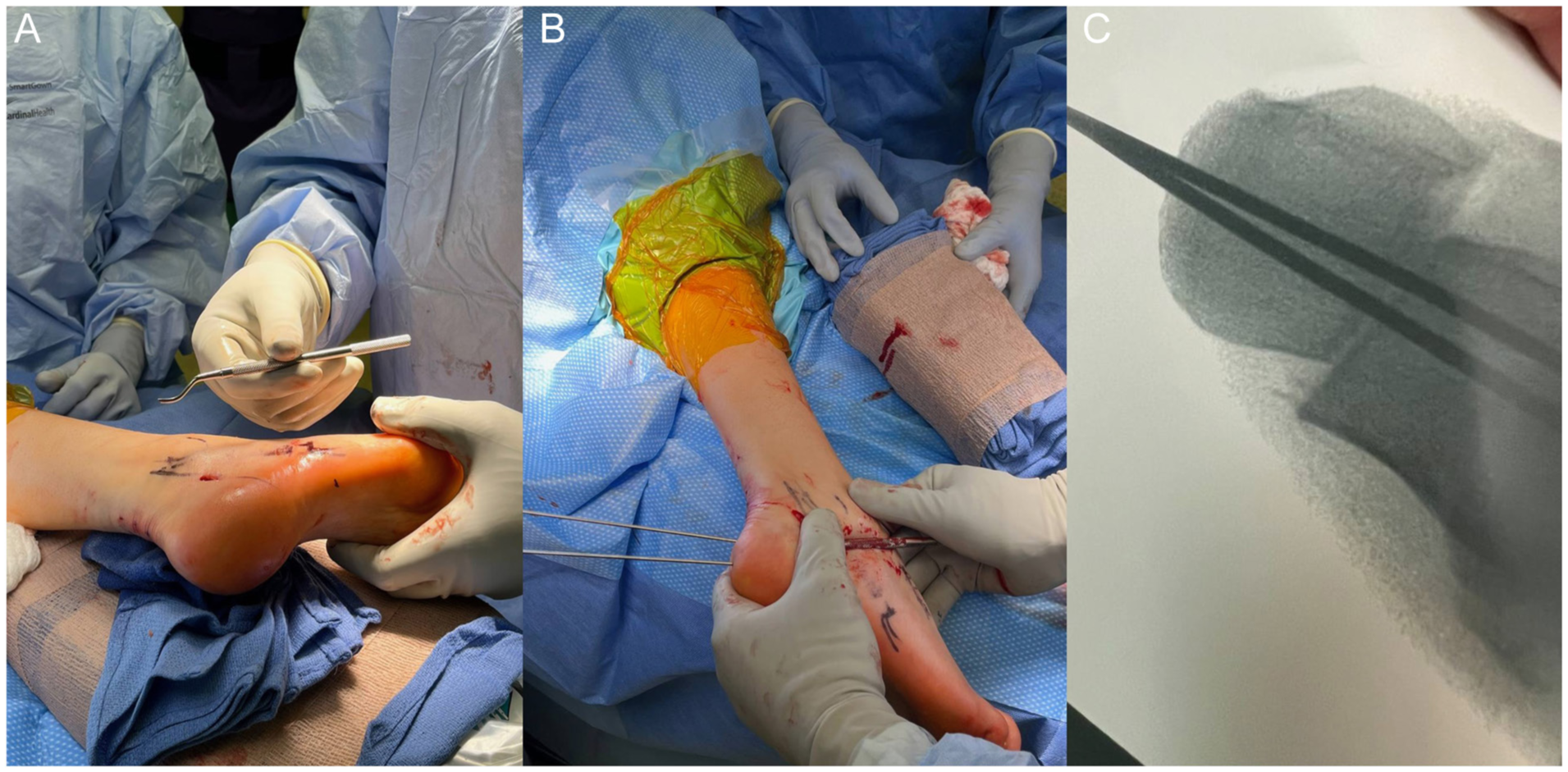
(A) Positioning in lateral decubitus with incision. (B) Dwyer-type lateral shift osteotomy using MIS technique. (C) Fluoroscopic view of Charcot-Marie-Tooth (CMT) reconstruction with MIS Dwyer-type, lateral shift osteotomy.
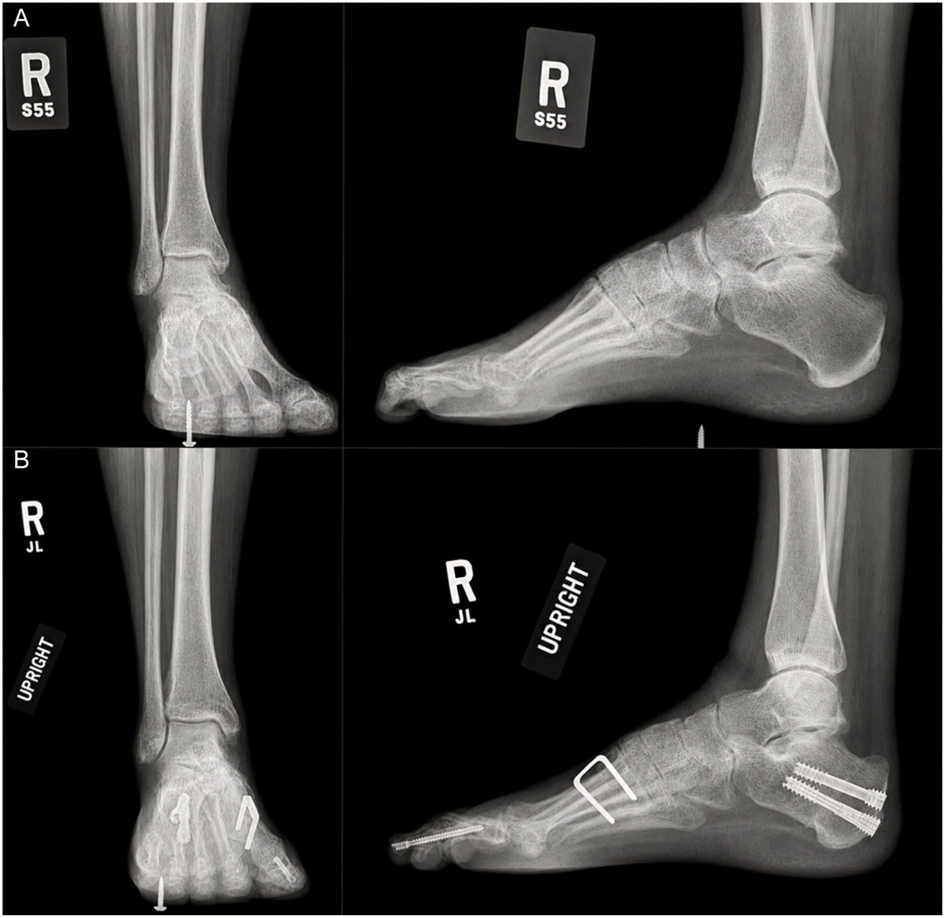
(A) Preoperative and (B) final postoperative radiographs in a patient with Charcot-Marie-Tooth who underwent an MIS Dwyer-type, lateral shift calcaneal osteotomy.

Surgical incision 2 weeks postoperation in a patient who underwent an MIS medializing osteotomy.
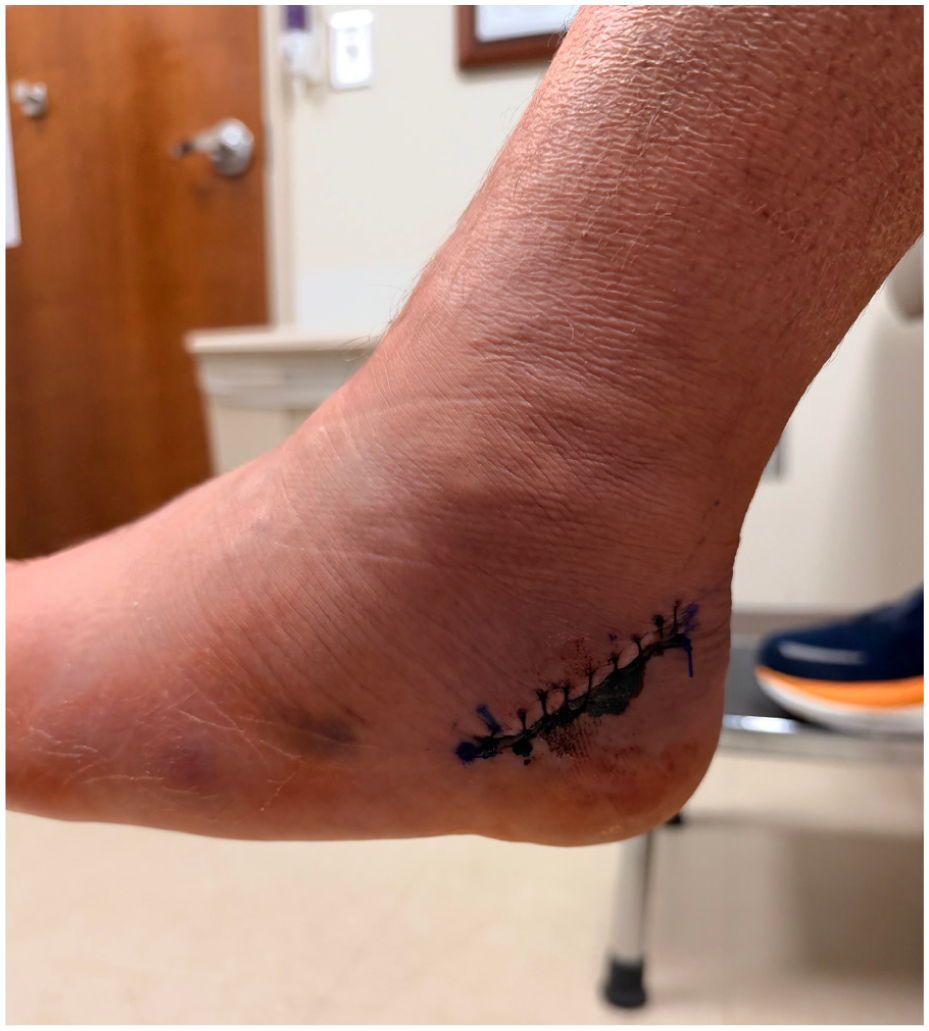
Surgical incision in a patient who underwent an open medializing osteotomy.
There were significantly more Cotton osteotomies (P < .001), posterior tibial tenotomies (P < .001), gastrocnemius recessions (P < .001), and Kidner procedures (P = .032) in the open cohort. Meanwhile, there were more first metatarsal dorsiflexion osteotomies (P < .001), Achilles lengthening (P = .02), posterior tibial tendon transfers (P = .001), extensor hallucis longus tendon transfers (P < .001), spring ligament repairs (P = .032), hallux IP fusions (P = .001), plantar fascia releases (P = .002), and peroneal longus to peroneal brevis transfers (P < .001) in the MIS cohort.
Primary and Secondary Outcomes
Patient medical records were reviewed for mention of postoperative complications in their orthopaedic foot and ankle clinic notes after surgery. Postoperative complications identified included infection, wound dehiscence, nerve damage, hardware failure, reoperation, or nonunion. Complications such as superficial wound dehiscence, superficial infection, transient neuritis, or painful hardware were considered minor, whereas complications such as nonunion, revision, deep dehiscence, deep infection, or persistent neuritis requiring neurectomy were considered major. Complications were only included in the current analysis if it pertained to the calcaneal osteotomy and/or associated incision over the lateral calcaneus. These measures served as primary outcomes for the current analysis.
The amount of calcaneal displacement achieved in each patient was measured on intraoperative fluoroscopic images via an axial heel view following shift of the calcaneus. Measurements were oriented off known screw size and fluoroscopy magnification, using the equation: magnification = apparent screw diameter ÷ true screw diameter. Standard measurement tools within our PACS imaging package were used (Vue Motion Phillip). Two independent investigators performed all measurements twice, with a 1-week period between measurements.
Data Analysis
Following data collection, patients were separated into 2 cohorts based on the surgical technique used in their intervention (MIS vs open calcaneal osteotomy). Continuous data were compared between MIS vs open cohorts on an independent sample t test; categorical data were compared using a χ2 analysis.
A Shapiro-Wilk test for normality was performed, demonstrating that many of our continuous data were not normally distributed within cohorts. Therefore, subgroup comparisons could not be reliably reported.
A false discovery rate (FDR) adjustment was performed using the Benjamini-Hochberg procedure to better interpret our multiple analyses. For radiographic measurements, intrarater and interrater intraclass correlation coefficients, mean absolute differences, and Bland-Altman 95% limits of agreement (LoA) were calculated to evaluate the reliability and reproducibility of our measurements. All statistical analyses were completed on IBM SPSS Statistics 29 (IBM Corp) software. All P <.05 were considered significant for each comparison.
Results
For treatment of various hindfoot deformities, patients who underwent an MIS calcaneal osteotomy were on average 9 years younger at the time of surgery in comparison to those who underwent open calcaneal osteotomy (P = .001). Although there were significantly more medializing calcaneal osteotomies in the open calcaneal osteotomy cohort (P = .003), there was no difference in the sex distribution or laterality of surgery between cohorts (respectively, P = .188 and .294) (Table 1).
Patient Demographic Summary.
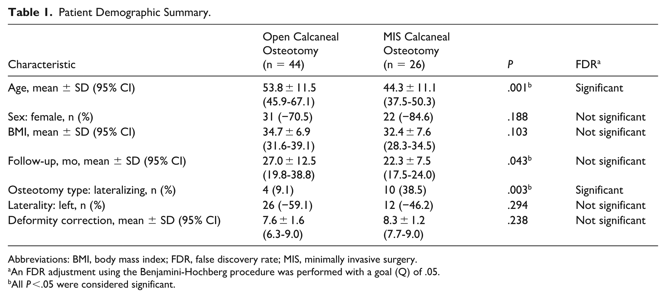
Abbreviations: BMI, body mass index; FDR, false discovery rate; MIS, minimally invasive surgery.
An FDR adjustment using the Benjamini-Hochberg procedure was performed with a goal (Q) of .05.
All P <.05 were considered significant.
A mean calcaneal displacement of 8.1 ± 3.0 mm and 9.2 ± 2.5 mm was achieved through the open and MIS approaches, respectively (P = .079). A mean absolute difference of 0.46 was observed between authors’ measurements, with 95% upper LoA of 2.08 and lower LoA of 1.16. Intrarater ICC was 0.98, whereas interrater ICC was 0.88, indicating excellent and good reliability, respectively.
The additional procedures performed among our cohorts are summarized in Supplemental Table 1. Although the specific procedures performed among patients who underwent an open calcaneal osteotomy vs MIS calcaneal osteotomy varied, there was no statistical difference noted in the mean number of procedures performed in each cohort (P = .974). The open cohort had a mean of 3.9 ± 1.5 (95% CI, 3.4-4.3) additional procedures; the MIS cohort had a mean 3.8 ± 2.5 (95% CI, 2.8-4.8) additional procedures.
Complications
Major
Patients who underwent open calcaneal osteotomy had an 11.4% (5/44) rate of major complications. One of the 44 patients (2.3%) experienced an isolated wound infection with significant lateral wound breakdown. This patient required an irrigation and debridement in the operating room with closure of the wound. Another patient had 2 major complications (4.6%) and was diagnosed with a calcaneal nonunion and sural neuritis. They underwent a revision open calcaneal osteotomy and a sural neurolysis. Separately, 2 patients (4.6%) returned to the operating room for sural neurolysis for persistent sural neuritis. Both patients underwent hardware removal at the time of revision procedure. One of these patients was also diagnosed with wound dehiscence prior to reoperation, which was irrigated and debrided during revision.
Patients in the MIS calcaneal osteotomy cohort had an 11.5% rate of major complications (3/26). All 3 required reoperation. One was diagnosed with a nonunion at 12 months following their original operation, and this was revised. One patient had a surgical wound dehiscence and postoperative infection at one of the calcaneal screw sites and underwent an incision and debridement with wound closure. One patient developed sural neuritis that initially resolved with corticosteroid injections but later recurred; patient underwent operative sural neurolysis.
Minor
Minor complications were observed in 20.5% (9/44) of the open calcaneal osteotomy cohort. Eight (18.2%) had a minor, superficial postoperative wound dehiscence over their lateral calcaneal incision that was treated with extended nonweightbearing and frequent wound checks. Of these patients, 4 were treated for presumed concomitant wound infection that resolved with a course of oral antibiotics. One of the 44 patients experienced painful hardware, which was removed. In the MIS cohort, minor complications were observed in 1 of 26 patients (3.8%). This patient experienced painful hardware, which resolved following removal.
The rate of total postoperative wound complications (including both minor and major wound complications) was higher in the open cohort than the MIS cohort (P = .036); however, on FDR this was found to be not significant. Otherwise, no differences were found when comparing the rate of minor (P = .055), major (P = .621), or total overall complication rates (P = .129) between open vs MIS calcaneal osteotomy cohorts (Tables 2 and 3).
Summary of Postoperative Outcomes: Specific Complications. a
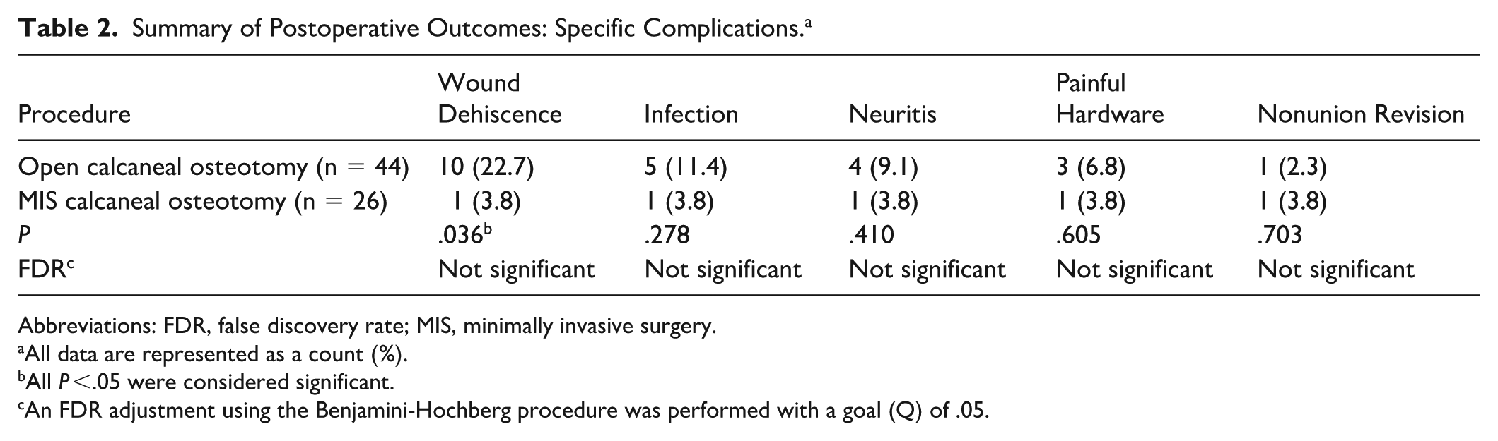
Abbreviations: FDR, false discovery rate; MIS, minimally invasive surgery.
All data are represented as a count (%).
All P <.05 were considered significant.
An FDR adjustment using the Benjamini-Hochberg procedure was performed with a goal (Q) of .05.
Summary of Postoperative Outcomes: Minor vs Major Complications. a
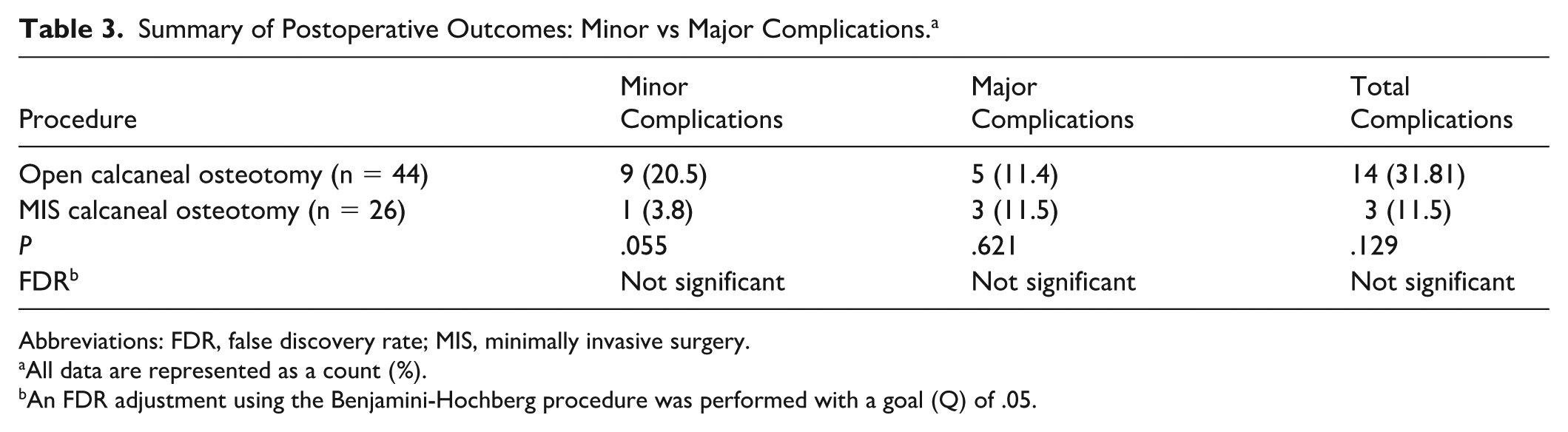
Abbreviations: FDR, false discovery rate; MIS, minimally invasive surgery.
All data are represented as a count (%).
An FDR adjustment using the Benjamini-Hochberg procedure was performed with a goal (Q) of .05.
Discussion
Patients who underwent an MIS calcaneal osteotomy for surgical correction of a hindfoot deformity had no significant difference in calcaneal correction compared to those who underwent open osteotomies, but with fewer minor wound complications, though this difference did not reach significance after FDR adjustment. Both cohorts demonstrated significant improvements in patient-reported function and mobility scores at greater than 1-year follow-up. A statistically significant improvement in PROMIS pain scores after FDR adjustment was observed in the MIS cohort (P < .001), whereas the improvement observed in the open cohort did not survive FDR correction (P = .027). Findings of the current study suggest that both the open and the MIS calcaneal osteotomy are effective interventions for hindfoot deformity correction, although the MIS procedure may be associated with fewer wound complications.
Over the past decade, various MIS techniques have exhibited safety and efficacy in foot and ankle orthopaedics while circumnavigating many complications associated with open procedures, such as wound dehiscence and postoperative infection.1,8,12 However, many surgeons have cited reasons to pause before implementing these novel techniques. In the setting of calcaneal osteotomies specifically, it has been previously suggested that MIS techniques may be associated with an increased rate of nonunion and postoperative osteonecrosis, secondary to thermal injury.4,18 Namely, Coleman et al 4 found a unique occurrence in their report of MIS calcaneal osteotomies. In their first 116 MIS calcaneal osteotomies, they had a less than 1% nonunion rate, comparable to this study; however, in the next 43 cases, the rate went up to 28%. The senior author did not change his technique and could not find a clear explanation for this observation. He speculated that his burr technique was susceptible to some degree of change, possibly secondary to calibration of the driver hardware, leading to a heat necrosis phenomenon and nonunion in higher-risk patients. 5 MIS calcaneal osteotomies included in the current study, and previous retrospective cohort studies by Kheir et al, 13 Gutteck et al, 10 and Kendal et al 12 were all performed using a high-torque, low-speed Shannon burr with copious, continuous irrigation in standard MIS fashion. Although this technique is similar to that reported by Coleman et al, there was no significant difference in the union rate observed between MIS vs open cohorts in any of the other aforementioned studies, which also align with our current data.
MIS calcaneal osteotomies have also been hypothesized to carry a higher risk of neurovascular compromise due to limited intraoperative visualization.4,5,9,14,18 Contrary to these ascertainments, we observed comparatively fewer nerve injuries (3.8% vs 9.1%), albeit not statistically significant, in those who underwent MIS osteotomies. While the current retrospective study adds to this ongoing debate, we are not the first to report these findings. Gutteck et al 10 reported fewer revision procedures and sural nerve injuries in patients who underwent MIS calcaneal osteotomies in comparison to open calcaneal osteotomies (0% vs 15.5%, P < .001). Furthermore, Kheir et al 13 reported no neurovascular complications in any of their 30 MIS calcaneal osteotomies. Similarly, Kendal et al 12 observed no postoperative neuropathy in their 32 MIS cases, although three patients in the open cohort did develop sural neuropathy.
Existing literature has offered limited descriptions of outcomes following MIS calcaneal osteotomies in comparison to open calcaneal osteotomies. These previous studies have reported significant improvements in patient-reported outcomes in addition to low complication rates.3,10,12,13,21 In the retrospective study by Waizy et al, 21 a statistically significant improvement in FAOS scores was observed in patients who underwent open calcaneal osteotomies and patients who underwent MIS calcaneal osteotomies; although exact means and/or P values were not provided by authors of this study, they describe no difference in patient-reported outcomes between cohorts. Similarly, Gutteck et al 10 found that both the open and MIS cohorts improved in AOFAS scores (64 ± 4.2 to 92 ± 8.6 vs 68 ± 5.1 to 94 ± 9.3, respectively) and VAS scores (6 ± 2.8 to 1 ± 0.7 vs 5 ± 1.3 to 1 ± 0.7, respectively). No significant differences were found between cohorts (P = .089).
The current retrospective study has inherent limitations, including the inability to match cohorts in terms of procedure type (lateralizing vs medializing), patient sex, patient age, or patient comorbidities. Additional procedures performed at the time of calcaneal osteotomy are a standard, necessary component of achieving effective deformity correction; however, it is important to recognize that this may limit the specificity of our analysis. Our cohorts reflect temporal shifts in our study surgeons’ standard practice; all patients who would have previously been offered an open osteotomy were offered the MIS procedure regardless of deformity size. Nevertheless, we did observe significantly more lateralizing osteotomies in the MIS cohort in comparison to the open cohort (P = .003); the open cohort was also older at the time of surgery (P = .001). Although unlikely, differences in patient demographics between cohorts may have influenced the complication rates observed. Although there was no difference in the number of additional procedures performed between cohorts, the specific additional procedures performed in open vs MIS osteotomies varied. Multiple continuous variables were not normally distributed among our data; we were unable to compare subgroups to better define these interactions. Further, all patients included in the current analysis underwent surgery performed by one of 2 fellowship-trained, orthopaedic foot and ankle surgeons with significant MIS experience; this may limit the generalizability of trends we reported.
Despite these limitations, we were able to describe a significant reduction in wound complications in the MIS cohort in comparison to the open cohort. Future expansion of the current study may allow for prospective or longer-term comparisons among these procedures. Still, this retrospective review may help guide surgeons in their decision-making process for hindfoot correction.
Conclusion
The findings of this retrospective cohort study suggest that both open and MIS calcaneal osteotomy, performed as part of a broader surgical reconstruction for hindfoot deformity, are associated with comparable rates of major complications and similar degrees of calcaneal correction. Although the MIS cohort demonstrated fewer minor wound complications than the open cohort (3.8% vs 20.5%), this difference did not reach significance after FDR adjustment and should be interpreted with appropriate caution given the study’s sample size. Patients in both cohorts experienced meaningful improvements in physical function and mobility at >1 year following surgery. Improvement in pain scores reaching statistical significance after FDR adjustment was observed in the MIS cohort; a similar trend was present in the open cohort but did not survive correction for multiple comparisons. Taken together, these data support the continued use of both techniques and suggest that MIS calcaneal osteotomy may be associated with a more favorable minor wound complication profile. This is a finding that warrants prospective investigation in larger cohorts.
Supplemental Material
sj-docx-1-fai-10.1177_10711007261443308 – Supplemental material for Complications After Minimally Invasive vs Open Calcaneal Osteotomy: A Retrospective Study
Supplemental material, sj-docx-1-fai-10.1177_10711007261443308 for Complications After Minimally Invasive vs Open Calcaneal Osteotomy: A Retrospective Study by Sarah Hall Kiriluk, Dylan Crawford, Preston Harrison, Rodrigo Encinas, John O’Keefe, Harley T. Davis, Moawiah Mustafa, J. Benjamin Jackson and Tyler A. Gonzalez in Foot & Ankle International
Supplemental Material
sj-pdf-1-fai-10.1177_10711007261443308 – Supplemental material for Complications After Minimally Invasive vs Open Calcaneal Osteotomy: A Retrospective Study
Supplemental material, sj-pdf-1-fai-10.1177_10711007261443308 for Complications After Minimally Invasive vs Open Calcaneal Osteotomy: A Retrospective Study by Sarah Hall Kiriluk, Dylan Crawford, Preston Harrison, Rodrigo Encinas, John O’Keefe, Harley T. Davis, Moawiah Mustafa, J. Benjamin Jackson and Tyler A. Gonzalez in Foot & Ankle International
Footnotes
ORCID iDs

Ethical Considerations
Ethical approval for this study was obtained from institutional review board at Prisma, University of South Carolina (2170226-2).
Funding
The authors received no financial support for the research, authorship, and/or publication of this article.
Declaration of Conflicting Interests
The authors declared the following potential conflicts of interest with respect to the research, authorship, and/or publication of this article: J. Benjamin Jackson, III, MD, MBA, reports consultant for Synthes. Tyler A. Gonzalez, MD, MBA, reports royalties from Treace, Vilex, Enovis; consultant for Treace, Vilex, Enovis, Conmed, Theramicro; stock or stocks options, Treace. Disclosure forms for all authors are available online.
Supplemental Material
Supplementary material is available online with this article.
References
Supplementary Material
Please find the following supplemental material available below.
For Open Access articles published under a Creative Commons License, all supplemental material carries the same license as the article it is associated with.
For non-Open Access articles published, all supplemental material carries a non-exclusive license, and permission requests for re-use of supplemental material or any part of supplemental material shall be sent directly to the copyright owner as specified in the copyright notice associated with the article.
