






Abstract
Introduction
Although rare, Kaposi sarcoma is the most common malignant neoplasm associated with human immunodeficiency virus (HIV) infection. Several agents have now been approved in the treatment of this malignancy and are used with varying degrees of success.
Case report
We present a unique case of a 64-year-old man with well-controlled HIV infection who developed necrotizing leg gangrene from invasive cutaneous Kaposi sarcoma. He responded very well to systemic chemotherapy, thereby avoiding limb amputation.
Management and outcome
Pegylated liposomal doxorubicin (PLD) at a dose of 20 mg/m2 every 3 weeks was utilized, with a near-complete response after six cycles of therapy. The patient continues to receive maintenance treatment with PLD. His HIV infection remains in excellent control, with a high-normal CD4 T-cell count. Periodic echocardiogram evaluations have not shown any decline in left ventricular ejection fraction (LVEF) over time.
Conclusion
Most patients with Kaposi sarcoma achieve partial responses to treatment with PLD. Our case illustrates that near complete and complete responses are possible with this agent, leading to potential limb salvage in necrotizing gangrene.
Keywords
Introduction
Kaposi sarcoma is a soft tissue tumor that arises from the endothelial cell lining of blood and lymphatic vessels. Although rare, Kaposi sarcoma is the most common neoplasm associated with human immunodeficiency virus (HIV). Herein, we report a patient with well-controlled HIV disease who presented with recurrent Kaposi sarcoma manifesting as severe necrotizing lower extremity gangrene and describe his spectacular response to pegylated liposomal doxorubicin (PLD).
Case presentation
A 64-year-old man with well-controlled HIV disease and known history of Kaposi sarcoma presented to the emergency department (ED) with intractable left lower extremity pain in January 2021.
The patient was diagnosed with HIV in 1988 and has been on antiretroviral therapy for over twenty years. Other pertinent history included prothrombin 20210A gene mutation with recurrent deep vein thrombosis (DVT) and pulmonary emboli. He was first diagnosed with Kaposi sarcoma in 2010, when he presented with a small elevated, erythematous lesion at the medial aspect of his left knee. At the time of diagnosis, his HIV viral load was undetectable and CD4 T-cell count was above 600/mcl. He underwent serial monitoring by dermatology every six months with interval excisions of Kaposi sarcoma lesions. For the last decade, this surveillance had been sufficient to keep his seemingly indolent Kaposi sarcoma lesions well-managed. Of note, the patient has never had any evidence of visceral Kaposi sarcoma.
In 2019, he developed several subcutaneous skin lesions, featuring violaceous, indurated nodules of the posterolateral aspect of the calf (Figure 1(a)). Aside from aesthetic concerns, the patient was otherwise asymptomatic. He subsequently received 10 cycles of PLD, with a near-complete resolution of the lesions (Figure 1(b),(c)). He was followed closely after this therapy, without any evidence of recurrent or new skin findings.
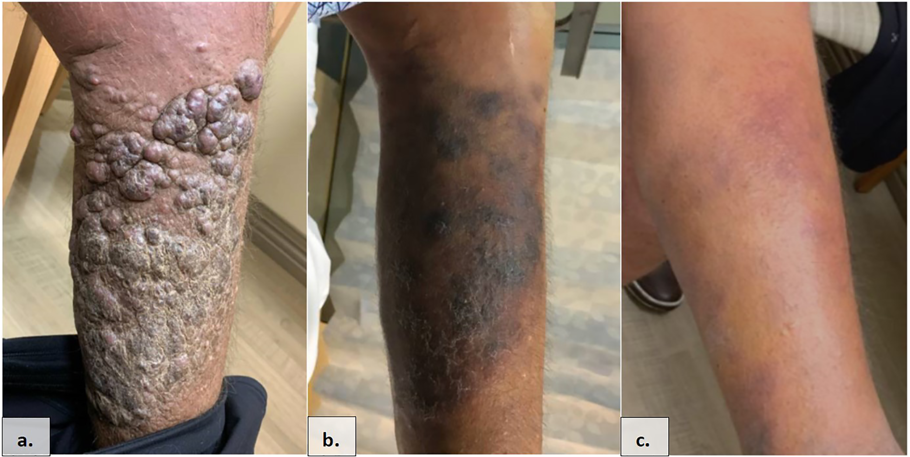
Kaposi sarcoma of the lower extremity (
Six months later, however, the patient noticed increased erythema, pain and swelling of his left ankle. He was evaluated outpatient and underwent several courses of antibiotics for a presumed cellulitis. Despite antibiotic use and supportive measures, his condition continued to worsen. During this time, he had been followed closely by a podiatrist who informed the patient that amputation was likely imminent.
He ultimately presented to the ED for intractable left leg pain. The pain had made it nearly impossible for him to bear weight on the extremity without appreciable discomfort. Previously independent at baseline, he now required a walker to perform even basic activities. What began as non-healing superficial ulcerations, rapidly transformed into blackened necrosis associated with intermittent, though prominent, purulent and serosanguineous drainage with global edema.
Although purulent drainage was not present at the time of our clinical exam, there was a 3+ nonpitting edema of the left foot extending to the mid-calf level. Blackened necrosis extending from the medial heel to the plantar surface of the foot was also present measuring approximately 10 × 12 cm (Figure 2(b), 2(e)). There was no foul-smelling odor. The necrotic area had a large 7 cm in length fissure with deep, 0.5 cm extension into the subcutaneous tissue. Exam was limited by exquisite tenderness on palpation and with movement.

Two views of the patient's left foot demonstrating rapid progression of his kaposi sarcoma cutaneous lesions with associated blackened necrosis. (
Magnetic resonance imaging (MRI) was not indicative of an abscess or osteomyelitis but did reveal soft tissue thickening. Probe-to-bone test was negative. DVT and occlusive phenomena were excluded with imaging via Doppler ultrasound and MRI, given his history of hereditary thrombophilia.
Podiatry and oncology were consulted during his admission for further evaluation. The podiatrist recommended amputation as definitive management with the necrotic process appearing unsalvageable. In the interim, the patient underwent incision and drainage of the left foot with an attempt to resect all nonviable subcutaneous tissue. He tolerated the procedure well and was subsequently fitted with a negative pressure wound therapy device in the form of a wound vacuum-assisted closure (wound VAC) to augment healing and prevent infection. His oncologist opted to begin immediate systemic therapy as a last-ditch effort to avoid amputation. He was also evaluated by vascular surgery for vessel mapping and determining the adequacy of blood flow, a necessary step prior to planned amputation.
Management and outcome
Two weeks following his inpatient debridement, he was evaluated as outpatient by oncology. He was found to have large exophytic and ulcerating tumor-like lesions expanding in the area of debrided necrosis (Figure 3(d)). There was also sanguineous drainage and pain, both exacerbated by resting his left leg in a gravity-dependent position.

Marked response to pegylated liposomal doxorubicin (PLD) over an 8-week period captured from 3 different angles of the left foot. (
Initially, pomalidomide was considered but not pursued as this agent can trigger hypercoagulability, a relative contraindication given this patient's prothrombin 20210A gene mutation and history of recurrent venous thromboembolism (VTE). With this important consideration, it was decided to restart patient on PLD at a dose of 20 mg/m2 every 3 weeks.
Despite some skepticism that this therapy would suffice to any considerable degree, the patient underwent rapid regression of Kaposi sarcoma lesions, with significant clearing after just three cycles of therapy and a near-complete resolution after six cycles (Figure 3). Immediately prior to therapy, he had been undergoing serial, biweekly debridement of the left foot lesions by podiatry. Given the marked clinical improvement, debridement was no longer indicated.
As of today, the patient has completed twelve cycles of PLD with a markedly restored quality of life, having been lifted from the debilitation of severe pain. He progressively transitioned from walker-dependency to a support cane, and ultimately was able to stand and walk freely on his own. With his independence restored, he was rescued from the brink of amputation, an experience that would have forever changed his life.
Discussion
There are four classifications of Kaposi sarcoma, where acquired immunodeficiency syndrome (AIDS)-related Kaposi sarcoma (Kaposi sarcoma-AIDS; also called epidemic Kaposi sarcoma) is the most common type diagnosed in the United States. The other 3 types include traditional (classic), African (endemic), and immunosuppression-related (iatrogenic) Kaposi sarcoma.
It was not until 1994 when human herpes virus type 8 (HHV-8) was shown to have a causal relationship with the development of Kaposi sarcoma in the setting of HIV. 1 Indeed, it is well-understood that all subtypes of Kaposi sarcoma are caused by HHV-8 which likely requires some degree of immunosuppression to take up residence in a host from subsequent research, HHV-8 seems to be necessary but not sufficient for developing KS. 2
Reflecting on the presented case, one should wonder how a severe manifestation of Kaposi sarcoma can occur in the setting of well-controlled HIV disease on anti-retroviral therapy with a normal CD4+ T-cell count and undetectable viral load. Perhaps, there is an inherent level of immune vulnerability that endures after HIV infection, irrespective of treatment or CD4+ T-cell count.
Nevertheless, multiple studies have shown a direct correlation between high HIV viral loads and worse morbidity and mortality from Kaposi sarcoma. 3 That is, poorly controlled HIV is associated with more severe manifestations of Kaposi sarcoma including visceral lesions, possibly even presenting as gastrointestinal or pulmonary hemorrhage.4,5
With such varied manifestations of Kaposi sarcoma, the approaches to treatment must also represent a varied and personalized approach, tailored to the specific disease manifestation, coexistent comorbidities and patient characteristics. Prior to effective antiretroviral therapy, the AIDS Clinical Trial Group (ACTG) of the National institutes of Health (NIH) established staging criteria to risk stratify Kaposi sarcoma in the setting of HIV disease. Although established in 1989, this criteria remains the most utilized prognostication method. It takes 3 characteristics into consideration (tumor burden, CD4 count and systemic symptoms) to stratify patients as having “good risk” or “poor risk” Kaposi sarcoma which is subsequently used to guide therapy.5,6 As opposed to cure, the goal of therapy is tumor remission along with symptom palliation and monitoring and prevention of recurrence.
Systemic chemotherapy is indicated for patients with either widespread, symptomatic, rapidly progressive disease or visceral manifestations. 6 There are currently several single-agent systemic chemotherapy options available, with different response rates and toxicity profiles. In particular, PLD was approved by the FOOD AND DRUG ADMINISTRATION (FDA) in 1995 for the treatment of AIDS-related Kaposi's sarcoma, refractory to conventional chemotherapy. 7 This agent is now the preferred 1st-line treatment for advanced Kaposi sarcoma. PLD is associated with response rates ranging from 45–60% and a median progression free survival of approximately 5 months. 8
PLD is a liposomal formulation of the cytotoxic anthracycline antibiotic doxorubicin, which acts by inhibiting DNA synthesis and by compromising DNA repair mechanisms.9,10 Specifically, it intercalates into DNA, resulting in inhibition of DNA synthesis and function. It also inhibits topoisomerase II thereby creating torsion of the DNA helix causing strand breakage. Protected from chemical and enzymatic degradation, this liposomal formulation has augmented bioavailability and better delivery to the tumor environment.7–10 Moreover, pegylation confers an improved toxicity profile with the most significant feature being a reduced rate of cardiotoxicity. 7 Nevertheless, cardiotoxicity remains a prominent and feared adverse effect. Treated patients require left ventricular ejection fraction monitoring with serial echocardiograms before, during and long after therapy. 9
This case report emphasizes the importance of a tailored regimen and thorough understanding of pharmacodynamics of chemical agents utilized in oncology. Furthermore, a multidisciplinary approach to managing patients with chemotherapy-sensitive malignancies cannot be overemphasized. 5
In summary, this case describes a patient who presented with necrotizing gangrene of the lower extremity for which amputation of the extremity would have been imminent under any other circumstance. In our patient, PLD led to a remarkable clinical response, thereby completely reversing the gangrene caused by rapidly progressive Kaposi sarcoma and salvaging the affected limb.
Footnotes
Consent statement
Informed consent for this publication was obtained from the patient before the submission of this case report.
Declaration of Conflicting Interests
The author(s) declared no potential conflicts of interest with respect to the research, authorship, and/or publication of this article.
Funding
The author(s) received no financial support for the research, authorship and/or publication of this article.
