






Abstract
Kaposi sarcoma is a malignant neoplasm arising from the endothelial cell lining of blood and lymphatic vessels. Herein, we discuss etiopathogenesis, clinical presentation, diagnostic criteria, updated guideline-based approach to its management and newer experimental approaches. Given its efficacy and side effect profile, pegylated doxorubicin is the currently preferred first-line therapy in advanced disease. Paclitaxel remains an alternative first-line option. At the time of relapse, patients can be retreated with the same agents as they often maintain their clinical efficacy. New therapeutic options are on the rise, with pomalidomide being approved in 2020 as a second-line therapy. Optimal control of retroviral infection in human immunodeficiency virus (HIV) positive is instrumental in preventing disease occurrence in most patients. Suppressing human herpes virus type 8 (HHV-8) infection might also play a role in controlling Kaposi sarcoma growth, yet clinical trials are lacking. Unraveling the molecular and genetic intricacies of Kaposi sarcoma's pathogenesis might allow for the emergence of novel and effective therapeutic strategies. Clinical trials are currently underway to establish potential roles for various targeted agents, immune checkpoint inhibitors (ICIs) and experimental agents in the treatment of advanced Kaposi sarcoma.
Keywords
Introduction
Kaposi sarcoma (KS) is a soft tissue tumor that arises from the endothelial cell lining of blood and lymphatic vessels. Although rare overall, it is the most common neoplasm associated with acquired immunodeficiency syndrome (AIDS). Nearly 150 years after its discovery, researchers have yet to establish the exact pathophysiologic basis of this disease.
Herein, we discuss a patient with human immunodeficiency virus (HIV) disease and advanced Kaposi sarcoma, who responded very well to systemic treatment with pegylated doxorubicin. Further, we discuss etiopathogenesis of Kaposi sarcoma, clinical presentation, and diagnostic criteria. We also present an updated guideline-based approach to its management, with a particular emphasis on AIDS-associated Kaposi sarcoma.
Clinical vignette
A 64-year-old gentleman with a history of well-controlled HIV infection (undetectable viral load, normal CD4+ T-cell count) presented to the emergency department with severe left lower extremity pain and swelling. One year prior, he developed Kaposi sarcoma of the lower extremities, which was successfully treated with 10 cycles of pegylated doxorubicin (PLD).
Prior to his current presentation, he received two courses of antibiotics to treat presumptive cellulitis of the left foot. Despite this therapy, his left foot lesions progressed to necrosis in a matter of weeks, with prominent drainage, swelling, and severe pain.
On the initial examination, there was 3 + non-pitting edema of the left foot extending to the mid-calf. Necrosis extending from the medial heel to the plantar surface of the foot was also present, measuring approximately 10 × 12 cm (Figure 1(a)). The necrotic area had a large, 7 cm in length fissure with deep extension into the subcutaneous tissue.

Marked response to pegylated liposomal doxorubicin (PLD) over an eight-week period captured from the plantar surface of the left foot. (a) Prior to the start of therapy, (b) two weeks into therapy and (c) eight weeks into therapy.
Podiatry was consulted during his admission and recommended amputation as definitive management, with the necrosis appearing to have progressed to an “unsalvageable” degree. In the interim, the patient underwent serial debridement of the left foot to resect all nonviable tissue. Paradoxically, debridement seemed to stimulate tumor growth, characteristic of evolving Kaposi sarcoma vascular lesions. Eventually, large, exophytic and ulcerating tumoral lesions developed in the debrided areas (Figure 1(b)).
The patient was evaluated by medical oncology, who opted to begin systemic therapy immediately as a last-ditch effort to avoid amputation. Single-agent PLD at 20 mg/m2 every three weeks was restarted without delay. Despite some skepticism that this therapy would generate a significant treatment response, the patient experienced dramatic regression of the Kaposi sarcoma lesions, with significant clearing after just three cycles of therapy (Figure 1(c)). Remarkably, the patient's Kaposi sarcoma entered a near-complete remission after six cycles of PLD, with a resolution of pain and a markedly restored quality of life.
Historical background
Discovered by Hungarian professor Moritz Kaposi in 1872, Kaposi sarcoma was initially referred to as an indolent cancer of unknown etiology that portended a poor prognosis. It was believed to have a predilection for men of Eastern European descent. In the 1950s, Kaposi sarcoma became one of the most common neoplasms throughout central Africa, affecting men, but also women and children. A surge of Kaposi sarcoma cases foreshadowed the AIDS epidemic in the United States in the early 1980s. After the first three cases became manifest, this malignancy became known as an AIDS-defining disease by the Centers for Disease Control (CDC) in 1981. 1 It was not until 1994 that human herpes virus type 8 (HHV-8) was shown to have a causal relationship in the development of Kaposi sarcoma.
Epidemiology
There are four subtypes of Kaposi sarcoma, out of which AIDS-related Kaposi sarcoma (also called epidemic Kaposi sarcoma) is the most common subtype observed in the United States. This malignancy can also affect immunocompromised HIV-negative patients, where certain elderly and children in HHV-8 endemic areas as well as transplant recipients are at the greatest risk. 2 The other three subtypes include traditional (classic), African (endemic), and immunosuppression-related (iatrogenic). Although the above four epidemiologic subgroups are well-characterized, there is wide variability in disease manifestation within each group.
AIDS-related (epidemic) Kaposi sarcoma
Acquiring Kaposi sarcoma in the setting of HIV infection is considered AIDS-related Kaposi sarcoma and represents an AIDS-defining illness. Between the late 1980s and early 1990s, epidemic Kaposi sarcoma was associated with considerable morbidity and mortality, with a five-year survival rate of only 12%. However, with the advent of effective antiretroviral therapy (ART), the incidence and associated mortality from Kaposi sarcoma have declined significantly, with the current survival rate ranging from 68% to 95%. Nevertheless, epidemic Kaposi sarcoma prevalence remains nearly 500-fold in HIV/AIDS positive compared with the general population. With a disproportionate impact observed in men who have sex with men (MSM), patients continue to face extensive morbidity. Furthermore, the degree of morbidity correlates with the stage of HIV disease, with a more aggressive presentation (such as lymphadenopathy, mucosal, and visceral involvement) more commonly observed in advanced immunosuppression. 2
Classic Kaposi sarcoma
Typically indolent, classic Kaposi sarcoma follows a protracted clinical course. It most commonly presents with slow-growing lower extremity cutaneous lesions. Nearly 15 times more common in males than females, it is mostly diagnosed in elderly men of Mediterranean, Italian, Greek, or Jewish ancestry. 3
Iatrogenic Kaposi sarcoma
Iatrogenic Kaposi sarcoma typically occurs in the setting of induced immunosuppression, as that required for organ transplantation, rheumatologic conditions or certain chemotherapy. As such, Kaposi sarcoma is nearly 200 times more prevalent after organ transplantation than in the general population. 4 Importantly, iatrogenic Kaposi sarcoma can be distinguished from HIV-associated Kaposi sarcoma by its onset. Unlike HIV-associated, this entity characteristically manifests months to years after initiating an immunosuppressive regimen and tends to resolve shortly after discontinuation or dose reduction of these agents. 4
Endemic Kaposi sarcoma
Endemic Kaposi sarcoma is largely observed in sub-Saharan Africa, with an overwhelming propensity for the pediatric and young adult population. In fact, it is among the most diagnosed cancers in Malawi, Uganda, Zimbabwe, and Swaziland. 5 It often has an aggressive presentation, with the invasion of viscera, lymph nodes and bone.4,5
Etiopathogenesis
The exact pathophysiology of Kaposi sarcoma is yet to be elucidated. Multiple molecular pathways are involved, which seem to unite toward a common endpoint with the production of viral progeny and disruption of proper host cell signaling. Ultimately, there is inhibition of the essential tumor suppressors p53 and retinoblastoma (Rb), along with transforming growth factor-beta (TGF-beta), the very proteins that regulate the balance between normal proliferation and oncogenesis.3,5,6
To further understand the Kaposi sarcoma etiopathogenesis, it is imperative to examine the molecular landscape of an HHV-8 infected host cell. It is now understood that all subtypes of Kaposi sarcoma are caused by HHV-8, a large double-stranded DNA virus of the herpes virus family. Although necessary, HHV-8 is not solely sufficient for developing Kaposi sarcoma. This virus appears to require some degree of immune compromise to take up residence and transform a host cell into a malignant cell.
At the cellular level, HHV-8 seizes the molecular machinery of endothelial and lymphatic host cells to supply a continuous reservoir of viral progeny. 4 It is this reprogramming of normal cell function that creates a favorable environment for transformation into the aberrant lymphangiogenesis that we call Kaposi sarcoma.
Many distinct pathogenetic nuances distinguish Kaposi sarcoma from other neoplasms. Generally, the hallmark of advanced neoplasia is a propensity towards distant metastasis through the vasculature or lymphatics. Kaposi sarcoma, on the other hand, displays almost exclusively paracrine behavior, causing local invasion rather than distant spread. 4
Another unique feature is the spindle-shaped cell, the hallmark cell type of Kaposi sarcoma tumors. This cell appears to be a chimera of multiple cell types. On its surface, are markers of lymphatic and endothelial cells including the vascular endothelial growth factor receptor (VEGFR), as well as markers of dendritic cells, macrophages, and smooth muscle cells. 4
Also unique is the latency-associated nuclear antigen (LANA), an essential viral protein expressed by all HHV-8-related neoplasms including Kaposi sarcoma. This protein is required for the replication and transcriptional modification of HHV-8 within a host cell. Given its ubiquity, LANA-specific monoclonal antibodies are now used for immunohistochemical diagnosis of an HHV-8 infection, where a positive result is virtually diagnostic. 5 Although clonality is well-established for most cancers, it is unclear whether Kaposi sarcoma represents a clonal malignancy, an oligoclonal process or a complex multifocal vascular proliferation. 5
Clinical presentation and staging
The morphologic variation of Kaposi sarcoma is intriguing. Histologic subtypes include anaplastic, lymphedematous, telangiectatic, hyperkeratotic, micronodular, granuloma-like, ecchymotic, and intravascular. 2 Mucosal and cutaneous lesions can present as papules, plaques, nodules, and/or bullae, all of which can regress, coalesce, or ulcerate. This vast morphologic spectrum of Kaposi sarcoma lesions can often present a diagnostic challenge.
Standard nomenclature for solid tumors such as TNM (tumor–node–metastasis) classification is not helpful for Kaposi sarcoma staging. Limited disease involving localized regions of the skin, lymph nodes, or oral mucosa is staged as T0. The presence of edema and ulceration, similar to that observed in our patient, is considered T1 disease, as is an extensive mucosal and visceral disease.
Kaposi sarcoma can affect every organ of the body, which can preclude a straightforward diagnosis and can even be mistaken for other diseases. For example, early-stage Kaposi sarcoma lesions can resemble bacillary angiomatosis, arteriovenous malformations, melanoma, or cutaneous vasculitis. 6 Kaposi sarcoma often presents with lymphedema, likely due to the involvement of lymphatic endothelial cells and lymph nodes, producing obstruction of lymphatic outflow. 3 However, lymphatic involvement is neither the exception nor the rule. Given this variability, a thorough history, physical examination and overall clinical assessment are indispensable, although histopathologic confirmation remains the gold standard for diagnosis.
The highly variable transcriptional profiling between any two Kaposi sarcoma neoplasms also creates a diagnostic conundrum and has precluded effective treatment options. Furthermore, it confounds our ability to identify exploitable targets by which to intercept the downstream neoplastic manifestations of an HHV-8-infected cell. 6
Approach to treatment
Although HIV-associated Kaposi sarcoma develops when CD4 + T-cell population declines below 200/µl, some patients experience de novo or recurrent lesions at a higher CD4 + T-cell count. 1 Reflecting on our patient, his Kaposi sarcoma reoccurred in the setting of a normal CD4 + T-cell count. Undoubtedly, HIV infection confers an inherent level of immune compromise, irrespective of T-cell count as an objective marker of immunity. Such deviation from optimal immune function might render patients with a history of HIV susceptible to HHV-8-related malignant transformation. Research surrounding Kaposi sarcoma occurrence in these circumstances is ongoing.
As HIV-associated Kaposi sarcoma is considered a disease of immune compromise, initial treatment should include initiation of antiretroviral therapy (ART), a means of recovering the diminished CD4+ T-cell population. In fact, HIV+ patients with T0 disease are initially treated with combination ART alone, and many patients respond well to this approach. A variety of local therapies are available for those who respond suboptimally to modern antiretrovirals. Systemic treatments in addition to ART are preferred for T1 disease or when a rapid disease resolution is desired. 1
Prior to the introduction of ART, the prevalence of Kaposi sarcoma in the U.S. was nearly 30% among patients diagnosed with AIDS. 7 Today, the estimated prevalence of these patients in the U.S. is only 4%. In 1987, zidovudine was the first antiretroviral drug approved by the FDA to treat AIDS. Shortly thereafter, there was a decreased incidence of AIDS-defining illnesses including Kaposi sarcoma (Figure 2). This medication had a dual effect on Kaposi sarcoma incidence. By treating HIV infection, it reenabled the immune system to defend against opportunistic infections. In addition, zidovudine was subsequently found to have inherent antiretroviral activity against HHV-8, which further led to a steep decline in Kaposi sarcoma cases.7,8
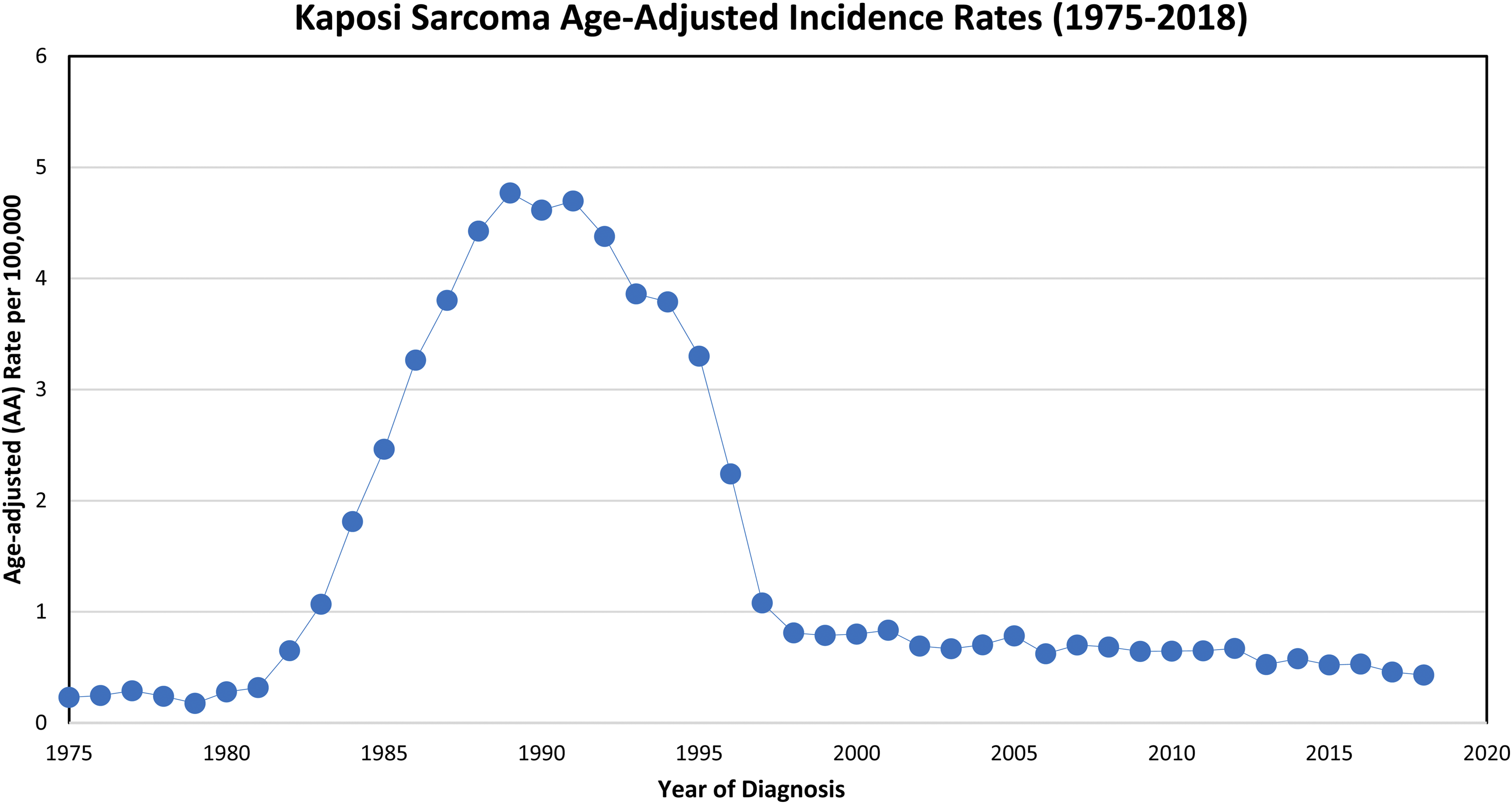
Age-adjusted incidence rates of Kaposi sarcoma between 1975 and 2018 among the SEER 9 areas: San Francisco, Connecticut, Detroit, Hawaii, Iowa, New Mexico, Seattle, Utah and Atlanta. Rates are per 100,000 and are age-adjusted to the 2000 US standard population. Created through the SEER Explorer database (seer.cancer.gov/explorer/application.html).
Other antiretrovirals have also been shown to have inherent activity against HHV-8 including nevirapine, an HIV non-nucleoside reverse transcriptase inhibitor (nNRTI), and antiviral agents used to treat cytomegalovirus (CMV) infection such as ganciclovir, foscarnet, and cidofovir. 5 In fact, studies have shown that early treatment of HHV-8 with ganciclovir or foscarnet is associated with a statistically significant reduction in the risk of developing Kaposi sarcoma.6,9,10 Today, nearly half of newly treated HIV + patients experience improvement in their Kaposi sarcoma lesions after achieving HIV viral load suppression with modern ART.
Paradoxically, some patients will respond to the initiation of antiretroviral therapy with Kaposi sarcoma progression, a transient phenomenon called Kaposi sarcoma immune reconstitution syndrome (IRIS). Although its etiology is unknown, this phenomenon characteristically occurs within 3–6 months of initiating ART.2,3,9 Management usually requires systemic therapy directed against Kaposi sarcoma. Importantly, ART should not be held or discontinued in the setting of IRIS unless life-threatening symptoms develop.2,3
Approaching literature-guided treatment of Kaposi sarcoma is a considerable challenge. This is, at least in part, owing to highly variable presentations as well as a lack of studies comparing head-to-head treatment efficacy. The approach to systemic treatment should be guided by patient characteristics and comorbidities, along with an understanding of pharmacodynamics and medication interactions. This approach should likewise foster a multidisciplinary effort, including input from pharmacists, medical and radiation oncologists, and HIV specialists.
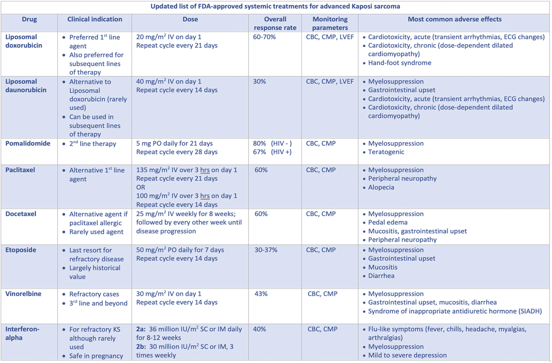
Initially, superficial and localized lesions are managed with clinical observation alone after initiation of ART, which is often sufficient to induce regression. 11 For lesions that are not aesthetically acceptable, alternative approaches include local therapies such as surgical removal, local radiation, and intralesional agents. The National Comprehensive Cancer Network (NCCN) has published updated guidelines for the systemic management of Kaposi sarcoma. We use these guidelines as a framework to discuss current systemic Kaposi sarcoma treatment for patients with AIDS-related, classic and iatrogenic subtypes (Table 1).
Various systemic therapies used in the treatment of advanced Kaposi sarcoma showing clinical indications, dosing schedule, overall response rates and most common adverse effects.
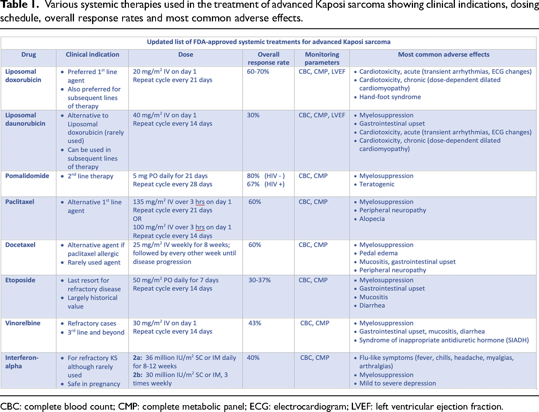
CBC: complete blood count; CMP: complete metabolic panel; ECG: electrocardiogram; LVEF: left ventricular ejection fraction.
Therapy for localized and superficial lesions
Local treatment options include argon laser photocoagulation, radiotherapy, surgery, cryotherapy, intralesional injections, and several topical agents. Topical agents were shown to be effective in some patients with superficial Kaposi sarcoma. The most commonly used is imiquimod 5% cream, administered triweekly for a total of 24 weeks. 12 It acts via stimulation of innate and acquired immune responses at the tumor site. Podophyllotoxin and retinoic acid derivatives may also be effective in some patients with superficial Kaposi sarcoma lesions. Alternatively, superficial lesions can be removed surgically, with cryotherapy or electrocautery.
The efficacy of radiotherapy is often inversely correlated with disease severity, where advanced skin lesions tend to respond suboptimally. External beam radiotherapy (EBRT) is highly effective in localized nodular KS, producing near-complete or complete initial treatment responses. However, this modality is ideal for lesions located above the level of dermis. Administering 6–8 Gy in 1 fraction is one of the more common dosing regimens. 13 Palliative EBRT can also be used in obstructive visceral Kaposi sarcoma lesions that are refractory to other systemic options. 13 However, it can lead to delayed wound healing, radiation-induced tissue inflammation and long-lasting fibrotic changes.
Intralesional chemotherapy is a form of localized treatment where a chemical agent is injected directly into the target lesions. This therapy has the benefit of limiting systemic absorption and toxicity. The most commonly used agent is intralesional vinblastine injections at a standard concentration of 0.1–0.5 mg/ml per 1 cm2 lesion, for a maximum dose of 2 mg daily. 14
Systemic therapies
In the 1980–90s, various systemic chemotherapy agents and combinations were used to treat advanced Kaposi sarcoma. Popular combinations included bleomycin–vincristine (BV), etoposide–bleomycin–vincristine (EBV), and doxorubicin–bleomycin–vinblastine (ABV). However, significant toxicity has limited the use of these combination regimens. Subsequently, doxorubicin and paclitaxel emerged as leading agents for advanced Kaposi sarcoma. Of note, the single agent’s docetaxel, etoposide, and vinorelbine have largely historical value in the management of this disease. The biologic response modifier interferon alpha (IFNa) is now rarely used due to its inconvenient daily administration schedule and significant side effect profile. Pegylated doxorubicin and pomalidomide are currently recognized as first and second line, respectively.
Therapy duration is determined by the degree of response to these systemic agents. Skin lesions are evaluated by their total number, anatomic location, and presence of nodularity. Many patients with T1 disease may require intermittent therapy for several years. Staging of visceral lesions with standard imaging, upper endoscopy, and bronchoscopy is usually employed as indicated.
Docetaxel
This antineoplastic agent acts at the M-phase of the cell cycle, promoting the assembly of tubulin dimers into microtubules and preventing their destabilization. Microtubules require constant flux, called dynamic instability, to facilitate the progression of the cell cycle. Docetaxel inhibits this vital process, resulting in the activation of the apoptotic pathway through the inhibition of B-cell lymphoma-2 gene (Bcl-2). 14
Docetaxel distributes widely to all body tissues with extensive (>90%) binding to plasma and cellular proteins. It is administered at a dose of 25 mg/m2 intravenously weekly for eight weeks, then every other week. 15 Although some studies showed that docetaxel may improve symptoms and even prolong survival in recurrent or refractory Kaposi sarcoma, this is overshadowed by its toxicity profile including severe myelosuppression, lower extremity edema, peripheral neuropathy and mucositis. 14
Paclitaxel
Paclitaxel is a semisynthetic taxane that, like docetaxel, disrupts the dynamic microtubule network required for mitosis and cell division. In the late 1990s, trials were undertaken to compare paclitaxel to various combinations of etoposide, bleomycin, and vincristine.16–18 Some patients received BV, and others were given EBV as a triple-agent regimen. Paclitaxel showed significantly improved outcomes compared to the other single and multi-agent regimens. The worst outcomes were observed in the etoposide group and in the triple-agent arm. This study was closed early given that paclitaxel was shown to be clearly superior to the other agents. 18
This study highlighted an important shift in Kaposi sarcoma treatment. Although multi-agent regimens such as BV, EBV and ABV were the previous standard of Kaposi sarcoma treatment, single-agent paclitaxel is at least noninferior with significantly reduced toxicity, morbidity and mortality. 18 This agent was approved for use in advanced Kaposi sarcoma in 1997. The recommended dose is 100 mg/m2 IV every two weeks or 135 mg/m2 every three weeks per the most updated NCCN guidelines.14,16–18 At relapse, retreatment with paclitaxel can be attempted.
The response rates to paclitaxel range between 56% and 71% with a progression-free survival (PFS) of 10–18 months and a two-year survival rate of nearly 80%.16–18 However, its use is somewhat limited by its toxicity profile, especially neurotoxicity and alopecia.16,17 In addition, severe and potentially fatal hypersensitivity can occur with this agent; premedication with steroids is now common practice. Although an older agent, paclitaxel, remains one of the most utilized first-line options in HIV+ Kaposi sarcoma. It is also the preferred alternative to liposomal doxorubicin in patients with congestive heart failure.
Etoposide
Etoposide is a derivative of a podophyllotoxin which exerts its antineoplastic affect in the late S and G2 phases of the cell cycle, stabilizing the DNA-topoisomerase II complex, and halting DNA synthesis. It rapidly distributes to all body fluids and tissues with 95% plasma protein binding.
The recommended starting dose is 50 mg per day orally for seven days out of a 14-day cycle. 19 Response rates of 30–37% have been reported. However, the toxicity can be severe including profound myelosuppression, intractable nausea, vomiting, and alopecia. 14 A recent study revealed that HIV-infected adults with clinically mild to moderate Kaposi sarcoma had worse outcomes after receiving etoposide in addition to anti-retroviral therapy as opposed to receiving antiretroviral therapy alone.19,20 Therefore, the use of this agent is generally discouraged in advanced Kaposi sarcoma.
Vinorelbine
As an alkaloid of vinblastine, vinorelbine exerts its antitumor activity at the M-phase of mitosis by inhibiting microtubule polymerization and thereby destabilizing the framework required for cell division. 14 This instability triggers the induction of the p53 tumor suppressor gene with downstream signaling of the apoptosis pathway. Owing to its wide distribution into most body tissues, with greater than 80% binding to plasma proteins, its toxicity profile includes dose-limiting myelosuppression, gastrointestinal toxicity, neurotoxicity, and syndrome of inappropriate antidiuretic hormone secretion (SIADH). Although not recommended as first-line therapy, vinorelbine has been shown to be effective in severe and aggressive classic and epidemic Kaposi sarcoma.21,22 The utilized dose is 30 mg/m2 every two weeks. 22
Interferon alpha
Interferon alpha (IFNa) is a biological response modifier with several antiviral and antiproliferative effects. It was approved for the treatment of Kaposi sarcoma in HIV disease in the pre-HAART era. This agent activates the innate immune system by augmenting the expression of major histocompatibility complex I (MHC I) molecules, thereby facilitating the presentation of viral peptides to antigen-presenting cells (APCs). 14 IFNa also activates CD8+ cytotoxic T-cells and primes macrophages for cell-mediated cytotoxicity. 23 It also inhibits angiogenesis and exerts antiproliferative effects on tumor cells through the inhibition of tumor cell protein synthesis. Originally, this agent was indicated in relapsed or refractory Kaposi sarcoma in combination with ART.23,24 It is typically administered at a dose of 30 million IU/m2 as a daily or triweekly subcutaneous administration. 24 Studies have shown that low doses of this agent are generally effective, achieving response rates up to 31%.23,24 However, the inconvenient dosing administration and its side effect profile including severe depression limit its use.
Pegylated liposomal doxorubicin
Nanotechnology and nanomedicines have revolutionized the field of oncology. They have allowed more effective targeting, delayed drug release and prolonged bioavailability, which offer less frequent dosing and reduced toxicity. Pegylated liposomal doxorubicin (PLD) was the first FDA-approved nanomedicine in 1995 when it was indicated for the treatment of AIDS-related Kaposi sarcoma refractory to conventional chemotherapy.25–28 Today, this agent is used as the preferred first line for the treatment of advanced or recurrent Kaposi sarcoma.
PLD is a liposomal formulation of the cytotoxic anthracycline antibiotic, doxorubicin, which acts by inhibiting DNA synthesis and by compromising DNA repair mechanisms. 14 Specifically, it intercalates into DNA, resulting in the inhibition of DNA synthesis and function. It also inhibits topoisomerase II thereby creating torsion of the DNA helix causing strand breakage.
Protected from chemical and enzymatic degradation, this liposomal formulation has augmented bioavailability and delivery to the tumor environment.25,26 Moreover, pegylation confers an improved toxicity profile, with the most significant feature being a reduced rate of cardiotoxicity. 25 Lack of alopecia and neurotoxicity favor the use of this agent over paclitaxel. Nevertheless, the risk of cardiotoxicity persists, and patients require serial echocardiograms before, during, and long after therapy. 25
This agent demonstrated an 85% overall survival when given in conjunction with ART in HIV-positive patients with advanced Kaposi sarcoma.27,28 Further randomized clinical trials have demonstrated a significant improvement in response rate with PLD (45–70%) along with a more favorable toxicity profile when compared to either single or combined agents, such as doxorubicin, bleomycin, and vincristine. 29 The recommended dose is 20 mg/m2 IV every three weeks. PLD has a U.S. FDA black box warning for cardiotoxicity at a cumulative dose above 550 mg/m2.
Liposomal daunorubicin
As with PLD, liposomal daunorubicin is a liposomal encapsulation of daunorubicin, thus protected from chemical and enzymatic degradation. It also exhibits increased uptake into tumor cells because the liposome allows increased penetration through the tumor vasculature with less plasma protein binding. 30 A randomized trial conducted in 1996 compared the efficacy of this agent with the efficacy of the three-drug regimen of ABV in advanced Kaposi sarcoma.30,31 Although this trial could not identify a statistically significant difference in response rates, liposomal daunorubicin–treated patients had considerably reduced systemic toxicity compared to the ABV arm. The suggested dosing was 40 mg/m2 IV every two weeks. However, liposomal daunorubicin exhibited a response rate of only 30%. These findings render this agent inferior to PLD in advanced Kaposi sarcoma.
Pomalidomide
Pomalidomide is an oral small-molecule cereblon E3 ligase modulator (CELMoD) agent which has been utilized for over a decade in multiple myeloma. It targets cereblon, an E3 ubiquitin ligase, leading to T-cell and NK-cell activation as well as co-stimulation and modulation of TNF-alpha, IL6, and VEGF.32,33 In addition to its antineoplastic and antiangiogenic properties, this agent is nearly 100 times more potent than other approved CELMoDs, such as lenalidomide or thalidomide.
An open-label trial conducted by the National Cancer Institute (NCI) showed a 67% overall response rate to pomalidomide among HIV-positive patients and 80% among HIV-negative patients.32,33 A median PFS of 10 months has been established. 32 Based on these findings, the drug was granted accelerated approval for the treatment of AIDS-associated Kaposi sarcoma refractory to antiretroviral therapy in 2020. 32
The recommended dose is 5 mg/day orally for 21 days out of a 28-day cycle. 33 Adverse effects include myelosuppression, GI symptoms, and a propensity towards thrombosis. Therefore, all patients are advised to receive prophylaxis with low-dose aspirin or low-molecular-weight heparin. Confirmatory studies on efficacy and safety of pomalidomide are currently underway in the U.S. and sub-Saharan Africa.
Ongoing clinical trials of approved and experimental agents
The current basic, translational and clinical research atmosphere in Kaposi sarcoma therapeutics shows great promise. The efficacy of several approved targeted agents, immune checkpoint inhibitors (ICIs), and novel cytokines is being explored in clinical trials.
As aberrant angiogenesis via the VEGF pathway has important implications in Kaposi sarcoma pathogenesis, agents that target this mechanism have been studied in clinical trials. Single-agent bevacizumab, a recombinant humanized monoclonal antibody that inhibits VEGF-A, showed a 31% response rate in a phase II trial. 34 In combination with PLD, the response rate was improved to 56%, at the expense of toxicity including hypertension and bleeding, resulting in discontinuation in several patients. 35
Tyrosine kinase inhibitors (TKIs) with anti-VEGF activity have also been studied. Response rates of 29% were documented with single-agent sorafenib. 36 Cabozantinib is currently in an early phase study in combination with the ICI nivolumab. Novel agents, including the anti-VEGFR TKI semaxanib, are currently being explored in the treatment of HIV+ Kaposi sarcoma.
Bortezomib, a proteasome inhibitor approved in plasma cell disorders, inhibits nuclear factor-kappa-B pathway. This agent showed a response rate of 60% in HIV+ Kaposi sarcoma in a small study; however, most responses were partial. 37 Consequently, the third generation oral proteasome inhibitor, ixazomib, is currently being investigated in this setting.
Several clinical trials are currently underway to examine the efficacy of currently available ICIs in the treatment of Kaposi sarcoma. The use of programmed death-1 (PD-1) inhibitors nivolumab or pembrolizumab exhibited a response rate of 67% in a small retrospective patient series with advanced HIV+ Kaposi sarcoma. 38 On the other hand, other studies of pembrolizumab have been discouraging. One study is currently investigating the combination of nivolumab with the CTLA-4 inhibitor ipilimumab in classical Kaposi sarcoma. Combinations of nivolumab–cabozantinib and nivolumab–pomalidomide are also being investigated in early trials.
In addition, the US NCI (National Cancer Institute) launched a recent study investigating pomalidomide in combination with PLD in patients with severe HIV+ and HIV− Kaposi sarcoma. Another NCI study is examining whether abemaciclib, a cell cycle inhibitor approved for advanced breast cancer, might be useful in Kaposi sarcoma treatment.
Interleukin 12 (IL-12) is a potent pro-inflammatory cytokine that exhibits antiangiogenic properties via induction of IFN-gamma production. This may lead to an increase in IFN-inducible protein 10, a downregulation of HHV-8 encoded vGPCR, essential in Kaposi sarcoma pathogenesis. Studies of IL-12 alone or in combination with PLD have demonstrated acceptable safety and efficacy in HIV+ Kaposi sarcoma. 39 Two studies are currently investigating the potential of a synthetic IL-12 protein alone and in combination with another experimental immunotherapy agent in the treatment of advanced Kaposi sarcoma.
Into the future
Over 150 years in the making, we have yet to fully elucidate the pathophysiologic mechanisms by which Kaposi sarcoma is able to subdue the immune system and trigger robust levels of tissue destruction. Approach to these patients should be equally robust, tailored to the specific disease manifestation, coexistent comorbidities and patient characteristics. As of today, pegylated doxorubicin, paclitaxel, and pomalidomide remain the most utilized systemic agents for the treatment of this malignancy. Optimal control of retroviral infection cannot be overemphasized in HIV+ patients. Targeting HHV-8 may also have a role in controlling the disease, but clinical trials are lacking. The efficacy of several approved targeted agents, immune checkpoint inhibitors (ICIs) and novel cytokines, alone or in combination with other agents, is currently being investigated. Uncovering the molecular and genetic steps of Kaposi sarcoma pathogenesis may eventually lead to new and effective therapeutic targets in this disease. With newly approved agents and experimental therapies currently on the rise, there is great potential for curative treatment for advanced Kaposi sarcoma in the near future.
Footnotes
Author’s Note
Informed consent for this publication was obtained from the patient before the submission of this case report.
Author Contributions
CD envisioned the report. CG and CD researched the literature. CG and CD performed data analysis and were instrumental in providing additional field-specific information which brought the final draft of the manuscript into fruition. All authors reviewed and approved the final version of the manuscript.
Declaration of Conflicting Interests
The author(s) declared no potential conflicts of interest with respect to the research, authorship, and/or publication of this article.
Funding
The author(s) received no financial support for the research, authorship, and/or publication of this article.
