






Abstract
Exaggerated placental site (EPS) is usually an incidental finding seen in curettings after an abortion. Placenta increta is, by definition, a disease that damages and destroys myometrium; however, prior literature has not paid sufficient attention to the role of myometrium in its pathogenesis and diagnosis. We present an unusual case of placenta increta in a hysterectomy performed for uterine perforation after curettage for the termination of pregnancy at 18 weeks. The initial histologic section of the implantation site suggested EPS. Actin stains showed degenerated inflamed muscle at the EPS-like site, keratin stains showed interstitial trophoblast in the zone of myometrial damage, and the wall of the corpus was grossly thinned under the placenta. The myometrial damage may have softened the wall, predisposing to uterine perforation by the curettage procedure.
Introduction
Exaggerated placental site (EPS) usually presents as an incidental microscopic finding in curettages after spontaneous or therapeutic abortion, but may also be seen in otherwise normal pregnancies.1,2 Histologically, it presents as increased invasive interstitial trophoblast of the inner third of the myometrium, sometimes with atypia, especially when associated with complete hydatidiform mole. 1 Cords, nests, and diffusely infiltrative individual trophoblast cells may be seen, 2 but there are no confluent masses of cells. Reliable quantitative histologic criteria are lacking, but nonmolar cases have a Ki-67 proliferativerate near zero. 2 EPS is benign and asymptomatic, with no risk of recurrence per se, but must be distinguished from trophoblastic tumors and placenta increta.1,2
Placenta accreta and increta often present as a morbidly adherent placenta, leading to postpartum hemorrhage. 1 In high-risk patients with prior Cesarean section and/or low implantation, they may be diagnosed by ultrasound or MRI, with emphasis on an abnormal retroplacental zone and chaotic intraplacental blood flow (blood lakes).3,4 In contrast to EPS, morbidly adherent placentas may have interstitial and endovascular trophoblast in the outer half of the myometrium, with a substantial risk of morbidity, or even mortality, and a definite risk of recurrence.5–7
Before the advent of diagnostic immunohistochemistry, true accreta was defined as adherence of villi to myometrium, with subjacent normal myometrium, while increta was defined as villous invasion of myometrium.8,9 This is still standard teaching. 1 However, it is becoming clear that using H&E stains alone limits the ability to identify trophoblastic cells. 2 Keratin (and other) immunostains now help to identify trophoblastic cells.1,2,4,6,10,11 According to the AFIP Fascicle: “In placenta accreta, it is a common misperception that well vascularized villi must directly abut smooth muscle,” and diverse experts have observed interstitial trophoblast between villi and muscle.3,5,11,12 Routine keratin stains in hysterectomies for morbid adherence recently suggested that placenta increta may actually be a disease of nonvillous trophoblast. 4
On the basis of our belief that myometrial damage is the sine qua non for the diagnosis of placenta increta, we present an unusual case where actin stains helped demonstrate retroplacental myometrial damage and inflammation in the same area where keratin stains confirmed an EPS-like zone of interstitial trophoblast.
Case Report
A 28-year-old woman had a curettage procedure at 18 weeks at another hospital, as part of a termination of pregnancy. She went home, developed pain and bleeding, and was rushed to Rochester General Hospital, where she underwent an emergency hysterectomy. There was no prior sonographic diagnosis of any abnormality. The uterus was perforated, accounting for the pain and bleeding. In the opened uterus, the placenta was firmly attached to the corpus. The section through the perforation site showed an acute hemorrhagic reaction to the instrumentation, consistent with the clinical history (Figure 1(a)). Neutrophils were seen in this area on higher magnification. Although the perforation passed through the implantation site in the inner myometrium, it also passed through normal appearing outer myometrium deep to the implantation site (Figure 1(b)).
(a) Section of perforation through the implantation site showing acute hemorrhagic reaction secondary to the instrumentation, consistent with the clinical history. Villi are on upper right. Endovascular and interstitial trophoblast in myometrium are on lower left. The perforation passed through the implantation site. (b) Deeper in the wall, the perforation site passed through normal outer myometrium. This myometrium was also normal on the MSA actin stain. Hemorrhage and acute inflammation as depicted in (a) are in upper left corner.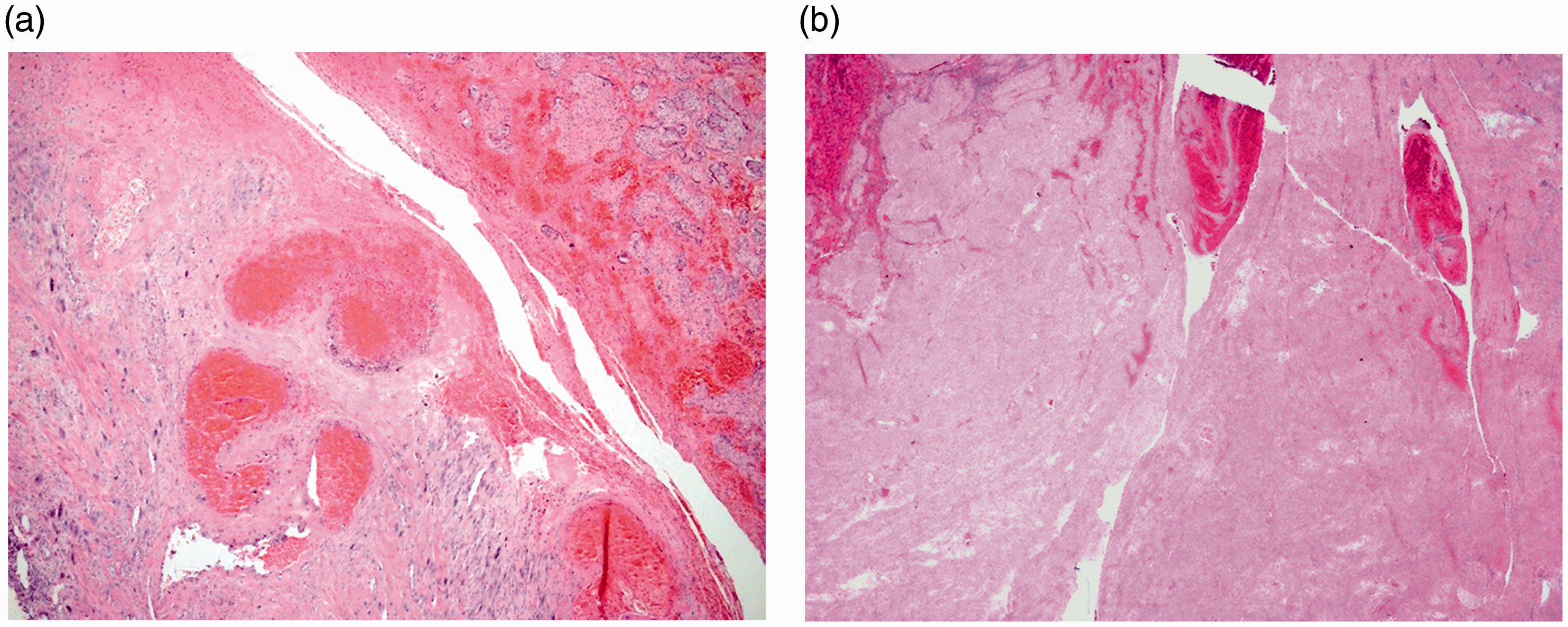
The initial slides suggested an EPS with prominent interstitial trophoblast with atypia (Figure 2) and foci of confluence.
10
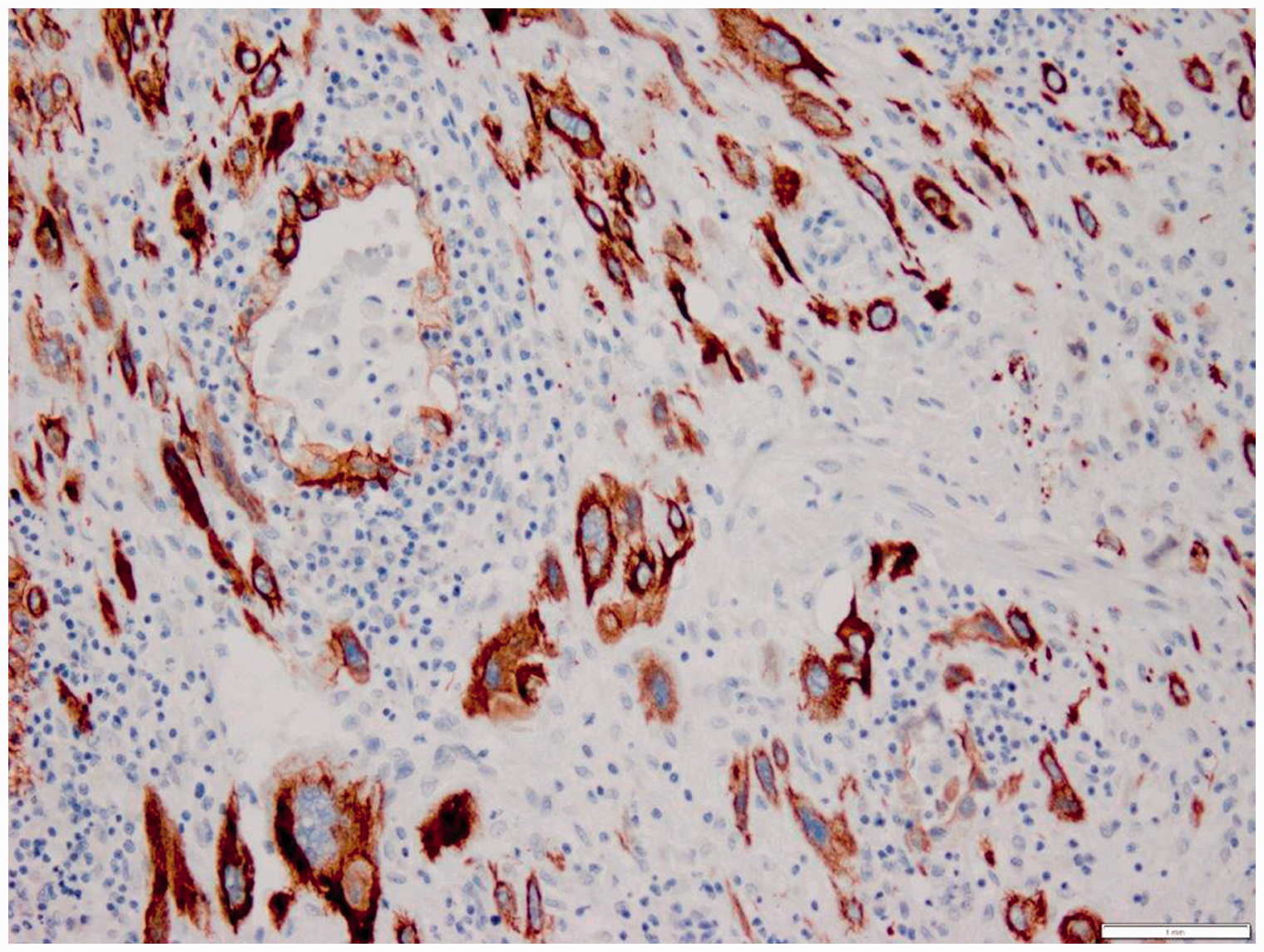
The villi appeared normal on both gross and microscopic examination. The intact decidua adjacent to the EPS-like area was chronically inflamed with dilated endometrial glands, which are characteristic of implantation sites.4,13 Decidua was focally deficient above the EPS-like area. Keratin stains (MAK6, high-molecular-weight cytokeratin) were positive in the interstitial trophoblast in the EPS-like area (Figure 3).
Section of implantation site showing prominent interstitial trophoblast with atypia, as seen in exaggerated placental sites in curettage specimens after spontaneous or therapeutic abortion.
1
It is not clear on H&E stain that muscle is being damaged. Keratin stain shows retroplacental interstitial trophoblast in the zone of muscle destruction and chronic inflammation.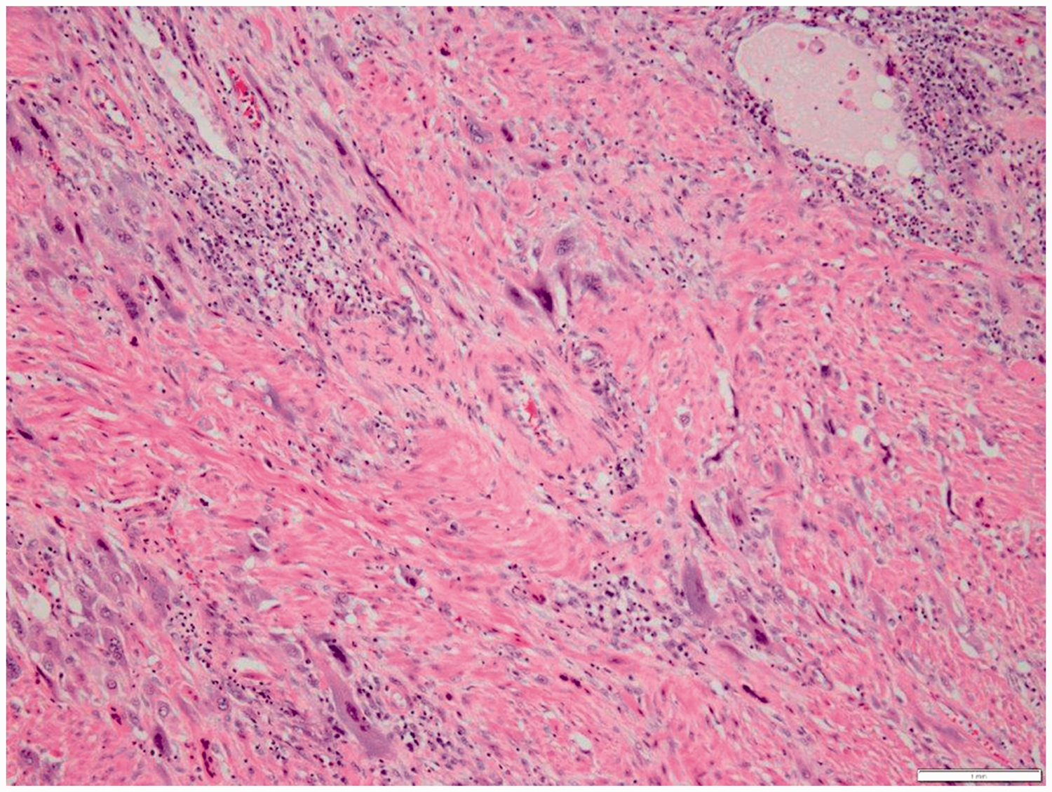
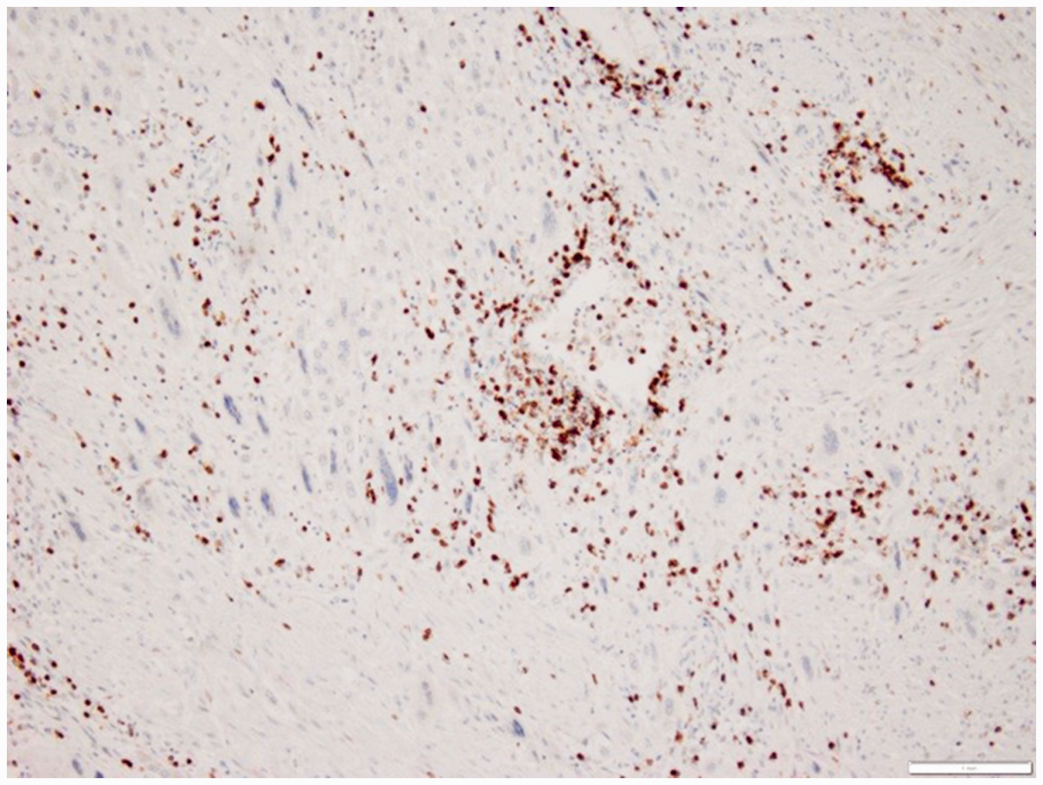
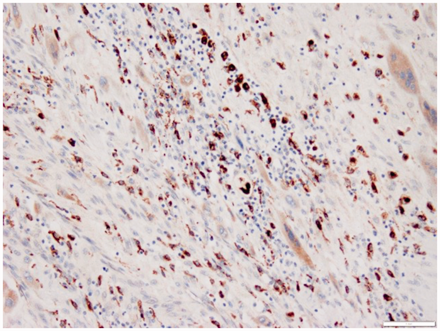
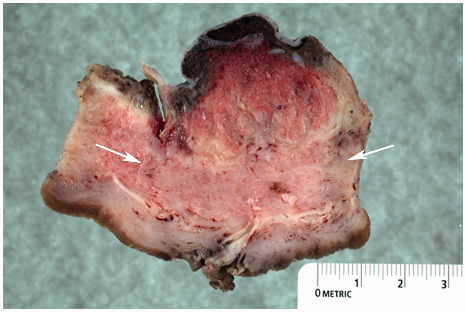
At first glance, the myometrium appeared normal in the EPS-like area (Figure 2), but MSA actin stain suggested considerable muscle destruction (Figure 4). The myometrium in this area was chronically inflamed, confirmed with immunostains for CD3 and CD68 (Figures 5 and 6). Both H&E and keratin stains showed endovascular trophoblast in dilated vessels deep to the interstitial trophoblast, as seen in placenta incretas.4–6 On reexamination of the gross specimen, a slice through the wall at the implantation site clearly demonstrated that the corpus myometrium was thinned by 5 mm under the placenta (Figure 7), and a diagnosis of second trimester placenta increta was made.
Actin stain shows retroplacental muscle destruction and chronic inflammation in the area simulating exaggerated placental site. This photograph is at lower magnification, to show that myometrium deep to the retroplacental zone is returning to a more normal pattern of actin staining. CD3 stain (T lymphocyte marker) shows the lymphoid element of the chronic inflammatory reaction at the site of muscle destruction. Lymphoid infiltrate was even more intense in decidua adjacent to the implantation site. CD68 stain (macrophage marker) shows the histiocyte element of the chronic inflammatory reaction at the site of muscle destruction. Some of the smaller or spindle shaped CD68+ cells may be mast cells. Gross photo taken after completion of histologic examination shows thinning of the wall under the implanted placenta (arrows) (2.4 cm), compared with adjacent myometrium (2.9 cm). This is away from the perforation site.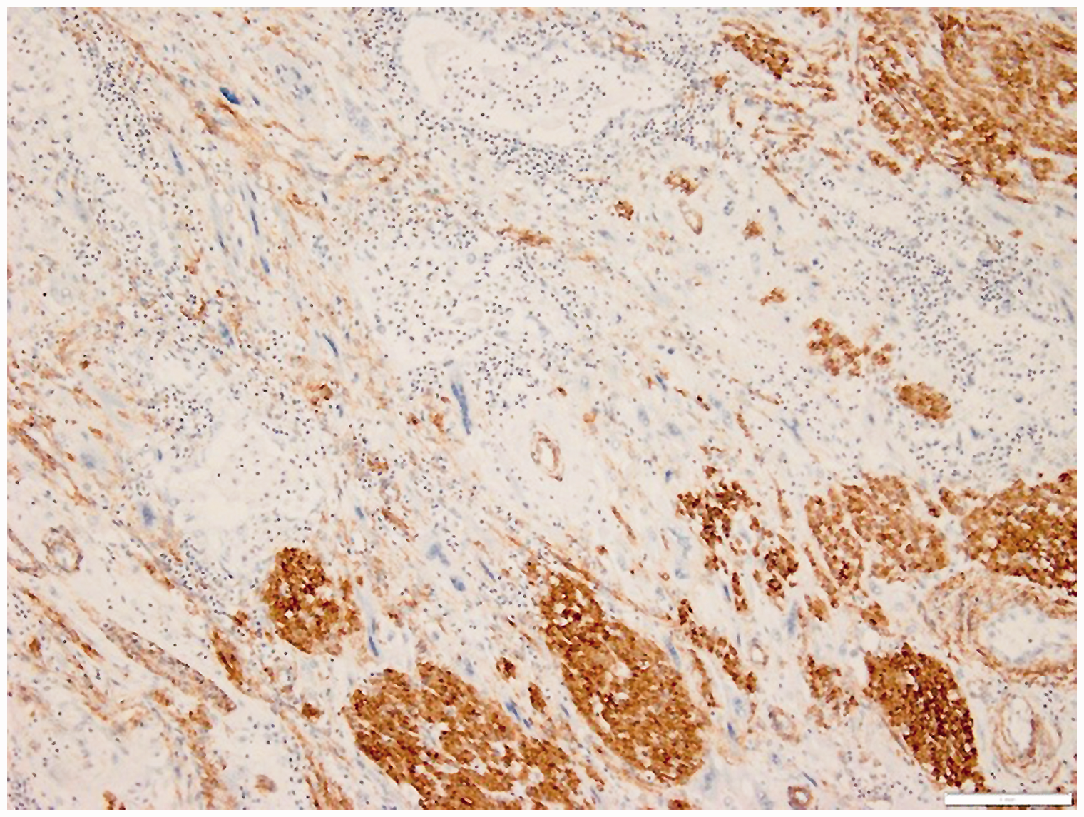
Discussion
Routine histologic sections have long suggested that direct implantation of villi onto or into myometrium explains morbid adherence, with loss of the normal cleavage plane in the decidua. However, it is now known that interstitial trophoblast provides a deep anchor for the placenta, using proteases to destroy basement membranes (and tissues).4,7,14–16 These proteases are regulated by antiproteases. 16 Although interstitial trophoblast appears to be composed of single or very small clusters of cells, appearing at first glance to be a less substantial anchor, it should be understood that a similar pattern is seen in tightly adherent protease-rich invasive carcinomas.4,17 The difference between normal anchoring and morbid adherence may, to some extent, reflect a disturbance in the protease–antiprotease balance.4,16 In the modern era, it is widely understood that “in placenta accreta, it is a common misperception that well vascularized villi must directly about smooth muscle.” 12 Diverse other experts have noted nonvillous trophoblast between villi and muscle,3,5,11 and there is now a general consensus that immunostains may help to identify trophoblastic cells.1,2,4,6,10,11
Decidua not only provides a deep anchor but also provides a substantial barrier to protect women from postpartum hemorrhage. 1 Although decidual deficiency is now recognized as a major factor in the pathogenesis and diagnosis of morbid adherence, a comprehensive review long ago suggested that decidual deficiency could not explain all morbid adherence.1,6,8,11 The concept of decidual deficiency still fails to explain many observations, including (1) increta of the corpus when the Cesarean section scar is in the lower uterine segment4,9; (2) wide variation in depth of penetration by increta, so it is not clear why every increta does not become a percreta 5 ; and (3) species variations wherein thinner decidua is not associated with deep invasion. 5 Given that trophoblast must normally invade deep decidua before accessing inner myometrium, it still seems likely that some decidual deficiency is secondary to placental invasion and destruction.1,5
Immunostains have proven useful not only for recognizing interstitial trophoblast at the invading front 4 but also for understanding the pathogenesis of morbid adherence. They have shown that depth of interstitial trophoblast and endovascular trophoblast is excessive in morbid adherence, as compared with normal implantation sites, with more tendency to confluence of interstitial trophoblast.6,10,11 No difference in measured thickness of interstitial trophoblast has been reported between second trimester and third trimester incretas. 6 Furthermore, no difference in thickness of interstitial trophoblast was noted in accreta versus increta versus percreta. 6 These findings suggest that neither increta nor percreta is due to excessive proliferation or invasion by nonvillous trophoblast,
We have previously written: Rather than the interstitial NVT moving down, we suggest that the myometrium may be moving up. This may be driven by myometrial tone,
18
with the greatest pressure exerted by the larger, more parallel myofibers of the outer third. Since myometrium is a complex and heterogeneous muscular tissue that can contract in various directions, uneven myometrial tone may at least partly explain the variable degree of myometrial destruction in incretas.
4
Although actin stains are often used to help identify basal plate myofibers in delivered placentas,1,19,20 it may be less widely appreciated that actin stains help to identify retroplacental myometrial damage in placenta increta.4,5 Khong and Robertson 5 identified retroplacental hyaline degeneration of myometrium with chronic inflammation many years ago, and routine use of actin stains helps to identify shriveled retroplacental myofibers, with associated chronic inflammation and edema, which may explain abnormal retroplacental zones on ultrasound in incretas. 4 Our previous work illustrated that retroplacental myometrial damage in first trimester increta may require actin stains to document shriveled myofibers with a substantial lymphoid infiltrate. 4 The findings in this second trimester increta are similar. Third trimester incretas may have more prominent edema that simulates autolysis at first glance, associated with chronic inflammation, but actin or trichrome stains are still helpful to identify shriveled myofibers that are strikingly unlike normal gestational hypertrophy of myocytes. 4 The most advanced stage of myometrial destruction we saw in increta was in a Cesarean scar pregnancy where interstitial trophoblast was apparently halted in its advance through the wall by a zone of marked globular elastosis in the C-section scar. 4 Adjacent to this scar was a large hypocellular zone with only a few residual myocytes and trophoblastic cells. 4 We believe that the demonstration of interstitial trophoblast at the invading front with keratin stains, combined with demonstration of myometrial damage at the invading front with actin stains, may facilitate the diagnosis of placenta increta. 4
Our previous work 4 confirmed standard teaching that myometrium is essentially normal in true placenta accreta. 1 More work is needed on Cesarean hysterectomies for true accreta, but our experience is that true accreta is primarily a disease of endovascular trophoblast that adheres to vascular wall smooth muscle within the decidualized endometrium. 4 This is harder to appreciate in postpartum hysterectomies. 4 We believe that using actin and trichrome stains helps to identify shriveled myofibers that favor a diagnosis of early increta over true accreta, 4 which should have essentially normal myometrium—by definition. 1 We believe that in the past, many early incretas have been classified as true accretas, because the subtle myometrial damage has not been obvious on routine H&E stains.
We feel that considerable further study of myometrial infiltration in normal implantation, in comparison to increta, merits consideration. Our current thinking is that when interstitial trophoblast first establishes its “foothold” in the inner myometrium, 1 it probably uses normal placental proteases to do so.15,16 Perhaps some degree of myometrial damage may be transiently demonstrable with actin stains in this phase of normal implantation. However, we suspect that myodestruction may continue throughout pregnancy in incretas, whereas it may cease significant activity after establishing its “foothold” in normal implantation. Antiproteases may play a role in these processes. 16
Despite normal interstitial trophoblast infiltration in the inner third of the myometrium, shear stress at the time of placental separation normally creates a plane of cleavage between decidua and myometrium.1,6 This may reflect that in normal implantation, the myometrium is sufficiently healthy to resist shear stress, while in placenta increta, damaged myometrium is unable to resist shear stress, causing basal plate myofibers to come out with the placenta.4,11,19,20
The clinical, gross, and immunohistochemical features of the present case suggest that a diagnosis of second trimester placenta increta could be made, despite the lack of any prior history of either decidual deficiency or sonographic/MRI features of placenta increta. Decidua was focally deficient, but this may have been due to exaggerated activity of the EPS-like interstitial trophoblast. This case had the typical retroplacental myometrial damage with chronic inflammation seen in placenta increta,4,5 although actin stains were necessary for microscopic diagnosis (Figure 4). This correlated with thinning of the wall on gross examination (Figure 7). Myometrial thinning is not the same thing as myometrial destruction by increta, since the lower uterine segment has a thinner myometrium; 1 but in this case the thinning is in the corpus. Keratin stains showed interstitial trophoblast at the invading front, as observed by diverse experts,3,5,11,12 which is consistent with the suggestion that placenta increta may be a disease of nonvillous trophoblast. 4 It is likely that retroplacental myometrial damage led to softening of the wall in this case, predisposing to perforation during the curettage procedure. This is of potential medicolegal significance. 21
Since both gross and immunohistochemical findings were characteristic of placenta increta, we concluded that the histologic appearance simulating exaggerated placental site did not constitute grounds to avoid a final diagnosis of placenta increta. Exaggerated placental site has never been reported to be associated with decidual deficiency or to cause myometrial thinning in the corpus.
With regard to pathogenesis, it is not clear whether the lymphoid infiltrate is to some extent a cause of myometrial damage at the invading front or a reaction to myometrial damage. The macrophages in the zone of retroplacental myometrial damage are of particular interest. As with pulmonary emphysema, smoking is a risk factor for morbidly adherent placentas. 22 When premature emphysema is due to alpha-1-anti-trypsin deficiency, 23 uninhibited macrophage proteases are thought to mediate destruction of lung parenchyma. The prominent macrophages at the invading front in this case of placenta increta raise the question whether a similar mechanism may be generally operative in placenta increta. More study is needed in this area.
Footnotes
Declaration of Conflicting Interests
The author(s) declared no potential conflicts of interest with respect to the research, authorship, and/or publication of this article.
Funding
The author(s) disclosed receipt of the following financial support for the research, authorship, and/or publication of this article: This work is supported by a grant for tissue research from The Genesee Hospital Foundation and Rochester General Hospital Foundation.