






Abstract
Objectives:
An abdominal aortic aneurysm (AAA) is a potentially life-threatening condition, the management of which has dramatically evolved over the past 2 decades with an increasing tendency toward endovascular repair (EVAR) rather than open surgical repair. Classically, contrast-enhanced multislice computed tomography (CT) angiography (CTA) is performed preoperatively for procedure sizing and EVAR planning. This entails voluminous contrast injection with risk of allergic reaction, nephropathy, and radiation exposure. Intra-vascular ultrasound (IVUS) has been increasingly used to guide EVAR procedures intraoperatively. The aim of this study is to investigate the accuracy of IVUS in sizing AAAs, device selection, and EVAR planning compared to the gold standard CTA.
Design:
This is a prospective observational study enrolling 10 patients who underwent standard infrarenal EVAR procedures performed for unruptured infrarenal AAAs over the course of 1 year. All patients had a preoperative CTA done upon which aneurysm sizing and device planning were performed, and the measurements obtained were compared to those obtained from intraoperative IVUS.
Methods:
All participating patients had unruptured infrarenal AAA, had no renal impairment, and had anatomical suitability for EVAR according to the instructions for use (IFU) of the device manufacturer. Primary endpoint was comparing anatomical measurements recorded by IVUS with those obtained from the preoperative CTA.
Results:
Mean age was 65.6 (±6.19), all patients were males and hypertensives and 4 (40%) had a positive family history for AAA. On comparing mean measurements taken by CTA and IVUS, there was no statistically significant differences with exception of maximal aortic diameter and aortic diameter at site of bifurcation (both p-values <.001). There were no statistically significant differences in length measurements between the 2 imaging modalities. Computed tomography angiography was more associated with neck thrombus detection, and IVUS was more associated with calcification detection.
Conclusion:
Although CT angiography is still the gold standard imaging modality for AAA, IVUS use is very beneficial in EVAR sizing and planning, in addition to intra-operative guidance of the procedure, saving the patient significant time, contrast administration, and radiation exposure, especially in patients with renal impairment and contrast allergy.
Clinical Impact
A preoperative CT angiogram is the gold standard required investigation for planning and sizing EVARs, with subsequent contrast injection entailing a risk of contrast induced nephropathy and allergic reactions. IVUS has been used as an adjuvant technique to guide EVAR stent graft deployment. However, our study concluded that it can also be reliably used in sizing and planning of the EVAR stent graft along with complementary non contrast imaging, especially in patients with high risk for contrast induced nephropathy and contrast allergy.
Introduction
An abdominal aortic aneurysm (AAA) is a potentially life threatening condition, that affects around 2% to 8% of patients in developed countries, with an annual incidence ranging from 0.4% to 0.67% in Western populations on average, or 2.5 to 6.5 aneurysms per 1000 person-years. 1
In the last 15 years, there has been a substantial clinical shift toward endovascular aneurysm repair of AAA. With these procedures, there is a significant amount of information that is required regarding aneurysm anatomy, sizing, and morphology prior to intervention. 2
Currently, both the European Society of Vascular Surgery and the Society of Vascular Surgery guidelines recommend performing a computed tomography (CT) angiography (CTA) for therapeutic decision making, device selection, and treatment planning.3,4 This tallies approximately 150 milliliters of contrast administration to the patient, after which an endovascular aortic aneurysm repair (EVAR) procedure is performed with a superadded mean of 114 milliliters of contrast (if the procedure is performed in a designated hybrid operating room [OR] suite) to 158 milliliters (if performed in an OR with a mobile c-arm). 5 Moreover, irradiation exposure is becoming more concerning to both patients and operators, both preoperatively and intraoperatively. Fusion technology and road-mapping have dramatically reduced the dosage of exposure; however, this can only be obtained in designated hybrid ORs which are overtly expensive, and not readily available on a large scale. 5
EVAR can be complicated by allergic reaction to contrast agents, in addition to contrast induced nephropathy and consequently renal impairment, with an estimated frequency of acute renal failure of 9%, an estimated frequency of chronic renal failure of 7%, a subsequent need for hemodialysis in 2% of patients, and up to 18% persistent clinically relevant renal functions deterioration after 1 year from intervention.6,7 In a study by Evelyn and her colleagues, the percentage of preoperative chronic kidney disease stages 1 to 5 were 16%, 42%, 31%, 6%, and 5%, respectively. 8
Consequently, finding a suitable replacement or adjunct imaging technique to plan and guide EVARs has been sought by many practitioners, especially for patients with history of renal impairment and allergy to contrast material, the most promising of which is the use of intravascular ultrasound (IVUS). Intravascular ultrasound has been used in guidance of peripheral arterial interventions owing to its ability to assess the plaque morphology, vessel diameter, presence of arterial dissections, and consequently, it helps in guiding of the selection of the most appropriate endovascular angioplasty technique, determination of the most appropriate stent size and assessment of the immediate technical outcome of the procedure. 9 Moreover, IVUS is becoming more popular in venous interventions, and many studies suggest its superior sensitivity in detecting stenotic lesions and planning landing zones for stenting compared to venography.10,11 In addition, IVUS has been widely used in aortic dissections, and is particularly useful in the positional confirmation of wire over which the device is to be deployed in the true lumen throughout the procedure, as well as checking proper alignment and coaptation of overlapping stent grafts. 12
Patients and Methods
This is a prospective observational cohort study that enrolled 10 patients who underwent standard EVAR procedure performed for unruptured infrarenal abdominal aortic aneurysms over the course of 1 year. Institutional review board and ethical committee approval was obtained prior to recruitment. Inclusion criteria were patients with unruptured infrarenal AAA who were willing to participate in the study, were indicated for intervention, 4 had no renal impairment (defined as serum creatinine less than 1.5), and with anatomical suitability for EVAR according to the instructions for use (IFU) of the device manufacturer. Patients with renal impairment, aneurysm rupture, those refusing to participate, or whose aneurysm anatomical measurements and configurations where outside the IFU of the device were excluded from the study. All patients had a preoperative multislice CT angiogram done upon which aneurysm sizing and device planning were performed through OsiriX MD (Pixmeo SARL, CH-1233, Bernex, Switzerland), a CE labeled, U.S. Food and Drug Administration (FDA)-approved class II Medical Device for diagnostic imaging in medicine. Volume rendering and centerline selection were done, followed by multiplanar views reconstruction. Diameters were measured in axial cuts and lengths were measured in stretch views. Measurements were made by the surgical team and confirmed by the EVAR device company representative (COOK MEDICAL LLC, Bloomington, IN, USA) prior to the planned intervention. Inter-rater concordance was not calculated statistically, as any minor discrepancies between measurements were addressed and a consensus was reached in team briefings prior to the procedure.
All procedures were done under fluoroscopy guidance in a Cath lab with IVUS assistance (Philips Systems, Visions PV .035, 1096 BC Amsterdam, Netherlands). In an attempt to minimize observer bias, a dedicated technician was provided by the IVUS company to perform the intraoperative measurements unaware of the preoperative CT angiogram measurements available with the operating team. The measurements were communicated to the operating consultant and the decision to change or proceed with the planned stent sizes according to the preoperative CT angiogram was left to the operating consultants, and none of them opted to change the operative and sizing plan. In order to minimize guidewire bias, all IVUS measurements were obtained over a stiff wire (Amplatz Super Stiff™—Boston Scientific, Marlborough, MA, USA). Various significant anatomical measurements were compared between IVUS and CTA (Table 1, Figure 1). Primary endpoint was comparing anatomical measurements recorded by IVUS to, and 220 those obtained from the preoperative CTA. The device used was the Zenith Flex® (COOK MEDICAL LLC, Bloomington, IN, USA). Sample size calculation was done using PASS program version 11, setting alpha error at 5%, and power at 80% result from previous study 13 showed that the mean contrast for computed tomography angiography was 123 +/- 50 while for IVUS it was 67 +/- 34. Based on that, the minimal needed sample is 10 cases.
Anatomical Measurements of Aneurysm Sizing Parameters.


Comparison of measurements taken by intra-vascular ultrasound (IVUS) and computerized topography angiography (CTA).
Results
Ten patients underwent IVUS-guided EVAR with a mean age of 65.6 (±6.19), all of them were males and hypertensives, 5 (50%) were diabetic, 8 (80%) had ischemic heart disease, none had renal or liver impairment, 2 (20%) had a previous history of stroke, 4 (40%) had a positive family history for AAA, and mean baseline serum creatinine was 1.09 (±0.23) (Table 2).
Patients’ Demographics and Baseline Clinical, Laboratory and Radiological Data.

Abbreviations: AAA, abdominal aortic aneurysm; CTA, computerized topography angiography; IVUS, intravascular ultrasound; SD, standard deviation; ALT, alanine transaminase.
With regard to aneurysm anatomical assessment, 7 patients (70%) had aortic neck thrombus and 4 patients (40%) had aortic neck calcification in CTA, while 4 patients (40%) had aortic neck thrombus and 5 patients (50%) had aortic neck calcification by IVUS, and mean neck angulation was 53.5 degrees (±3.89) (Table 2).
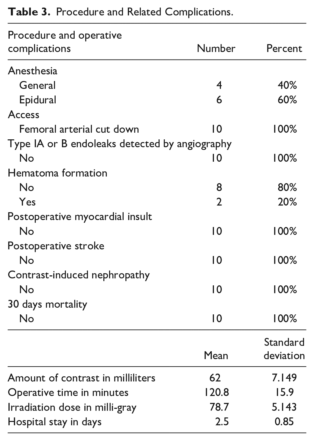
The procedure was done under general anesthesia in 4 patients (40%) and epidural anesthesia in 6 patients (60%), and all of the patients had a femoral vessel cutdown. Mean amount of contrast injected was 62 mL (±7.15), mean operative time was 120.8 minutes (±15.9), mean irradiation dose was 78.7 milli-gray (±5.143), and mean hospital stay was 2.5 days (±0.85) (Table 3).
Procedure and Related Complications.
Preoperative measurement with CT angiography showed that the mean D1 value was 27.95 mm (±2.9), mean D2 was 28.8 mm (±2.46), mean D3 was 64.8 mm (±15), mean D4 was 46.2 mm (±13.43), mean D5 was 9.8 mm (±0.62), mean D6 was 9.85 mm (±1.2), mean L1 was 14.6 cm (±0.56), mean L2 was 125.8 cm (±11.7), mean L3 was 192.2 cm (±21), mean L4 was 179.4 cm (±16.4), mean L5 was 66.4 cm (±21.5), mean L6 was 53.6 cm (±15), and mean neck angulation was 53.5 degrees (±3.89) (Table 4).
Comparing Mean Values of Measurements Taken by CTA and IVUS.
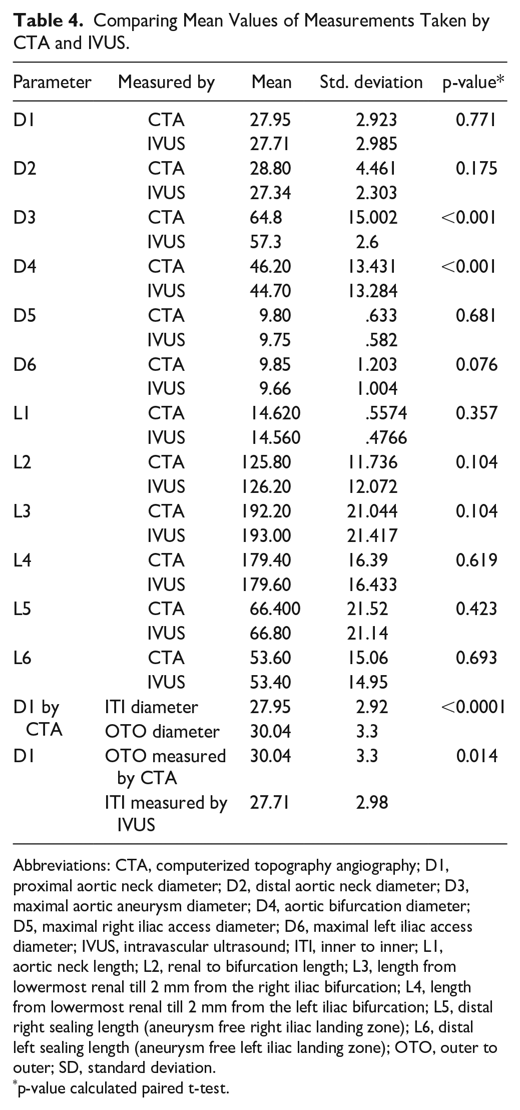
Abbreviations: CTA, computerized topography angiography; D1, proximal aortic neck diameter; D2, distal aortic neck diameter; D3, maximal aortic aneurysm diameter; D4, aortic bifurcation diameter; D5, maximal right iliac access diameter; D6, maximal left iliac access diameter; IVUS, intravascular ultrasound; ITI, inner to inner; L1, aortic neck length; L2, renal to bifurcation length; L3, length from lowermost renal till 2 mm from the right iliac bifurcation; L4, length from lowermost renal till 2 mm from the left iliac bifurcation; L5, distal right sealing length (aneurysm free right iliac landing zone); L6, distal left sealing length (aneurysm free left iliac landing zone); OTO, outer to outer; SD, standard deviation.
p-value calculated paired t-test.
Intra-operative measurement with IVUS showed that the mean D1 value was 27.71 (±2.98), mean D2 was 27.34 mm (±2.3), mean D3 was 57.7 mm (±3.68), mean D4 was 44.7 mm (±13.28), mean D5 was 9.75 mm (±0.58), mean D6 was 9.66 mm (±1), mean L1 was 14.5 cm (±0.48), mean L2 was 126.2 cm (±12), mean L3 was 193 cm (±15), mean L4 was 179.6 cm (±16.4), mean L5 was 66.8 cm (± 21.4), and mean L6 was 53.4 cm (±14.95) (Table 4).
On comparing mean values of measurements taken by CT angiography and IVUS, there were no statistically significant differences between D1, D2, D5, and D6 (p-values=0.77, 0.17, 0.68, and 0.08 respectively); however, there were statistically significant differences between measurements of D3 (maximal aortic diameter) and D4 (aortic diameter at site of bifurcation), with the mean D3 measurements 64.8 mm (±15) by angiography and 57.3 mm (±2.68) by IVUS (p-value <.001), and the mean D4 measurements 46.2 mm (±13.4) by angiography and 44.7 mm (±13.3) by IVUS (p-value <.001). There were no statistically significant differences in length measurements between the 2 imaging modalities with p=0.36, 0.1, 0.1 0.62, 0.42, and 0.69 for L1, L2, L3, L4, L5, and L6, respectively (Table 4, Figure 2).

Boxplot representation comparing mean measurements. CT, computerized topography; IVUS, intra-vascular ultrasound.
Computerized topography angiography was more associated with neck thrombus detection and IVUS was more associated with calcification detection (p=0.03 and 0.024, respectively) (Table 5).
Association Between Neck Thrombus and Calcification Detection in CTA and IVUS.
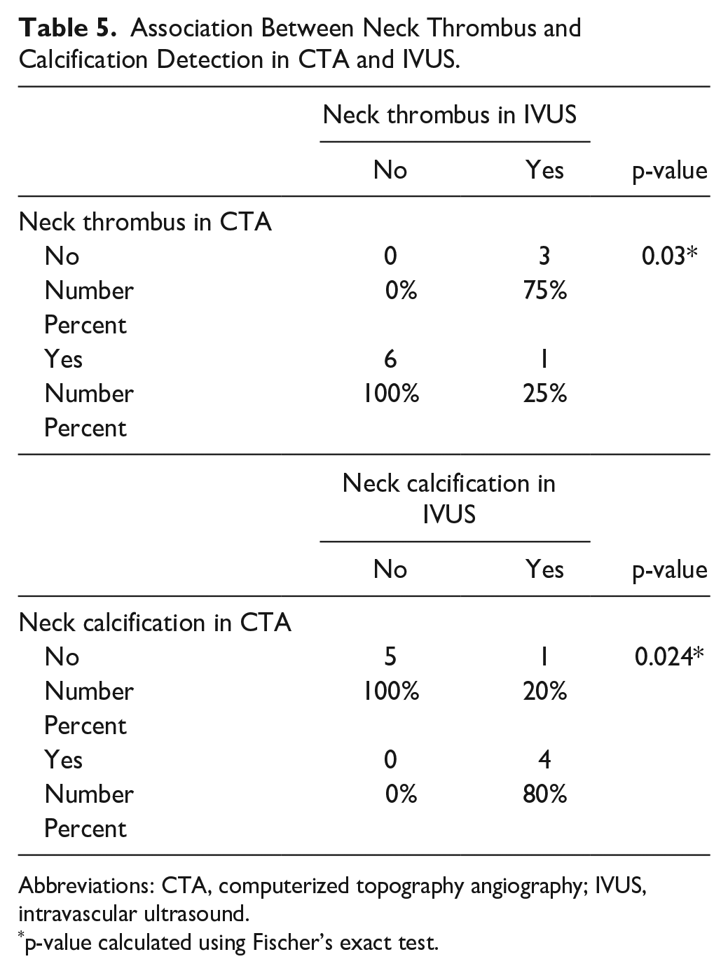
Abbreviations: CTA, computerized topography angiography; IVUS, intravascular ultrasound.
p-value calculated using Fischer’s exact test.
Mean outer to outer (OTO) D1 diameter measured by CTA (according to the device manufacturer’s IFU) was 30.04 mm (±3.3) and mean inner to inner (ITI) diameter measurement by CTA and IVUS were 27.95 mm (±2.92) and 27.71 (±2.98), respectively. There was a statistically significant difference between OTO D1 measurement using CTA and ITI measurement using CTA and IVUS with p-values of <.0001 and .014 and a mean difference of 2.09 and 2.33 mm, respectively (Table 4).
Discussion
CT angiography is the recommended gold standard for preoperative assessment, sizing and planning of EVARs, and classically, contrast angiography is the most dependable and popular imaging technique to guide and ascertain proper stent graft deployment intraoperatively. However, they both carry a superadded hazard of irradiation exposure, as well as contrast injection with subsequent risk of contrast-induced nephropathy and allergic reactions.
CO2 angiography has emerged as a replacement for iodine-based contrast in angioplasty for patients with peripheral vascular disease associated with renal impairment. Despite being used in certain centers, CO2 angiography shows limitations in EVAR; particularly in determining the proximal endograft landing zone (around 61% accuracy), especially in large aneurysms with scarce thrombotic apposition and a luminal volume greater than 95.9 mm3 (interquartile range [IQR]: 25.2). 14 Moreover, CO2 angiography needs special expertise and a learning curve, more operative time and radiation exposure, a 3-minute wait between injections to allow time for the complete absorption of the gas before a subsequent injection, in addition to an associated risk of bowel ischemia, and cannot be injected in the aorta above the level of the diaphragm for fear of embolization to the coronaries, cerebral, and spinal vessels.15,16 Furthermore, in spite of having some benefit in reducing contrast agents exposure during EVAR procedures, CO2 angiography has no role in aneurysm sizing and EVAR device planning, which is crucial for proper outcome, and exposes the patients to voluminous contrast and radiation. Consequently, IVUS use in EVAR planning and execution is of dire need, especially in patients with associated renal impairment.
Intra-vascular ultrasound is a critical technology that is extraordinarily useful during thoracic aortic stent graft surgery, especially in the presence of aortic dissections. Intra-vascular ultrasound technology provides detailed information about lesion morphology and precise visualization of vessel wall anatomy. This provides useful diagnostic information as well as aiding in treatment of aortic lesions. Current IVUS catheters operate in a high-resolution B-mode.
Intra-vascular ultrasound creates axial images perpendicular to the long axis of the catheter by transmitting sound waves covering 360 degrees around the tip of the catheter, which is delivered into the lumen of the vessel over a guidewire. This is achieved either mechanically or electronically. The mechanical system, produced by Boston Scientific Corporation, utilizes a flexible high-torque catheter with a quickly rotating ultrasound transducer located at the tip. Electronic systems, produced by Volcano Corporation (now acquired by Philips), utilize 64 miniaturized transducer elements located circumferentially around the tip of the catheter that are activated in sequence to produce an array of images. Information collected by IVUS transducers is reconstructed, displayed, and recorded visually on a separate console. This workstation facilitates the collection of measurements such as the diameter, circumference, and area of a vessel and allows for the capture of still images and video loops. 17
The most important is accurate assessment of the proximal and distal landing zones, particularly diameter and length measurements, as well as detection of arterial wall calcium and thrombus formation. Such assessment is generally performed preoperatively with the use of CTA, and this has become the imaging modality of choice. 18
However, in certain cases, IVUS can be used in addition to preoperative CTA. Intra-vascular ultrasound is particularly helpful in further assessing cases that are technically challenging on CT scan. In addition, IVUS can be used instead of CTA in patients who have renal impairment, for whom contrast material cannot be safely given, or in patients with an allergy to contrast agents. 19 In those patients, a preoperative non-contrast CT can be done to assess whether the aneurysm is anatomically eligible for EVAR or not, prior to proper intraoperative sizing by IVUS.
Within the same context, a pilot study conducted by G. Illuminati and his colleagues aimed at comparing IVUS assistance in EVAR to standard guidance of the procedure by angiography. In this study, 173 consecutive patients underwent EVAR, 69 procedures were IVUS-assisted with X-ray exposure limited to completion angiography for safety purposes because an IVUS probe does not yet incorporate a duplex probe (group A), and 104 were angiography-assisted procedures (group B). 20
They found that IVUS-assisted procedures required less volume contrast media than standard angiography-assisted procedures (60 ± 20 mL vs. 120 ± 40 mL, p<0.01). The mean duration of the procedure was comparable in the two groups (120 ± 30 min vs. 140 ± 30 min, p=0.07). No difference in renal clearance before and after the procedure was observed in either of the two groups (99.0 ± 4/97.8 ± 2 mL/min in group A and 98.0 ± 3/ 97.6 ± 5 mL/min in group B, p=0.28). There were no significant associations between postoperative mortality, morbidity, endoleaks, or arterial access complications between the 2 groups. 20
Another study by F. Pecoraro and his colleagues retrospectively compared 26 patients who underwent IVUS-assisted EVAR with propensity score matched 26 patients receiving classic contrast guided EVAR. The authors found a significant reduction of contrast medium volume (92 [vs. 51 ± 17] vs. 51 [20–68] mL; p=0.003) and radiation exposure including fluoroscopy time (12 [9–16] vs. 20 [12–25] min; p=0.001). No differences were observed in terms of glomerular filtration rate (86 [45–121] vs. 90 [38–117] mL/min; p=0.14) and operation time (176 [124–210] vs. 179 [120–210]; p=0.48) as well as survival (p=0.845) and freedom from re-intervention (p=0.834). 21
Both of these studies show the applicability and feasibility of the intraoperative use of IVUS in EVAR procedures, dramatically reducing the amount of contrast use without compromising accuracy of deployment or occurrence of complications.
Initial reports indicated that IVUS tended to undersize the aorta when compared with digital subtraction angiography and CT angiography. 22 However, when it comes to EVAR sizing and planning, our study showed no significant difference between measurements obtained by IVUS and those obtained through CTA, which are used for planning, sizing, and device selection, apart from D3 measurements (maximal aortic diameter) and D4 (aortic diameter at bifurcation), both of which do not significantly affect the size selection of the device, rather than confirming the indication for intervention (D3) and selecting the type of device used (D4).
The former can be explained by the fact that the IVUS catheter used in our study (10 MHz visions PV .035—Philips, the Netherlands—formerly Volcano, USA) has a maximal diameter of measurement of 60 mm, and the mean maximal diameter measured through CTA was 64.8 mm, which is beyond the measurement range provided by the catheter used. 23 The latter, however, is attributed to the general limitations of IVUS application, including sub-optimal calcification thickness measurement (despite better calcification detection) and thrombus detection compared to CTA, and the aortic bifurcation is a particular location for heavy calcification and thrombus loads. Recent and acute thrombus may not be clearly visualized by IVUS because of a high concentration of red blood cells and low fibrin deposition in these lesions. 24 With regard to calcium assessment, only the leading edge of calcium deposit is seen through IVUS, owing to the little penetration of the ultrasound beam through calcium. The apparent thickness of calcium reflection in grayscale IVUS is a function of transducer saturation by reflected ultrasound waves rather than anatomic thickness. 25
As regard the superior capabilities of detection of calcification using IVUS compared to CTA, despite being significant in coronary and peripheral arterial pathologies, calcification influence on sizing and planning of EVAR is imperative in the selection of access site as well as ascertaining the concordance of the degree of neck calcification with the intended device’s IFU to minimize stent migration. In our study, the superior calcium detection through IVUS did not signify any change in EVAR planning and execution, as opposed to coronary and peripheral arterial interventions, where calcification can influence intervention plan, access site, and equipment necessary for better outcome. Calcification can also be detected using non-contrast CT that is done during planning of the procedure. 26 The authors believe that the calcium load that is not detected using preoperative non-contrast CT would hardly be of significance in planning and execution of the EVAR procedure.
Thrombus load on the other hand is particularly significant in the assessment of aortic neck and access sites. Presence of extensive or circumferential thrombus hinders graft apposition and fixation, and may lead to type 1 endoleaks, in addition to the risk for distal embolization. Although IVUS is less sensitive in detecting aortic neck and access site thrombus, thrombus can be better detected using preoperative Doppler ultrasound prior to classifying the patient as having a suitable anatomy for EVAR according to the device manufacturer’s IFU. 26 All our patients had preoperative aortic and peripheral arterial duplex scans done prior to the non-contrast CTA, and no significant neck thrombus was detected in any of them.
In conclusion, clinically significant aortic neck thrombus or calcification comprising more than 50% of the neck diameter can be competently detected preoperatively using imaging modalities other than IVUS. Less degrees of calcification and/or thrombus load within the device IFU seldom affect the choice of EVAR device and plan.27,28
From our experience, several considerations need to be met while using IVUS for sizing and planning EVARs. With regard to diameter measurements, IFU of most commercially available EVAR devices reference OTO measurements in sizing. On the contrary, IVUS catheters measure ITI diameters only, which might not fall in line with the IFU for many of the EVAR devices. Although this would mainly affect the D3 measurements (maximal aortic diameter), it might be of significance in aortic necks with high thrombus load. This difference can be minimized by measuring the OTO diameter using a preoperative non-contrast CT, and considering the difference in measurements during device oversizing. Pertaining to length measurements, the type of guidewire used plays an important role. Length measurement using IVUS catheters can be done through pulling back the IVUS catheter and counting the 1 cm apart radio-opaque markers. This is highly affected by the degree of straightening of a tortuous aorto-iliac system, meaning that a straightened aorta using a super stiff guidewire would yield different length measurements than a tortuous one that is less straightened by a standard guidewire.
Additional drawbacks for using IVUS in EVAR sizing include inability of low-frequency catheters to visualize the entire circumference of the dilated aorta; consequently, branch visualization may be dependent on the path of the catheter within the aneurysm. 22 Moreover, although IVUS is useful post-deployment of the stent graft in assessing patency of the branch vessels and graft apposition to the neck, the lack of duplex function limits its use in the detection of endoleaks, necessitating a completion angiography post deployment of the stent to conclude the procedure. For these reasons, iodine-based contrast remains the most accurate and reliable guidance modality for EVAR. Our study advocates the use of IVUS in sizing and aortic measurements in patients with contraindication for contrast agent.
With regard to our study limitations, the sample size—although referenced by a sample size calculation—is considered a small number. Moreover, sampling method was convenience sampling without randomization, as all patients included in the study had both CTA and IVUS, and the paired measurements of both were compared in each patient individually. In addition, all patients included in our study had already fulfilled the eligibility criteria for EVAR according to the device manufacturer’s IFU. Intra-vascular ultrasound cannot be recommended as a standalone investigation to detect anatomical suitability of an aortic aneurysm for an EVAR; it can, however, be used to measure and select appropriate endograft size, as well as guide EVAR procedures in patients with contrast allergy or at high risk for nephropathy after deciding anatomical suitability for EVAR through a non-contrast CT scan.
Conclusion
Intra-vascular ultrasound use has been proven to be very beneficial in EVAR assistance intraoperatively, providing less contrast and radiation exposure, as well as providing more complementary anatomical data, such as branch vessels level, presence of dissections, detection of calcification, and ensuring graft apposition and patency. Our study suggests that preoperative measurements using IVUS could provide solid data regarding EVAR sizing and planning in addition to intra-operative guidance of the procedure, saving the patient significant time, contrast administration, and radiation exposure, and although it cannot replace CT angiography as the recommended gold standard imaging technique, it can be useful as an adjuvant modality in preoperative planning and intraoperative execution of the EVARs, especially in patients with contraindications for contrast injection.
Footnotes
Author’s Note
This research article was not presented in any of the international meetings or conferences so far.
Declaration of Conflicting Interests
The author(s) declared no potential conflicts of interest with respect to the research, authorship, and/or publication of this article.
Funding
The author(s) received no financial support for the research, authorship, and/or publication of this article.
