






Abstract
Introduction
With the rise in Caesarean deliveries, complications related to the procedure are increasingly encountered. Sonography has an indispensable role in the assessment of these complications and is often the first-line investigation of choice.
Discussion
Non-pregnant complications include Caesarean scar defects, scar endometriosis and malpositioned intrauterine devices. Complications related to future gestations include scar ectopic pregnancy, abnormal placentation and intrapartum uterine dehiscence or rupture. Key sonographic features of these conditions are illustrated. Pitfalls, mimics, limitations and indications for cross-sectional imaging are discussed.
Conclusion
Sound knowledge of the sonographic features of common non-pregnant and pregnancy-related complications of Caesarean delivery will facilitate accurate diagnosis, timely management and improved patient outcomes.
Introduction
Global data show a doubling in lower segment Caesarean delivery (CD) births in the past two decades from 16.0 million in 2000 to 29.7 million in 2015. 1 Although CD is potentially life-saving with valid elective indications, the procedure is not without risks. Compared to vaginal delivery, CD is associated with higher postpartum maternal morbidity including major puerperal and wound infections, thromboembolic events, 2 as well as high risks for fertility and future pregnancies in the long-term. 3 In addition, acute subfascial and bladder flap haematomas at the incision sites, Caesarean scar defects and scar ectopic pregnancies are complications unique to CD.
Imaging plays an important role in the assessment of these complications; in particular, ultrasound is indispensable in obstetrics and gynaecology as both a screening and diagnostic tool. Part 2 of this pictorial review illustrates the key sonographic features of common non-pregnant and pregnancy-related complications unique to and associated with CD. Sonographic pitfalls, mimics, limitations and indications for cross-sectional imaging are also discussed. All images were acquired at a single high-volume specialised women and children’s hospital.
Non-pregnant complications
Caesarean scar defects and dehiscence
The intact Caesarean section scar, if visible, is seen as a thin hypo- or hyperechoic line in the myometrium (Figure 1). Mild retraction of the anterior uterine border is considered nonpathological (Figure 1), whereas a defect in the posterior myometrial border constitutes a scar defect.
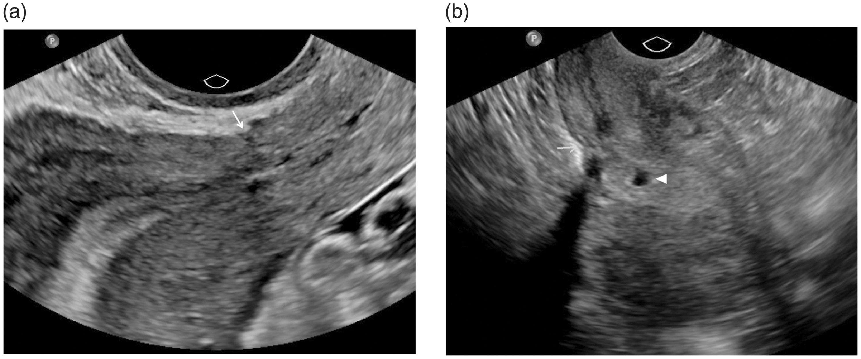
Intact Caesarean section scar. (a) Sagittal TVUS demonstrates an intact Caesarean section scar, seen as a thin hypoechoic line (arrow) without overlying myometrial thinning. (b) Sagittal TVUS in a different patient shows mild retraction at the anterior myometrial border at the Caesarean scar (arrow). A round intrauterine gestational sac outlined by hyperechoic decidua capsularis (arrowhead) is present in the decidualised endometrium.
A common complication is the development of a Caesarean scar defect (synonyms: scar niche, isthmocele), with reported prevalence ranging from 24% to 70% on transvaginal ultrasound (TVUS) and 56% to 84% on sonohysterography in women with one or more previous CD. 4 Scar defects are associated with abnormal postmenstrual spotting, scar pregnancies and uterine rupture in subsequent pregnancies. 5 Criteria for scar defect on sagittal TVUS include an anechoic space in the myometrium at the hysterotomy site with a depth of at least 2 mm and overlying residual myometrial thickness of less than 5 mm (Figure 2). 6 Gel sonohysterography and hysterosalpingo-contrast sonography with saline medium (HyCoSy) have higher sensitivity and can better characterise these defects (Figure 2). 5 Scar dehiscence is reserved for defects spanning 80% or more of the myometrium depth (Figure 3). 7
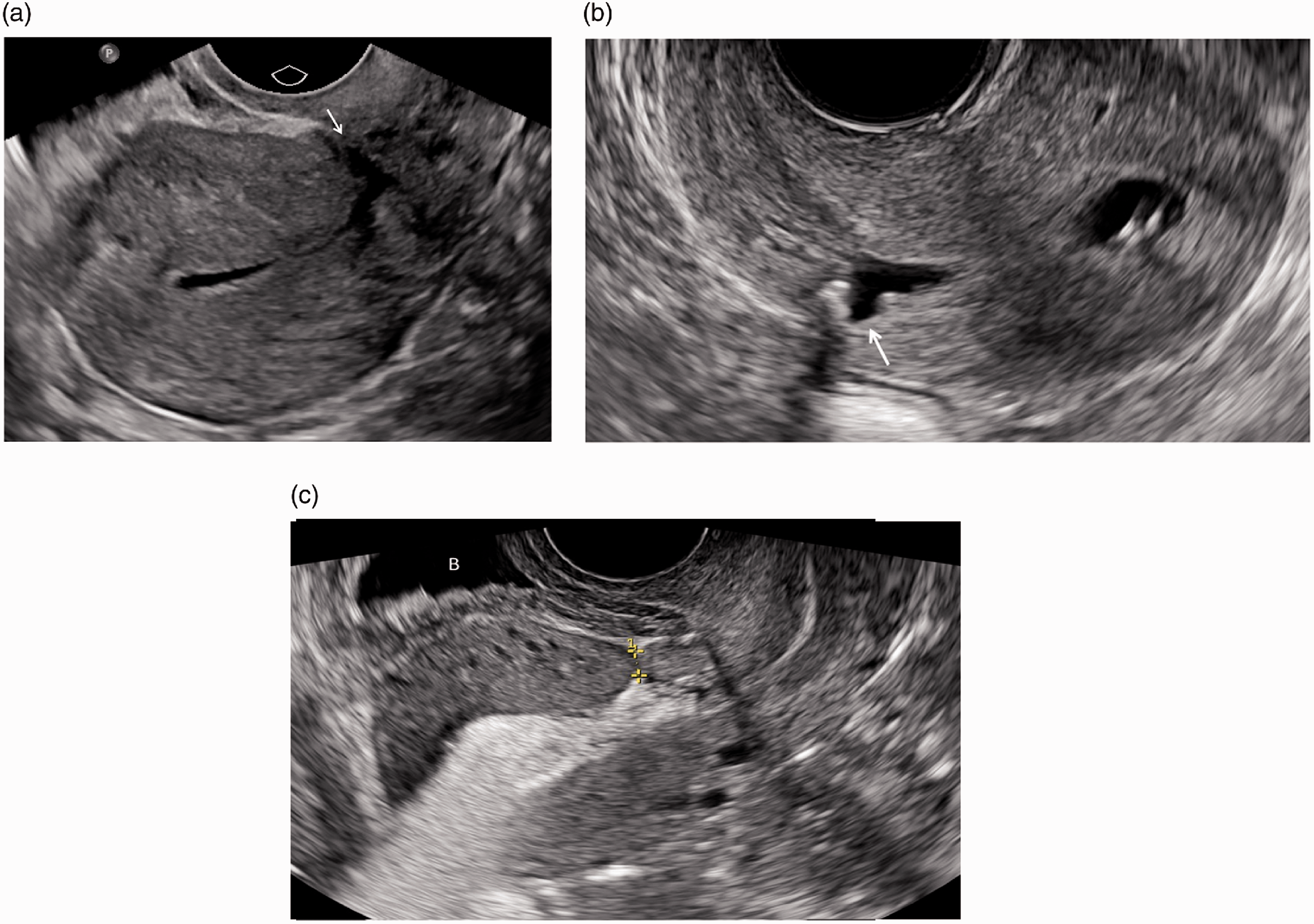
Caesarean scar defect. (a) Sagittal TVUS demonstrates a triangle-shaped scar defect (arrow). (b) Sagittal TVUS demonstrates a triangle-shaped scar defect distended with fluid (arrow). (c) Hysterosalpingo-foam sonography (HyFoSy) using ExEm® foam contrast shows a scar defect with overlying residual myometrial thickness measuring 4 mm (callipers). Other important sonographic measurements include length and depth of the niche and adjacent myometrial thickness.
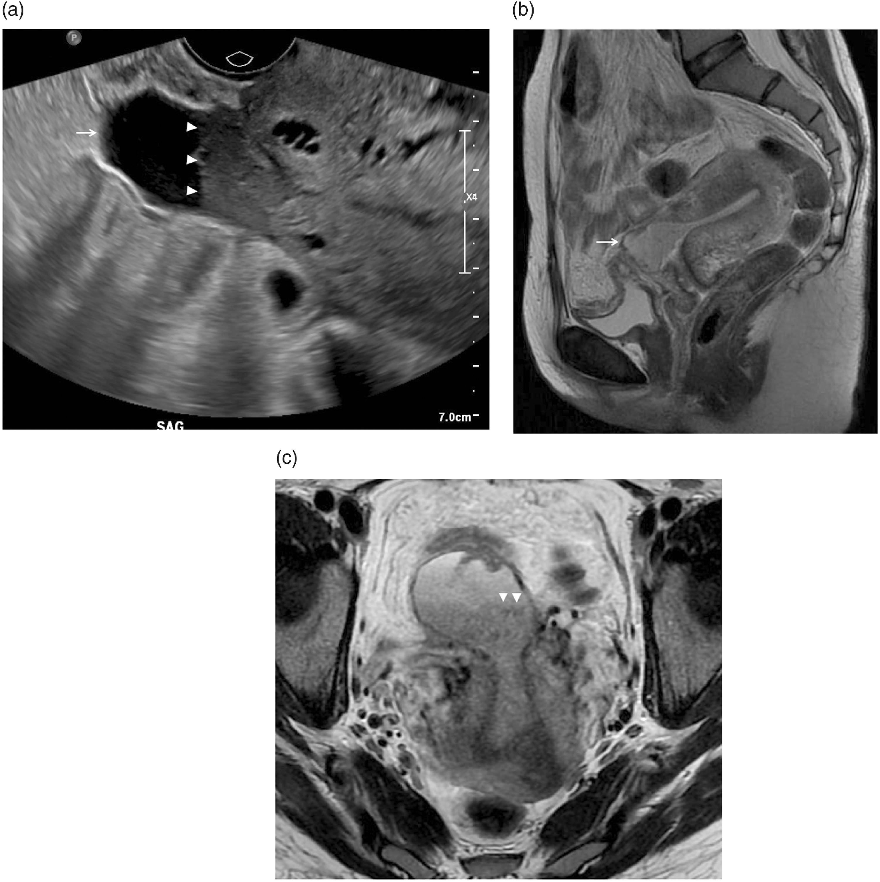
Caesarean scar dehiscence. (a) Sagittal TVUS demonstrates a cystic structure containing fluid-fluid level (arrowheads) in the region of the Caesarean scar covered by extremely thinned out myometrium (arrow) consistent with scar dehiscence containing blood products of varying ages. (b) Sagittal T2-weighted (T2W) MRI of the pelvis confirms that the fluid collection is continuous with the endometrium and only covered by thin T2W hypointense serosa (arrow), consistent with scar dehiscence. (c) Small field of view axial T2W MRI better demonstrates the fluid-fluid level (arrowheads). Scar dehiscence was confirmed intraoperatively. The patient was treated with excision of scar niche and uterine repair.
Abdominal wall scar endometriosis
Although scar endometriosis is a rare complication of open abdominal surgery, CD accounted for more than half the cases in several publications. 8 The most common presentation is catamenial pain and/or mass at the abdominal scar. 8 Ultrasound is the first-line modality for suspected anterior abdominal wall scar endometriosis and can exclude other focal lesions such as incisional hernias. Sonographic features are varied, but typical findings are of a solid inhomogeneous hypoechoic mass with irregular/ill-defined margins located in the skin, subcutaneous and/or muscle layer, and some vascularity is usually present (Figure 4). 9 Echotexture depends on composition of haemorrhage and fibrotic tissue. 9 Other reported sonographic findings include hyperechoic inflammatory ring, single vascular pedicle entering the mass and dilated peripheral feeding vessels. 9
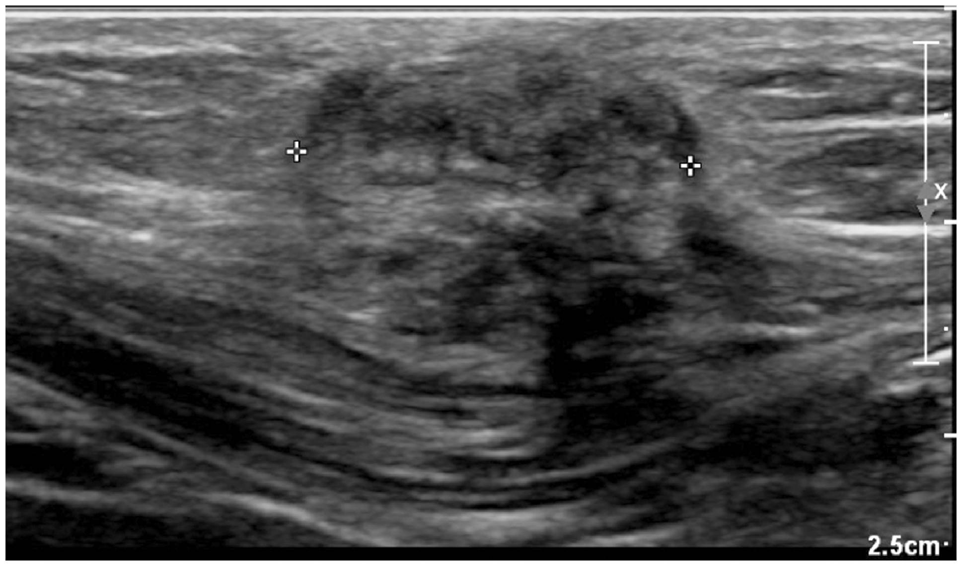
Abdominal wall scar endometriosis. High-resolution ultrasound of the abdominal scar in a patient with previous Caesarean delivery shows a heterogeneous hypoechoic solid nodule containing echogenic strands in the subcutaneous layer in the longitudinal plane (calipers).
Further evaluation with magnetic resonance imaging (MRI) may be required if sonographic features are inconclusive. In particular, scar endometriosis may be indistinguishable from hypertrophic surgical scars (keloids) and desmoid tumours on ultrasound. 9 Treatment is with wide surgical excision.
Displaced intrauterine contraceptive devices
CD is not known to increase risk of intrauterine contraceptive device (IUCD) complications (e.g. perforation, expulsion)10,11; however, IUCDs can occasionally become lodged within or perforate through the Caesarean scar defect (Figure 5), causing pain and/or abnormal uterine bleeding. Three-dimensional TVUS is increasingly replacing two-dimensional TVUS for IUCD assessment. Properly positioned IUCDs are located in the upper endometrial cavity with the arms extending laterally into the fundus.
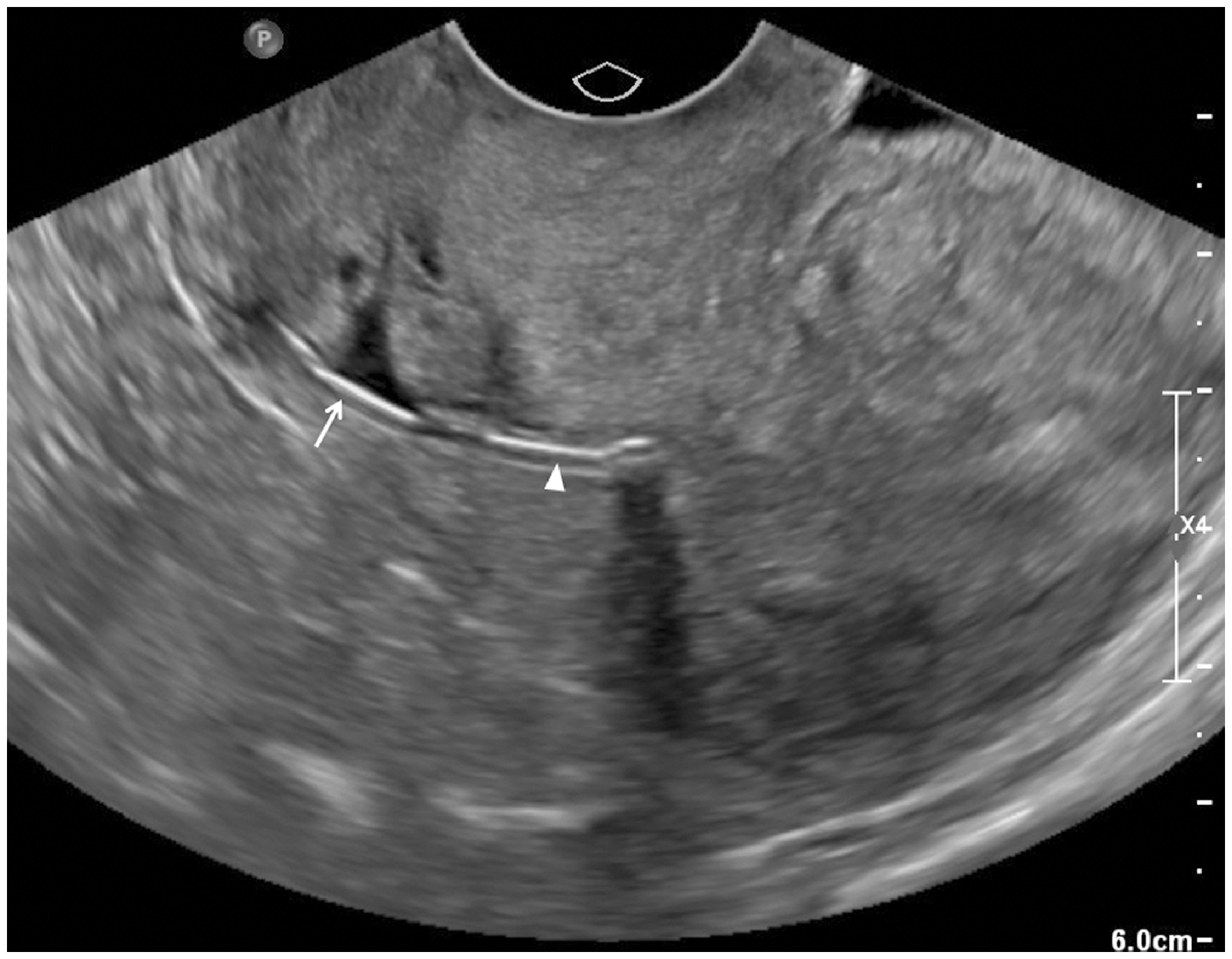
Displaced intrauterine contraceptive device. Sagittal TVUS for routine follow up of IUCD shows an echogenic arm of the device (arrow) lodged within the triangular anechoic Caesarean scar defect. The other arm is within the endometrial cavity (arrowhead). The stem of the IUCD is within the cervical canal but not imaged on this plane.
Pregnancy-related complications
Scar ectopic pregnancy
Caesarean scar pregnancy is a rare form of ectopic pregnancy involving implantation on or within the hysterotomy scar. The entity is associated with placenta accreta spectrum (PAS) and high risk of uterine rupture. TVUS is the primary diagnostic modality but may be supplemented by MRI in a minority. 12 Sonographic features of scar pregnancy include an empty uterine cavity, gestational sac implanted at the previous Caesarean scar with thin or absent myometrium between the gestational sac and bladder, and presence of high‐velocity, low‐resistance peritrophoblastic arterial flow (Figure 6). 12
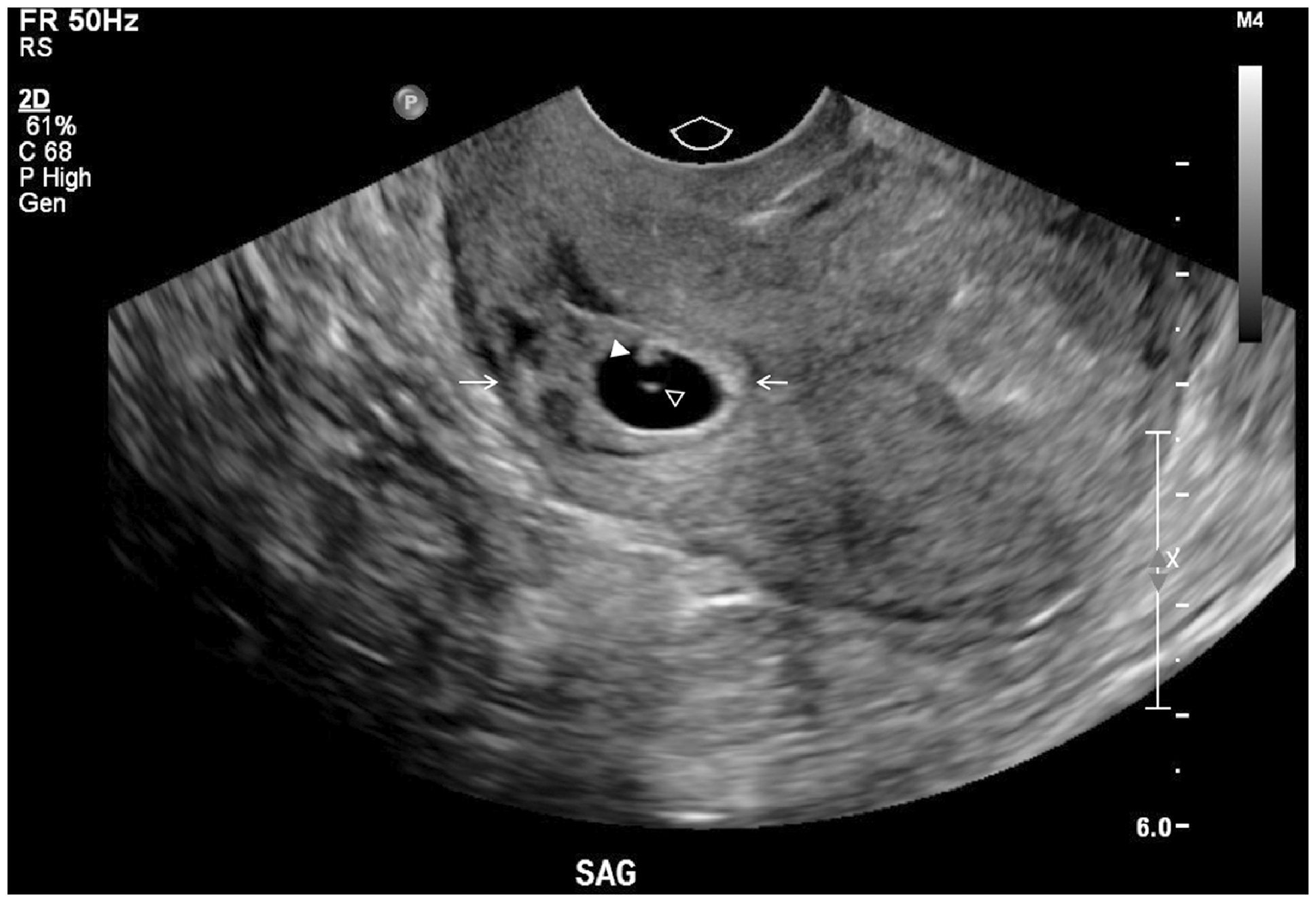
Scar ectopic pregnancy. Sagittal TVUS in a patient with two previous Caesarean deliveries shows scar pregnancy within the lower anterior myometrium (arrows). A 0.3 cm embryo (arrowhead) and yolk sac (empty arrowhead) are within the gestational sac.
Mimics of scar pregnancy include cervical ectopic pregnancy, low intrauterine pregnancy and miscarriage in progress. 13 Scar pregnancy is located at the level of the internal os, whereas cervical pregnancy is below the internal os within a ballooned/barrel-shaped cervical canal. 12 Distinguishing between scar pregnancy growing into the uterine cavity and a low intrauterine pregnancy is difficult, more so later in gestation. 14 For scar pregnancy and miscarriage in progress, the most useful sign is the sonographic ‘sliding sign’ which will be negative in scar pregnancy and positive in miscarriage in progress as the products of conception have detached from the implantation site. Additionally, miscarriage in progress will show an irregular elongated gestational sac with less florid vascularity (Figure 7).
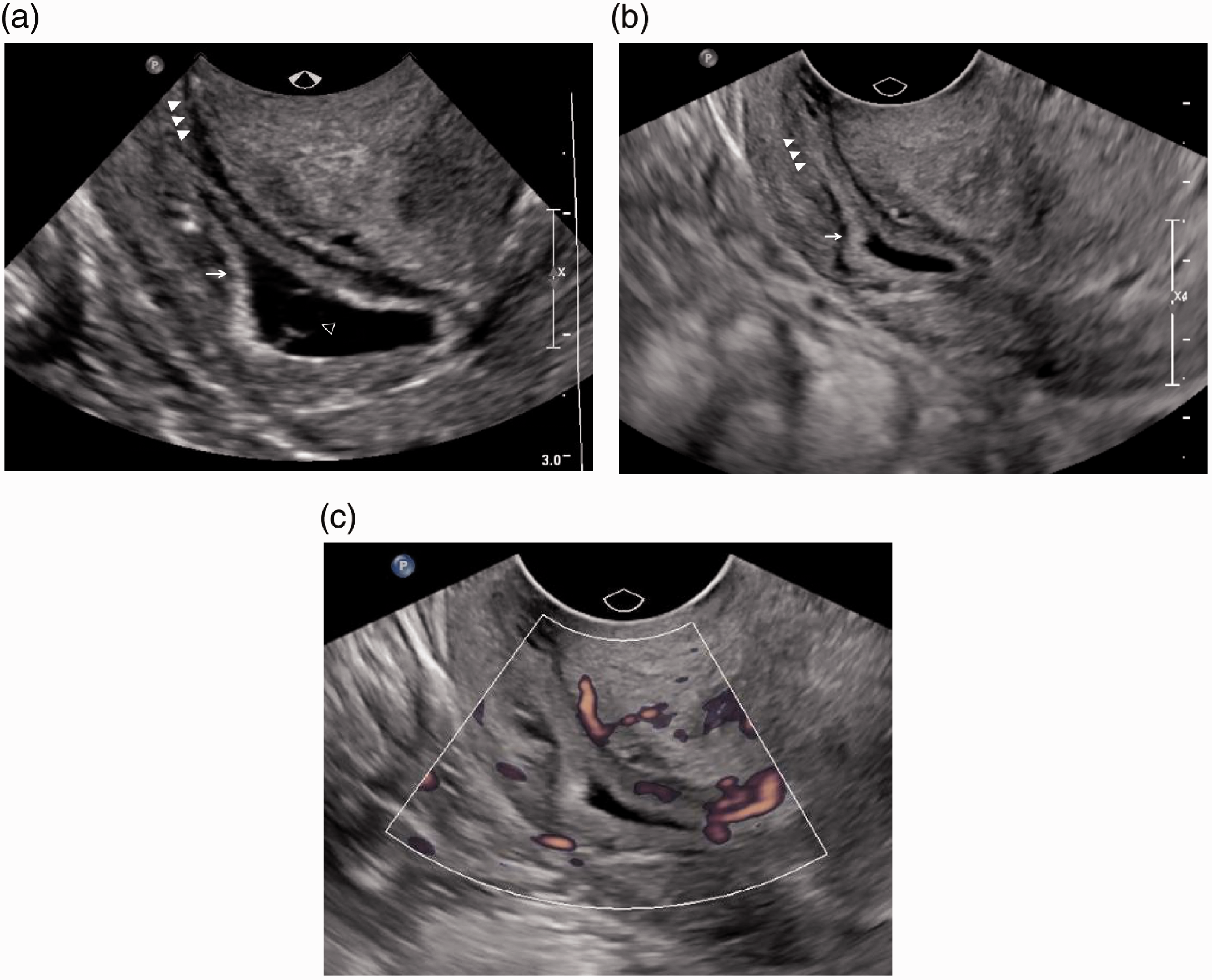
Miscarriage in progress. (a) Sagittal TVUS shows an elongated gestational sac (arrow) containing yolk sac (empty arrowhead) in the open endocervical canal (arrowheads). (b) Sagittal TVUS acquired the next day for follow-up shows further descent of the elongated gestational sac (arrow) within the cervical canal (arrowheads). The yolk sac is no longer seen. (c) Colour power angiography Doppler shows only mild vascularity around the gestational sac.
Placenta accreta spectrum and placenta previa
CD is an established risk factor for abnormal placentation with potential for life-threatening postpartum haemorrhage. An anterior low-lying placenta on antenatal US in a patient with previous CD is highly suspicious for placenta previa and/or PAS. 15 Thorough assessment of the placenta and follow-up TVUS at 32 weeks’ gestation are recommended to confirm a low-lying placenta (placenta less than 2 cm from the internal os) or placenta previa (placenta completely covering the internal os) (Figure 8). 15
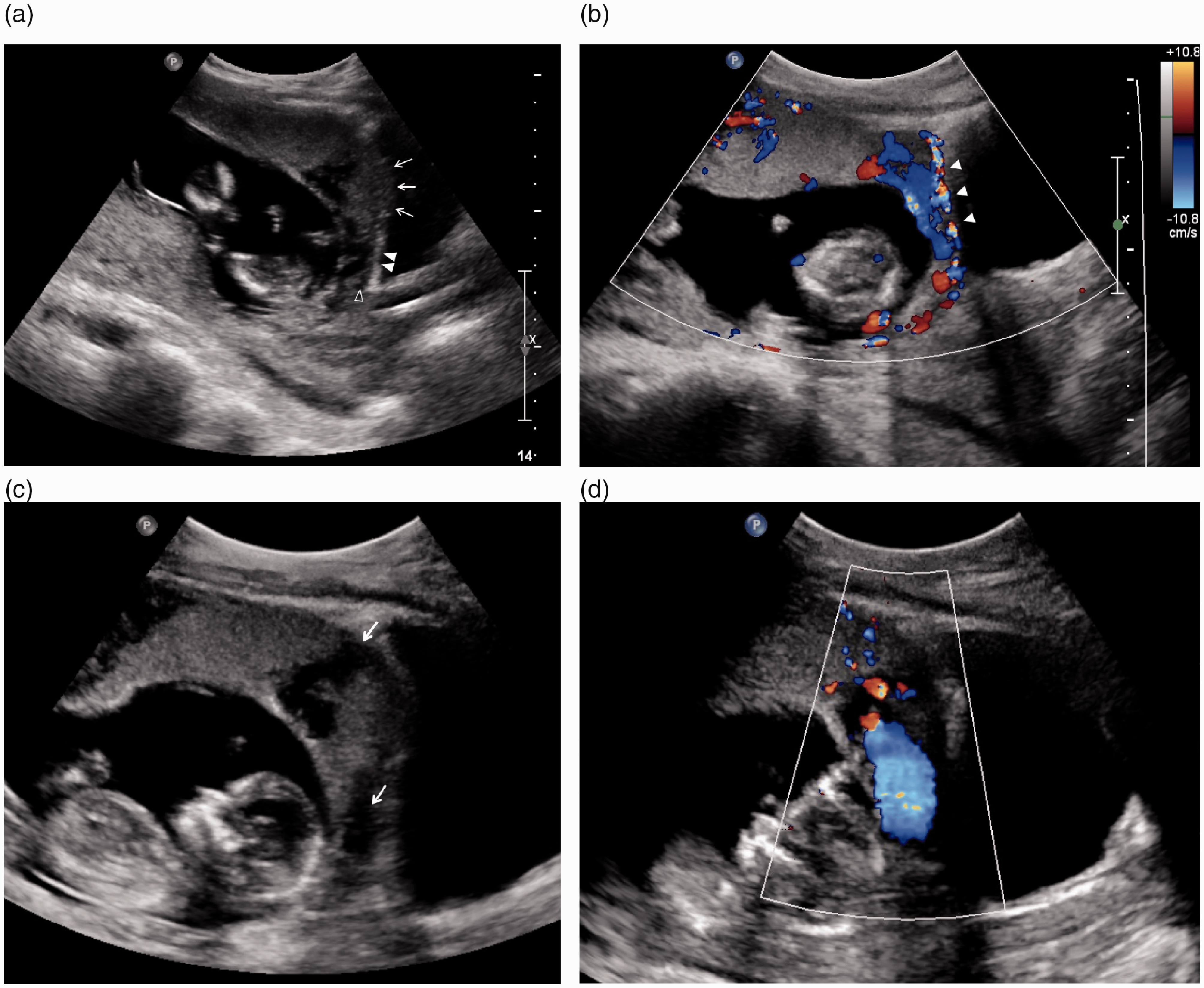
Placenta accreta spectrum and placenta previa. (a) Sagittal TVUS of a woman with previous Caesarean delivery shows an anterior low-lying placenta with focal exophytic bulge into the urinary bladder with loss of the retroplacental clear zone, no myometrium and bladder wall interruption suggestive of placenta percreta (arrows). The hypoechoic retroplacental clear zone (empty arrowhead) and hyperechoic bladder wall (arrowheads) are preserved more posteriorly. (b) Colour Doppler examination shows subplacental and uterovesical hypervascularity (arrowheads) in keeping with abnormally invasive placenta. (c) Sagittal transabdominal US of the same patient shows large and irregular lacunae (arrows). (d) High velocity flow is seen within the large lacunae on colour Doppler.
Sonographic features of PAS include loss of the hypoechoic retroplacental clear zone, retroplacental myometrial thickness less than 1 mm, placental bulge, focal exophytic mass, interruption of the hyperechoic bladder wall and abnormal placental lacunae on greyscale ultrasound (Figure 8). 16 Doppler features include placental and uterovesical hypervascularity, bridging vessels across and exiting the myometrium and placental lacunae feeder vessels (Figure 8). 16 MRI complements ultrasound in assessing depth and extent of myometrial invasion, especially in posterior placentation and suspected placenta percreta. 15 On MRI, myometrial invasion with loss of the normal T2-hypointense placental-myometrial interface, focal placental or uterine bulging and T2 dark intra-placental bands are suggestive of PAS and should be confirmed on at least two planes.
Intrapartum uterine dehiscence and rupture
Intrapartum uterine rupture is a rare but catastrophic event. Patients present with pain, vaginal bleeding and may be clinically unstable. The tear tends to occur at the CD scar and may be associated with trauma. Point-of-care ultrasound may be performed in the emergency setting but definitive diagnosis and management is with emergency laparotomy. Sonographic findings include haemoperitoneum, uterine wall defect, extrauterine fetal parts, protruding amniotic sac and anhydramnios.
Intrapartum uterine dehiscence, on the other hand, is often clinically occult. Sonographic findings include thinned myometrium, haematoma at the dehiscence and prolapsed amniotic sac or fetal parts (Figure 9).
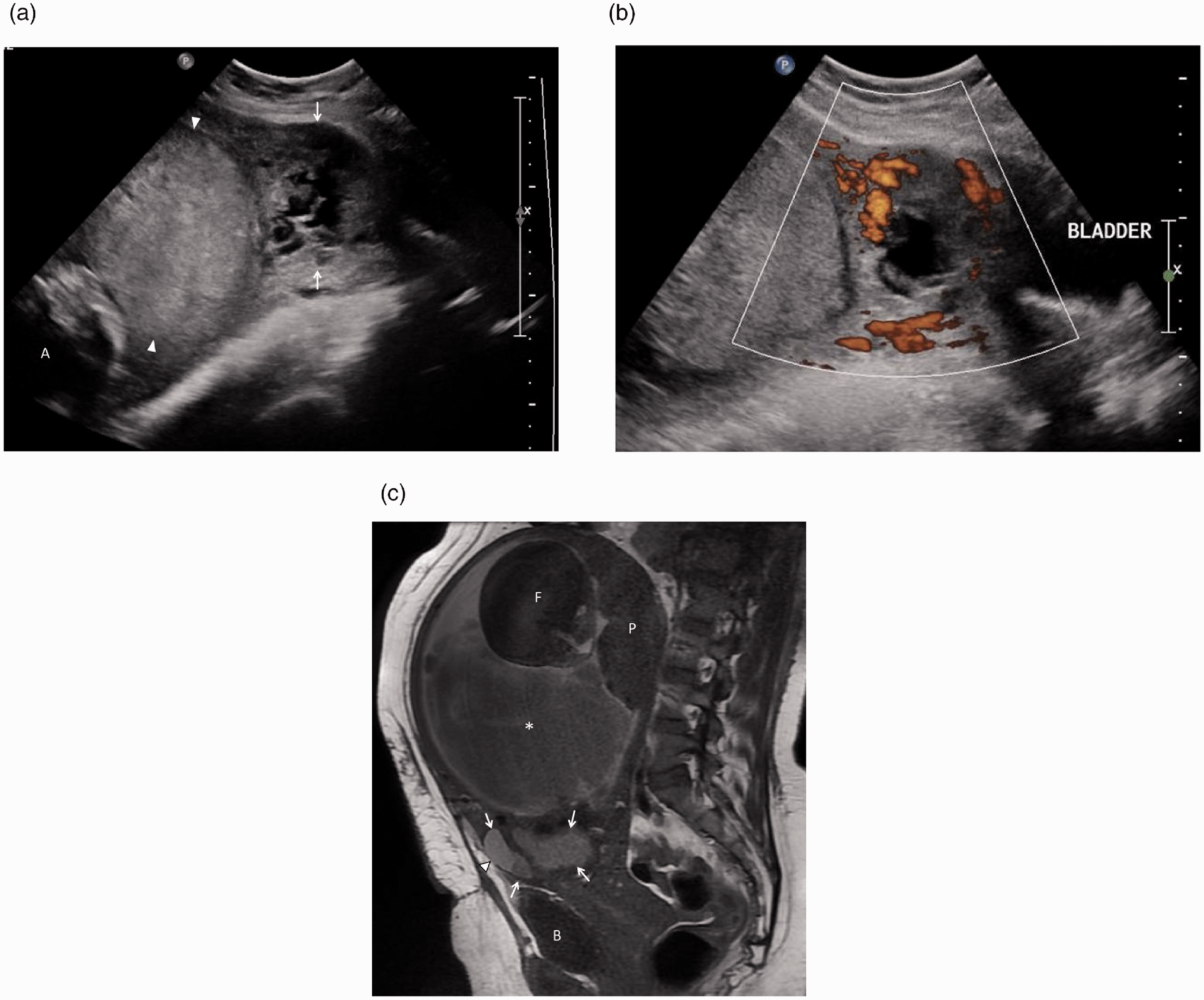
Intrapartum uterine dehiscence. (a) Sagittal TVUS shows complex mass in the expected location of the Caesarean scar suggestive of haematoma (arrows), which raised suspicion for the possibility of partial scar rupture. More superiorly, the hyperechoic avascular structure in the inferior gestational sac could represent retrochorionic or subamniotic haematoma (arrowheads). (b) Complex mass at the scar shows peripheral vascularity, probably uterine vascular channels. (c) MRI of the same patient was performed three weeks later. Sagittal T1-weighted MRI shows T1-hyperintense haematoma at the scar (arrows) with focal uterine dehiscence anteriorly (arrow head). MRI characterised the large haematoma as subamniotic (*). B: bladder; F: fetus; P: placenta.
Limitations of ultrasound and indications for cross-sectional imaging
Ultrasound has a limited role in the assessment of bowel adhesions, which are best assessed on CT. In addition to the previously discussed indications, cross-sectional imaging is also useful when ultrasound findings are inconclusive. The most appropriate imaging modalities for common non-pregnant and pregnancy-related CD complications are summarised in Table 1.
Imaging investigations for common non-pregnant and pregnancy-related complications of Caesarean delivery.
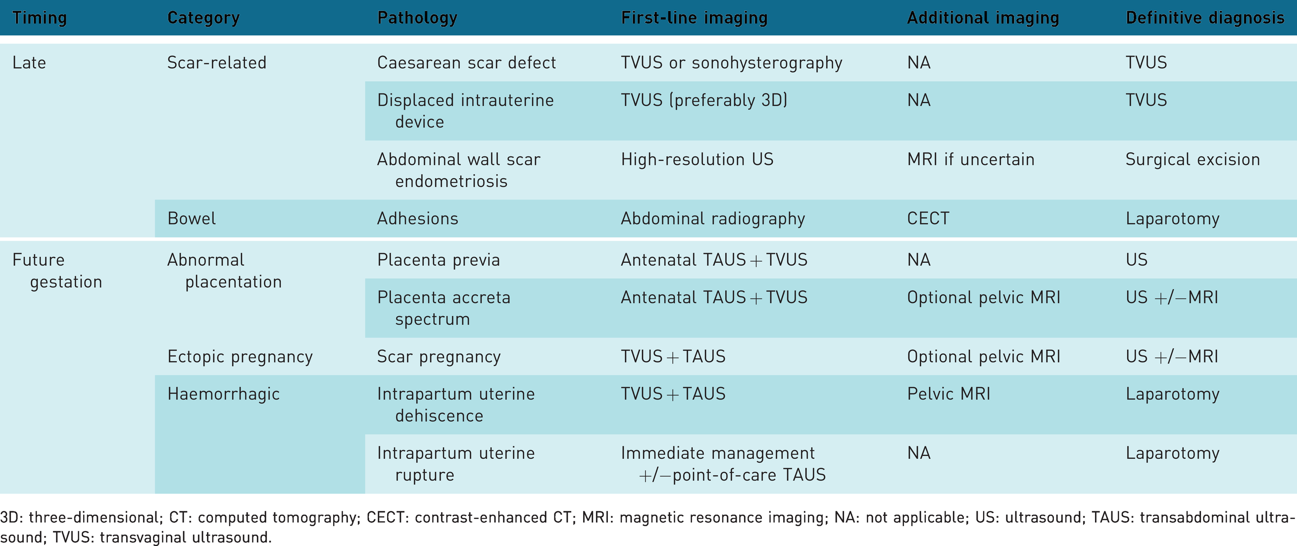
3D: three-dimensional; CT: computed tomography; CECT: contrast-enhanced CT; MRI: magnetic resonance imaging; NA: not applicable; US: ultrasound; TAUS: transabdominal ultrasound; TVUS: transvaginal ultrasound.
Conclusion
Ultrasound imaging plays an important role in the assessment of CD-related complications at various stages following delivery. Scar defects and IUCDs are best assessed on TVUS, while incisional endometriosis may require MRI for confirmation. During antenatal ultrasound, it is important to assess for abnormal placentation and scar ectopic pregnancies in all patients with previous CD. Sound sonographic knowledge will facilitate accurate diagnosis, timely management and improved patient outcomes.
Footnotes
Declaration of conflicting interests
The author(s) declared no potential conflicts of interest with respect to the research, authorship, and/or publication of this article.
Funding
The author(s) received no financial support for the research, authorship, and/or publication of this article.
Ethics approval
Ethics approval was waived by the Centralised Institutional Review Board at Singapore Health Services (SingHealth) for this educational pictorial review of anonymised patient images.
Guarantor
ET.
Contributorship
LCL and HELT conceived the study. ET, TSET and LCL researched literature. ET wrote the first draft of the manuscript. All authors reviewed and edited the manuscript. All authors approved the final manuscript and were in agreement of the submission.
Acknowledgments
The authors would like to thank all the sonographers at our institution.
