






Abstract
Purpose:
Placenta accreta spectrum encompasses abnormal placental attachment disorders characterised by progressive invasion into the uterine wall. Its incidence is rising due to increasing caesarean section rates and advanced maternal age, posing significant risks such as haemorrhage, uterine rupture and hysterectomy. Early and accurate diagnosis is critical for optimizing maternal and neonatal outcomes.
Findings:
Diagnosis of placenta accreta spectrum is possible in early pregnancy using transvaginal ultrasound. Key indicators include caesarean scar pregnancy, anterior myometrial thinning, placental lacunae, loss of the clear zone, bladder wall interruption and uterovesical hypervascularity. These findings facilitate early counselling and tailored management to mitigate emergency surgical complications. Evaluating the gestational sac’s relationship with the endometrial cavity in early pregnancy is essential in predicting pregnancy progression and associated risks, including uterine rupture or the need for caesarean hysterectomy. When transvaginal ultrasound provides limited views, transabdominal ultrasound may offer superior views to understand the relationship between the gestational sac and the endometrial cavity. Serial ultrasound monitoring is recommended in women with risk factors, particularly if they are symptomatic, to track pregnancy progression accurately.
Conclusion:
This pictorial review consolidates early gestational imaging findings to enhance diagnostic precision. It highlights standardised ultrasound criteria proposed by international consensus groups and explores magnetic resonance imaging as an adjunctive tool for cases with inconclusive ultrasound findings. By providing a comprehensive visual guide, this review aims to improve early detection, refine surgical planning and enhance maternal outcomes in placenta accreta spectrum management.
Introduction
Placental accreta spectrum (PAS) is a term used to describe a rare pregnancy condition in which there is abnormal trophoblast invasion of the placenta into the uterine myometrium. It can be divided into three categories: (a) placenta accreta, where the placental villi penetrate only to the surface of the myometrium; this is the most common; (b) placenta increta, where the placenta villi invade into the myometrium and (c) placenta percreta, where the villi invade beyond the myometrium to the uterine serosa and may involve adjacent organs such as bladder and parametrium.1,2
PAS is a potentially life-threatening condition that can result in significant maternal mortality and morbidity at any gestation. During early pregnancy, there is an increased risk of uterine rupture, bleeding after dilatation and curettage necessitating hysterectomy and extensive blood transfusions to manage life-threatening haemorrhage. 3 These complications contribute to prolonged recovery times and substantial physical and emotional distress for affected patients. In addition, PAS places a considerable burden on the healthcare system, which is multifaced, involving the direct costs associated with medical interventions and prolonged hospital stays 2 and also the indirect costs related to long-term consequences for maternal health and well-being, leading to indirect economic and social impacts.
Diagnosis of PAS in early pregnancy has the potential to reduce the impact on both patients and the healthcare system by allowing early recognition and prompting comprehensive management plans to minimize the need for emergency surgery and potentially reduce blood loss. For those patients who would choose to continue their pregnancy, it also provides an opportunity for appropriate counselling and surveillance to allow for a safer delivery by ensuring adequate surgical planning. 4
This pictorial review aims to identify and elucidate the early signs of PAS to enhance diagnostic accuracy and improve clinical outcomes. By providing a detailed visual guide, this review seeks to assist in recognizing early indicators of PAS, thereby facilitating timely and effective intervention.
Incidence
The worldwide incidence of PAS is rapidly rising. 5 However, despite the rising incidence, PAS is still rare (0.79–3.11/1000 births after prior caesarean delivery). 6 The rate in Australia is approximately 1 in every 2000 births. 1 The incidence of PAS will vary among diverse populations due to the differing prevalence of risk factors, 7 and is likely to be higher in populations with increasing rates of caesarean delivery and advancing maternal age. However, the overall rarity of PAS poses a challenge in defining and reaching a consensus on optimal criteria for diagnosis and management. 5
Risk factors
Previous caesarean delivery is the biggest risk factor for PAS. The risk increases with each subsequent caesarean section from 0.3% in women with one previous caesarean delivery to 6.74% in women with five or more caesarean deliveries. 8
Placenta praevia is another risk factor with PAS occurring in 3% of women diagnosed with placenta praevia and no prior caesarean deliveries. 2 The risk is further increased in the setting of placenta praevia and prior caesarean deliveries to 3%, 11%, 40%, 61% and 67%, for the first, second, third, fourth and fifth or more caesarean deliveries, respectively. 9
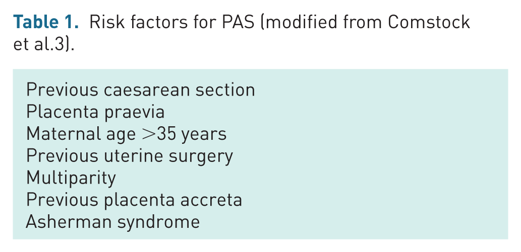
Other risk factors for PAS include advanced maternal age, multiparity, previous uterine surgery such as curettage or myomectomy, previous placenta accreta and Asherman syndrome.2,3,10 These risk factors are summarsied table 1. PAS can occur even in the absence of identifiable risk factors. Therefore, suspicion should be heightened if ultrasound findings suggestive of PAS are observed in women without apparent risk factors.
Risk factors for PAS (modified from Comstock et al.3).
Ultrasound findings of early placental accreta spectrum
Placental accreta spectrum and caesarean scar pregnancy
The finding of a caesarean scar pregnancy (CSP) may be one of the first signs of PAS on a first-trimester ultrasound as demonstrated in Figure 1. As caesarean section rates continue to rise, there is a corresponding increase in the incidence of CSPs being identified in women with previous caesarean deliveries. Several studies have described CSPs as a precursor of PAS.3,11–13 Timor-Tritsch et al. 11 explored the natural progression of CSP in ten women, diagnosed prior to 10 weeks’ gestation, who chose to continue their pregnancies. All ten patients had developed sonographic features of morbidly adherent placenta in the second trimester, underwent caesarean hysterectomy at delivery and placenta percreta was confirmed at histological examination. Based on this, the authors proposed that PAS is a continuum of CSP, eventually resulting in a morbidly adherent placenta that is classically seen with advancing gestation.

A case of a 40-year-old patient with one previous caesarean section who was diagnosed with a CSP at 6 weeks of gestation. She was asymptomatic at this time and chose to continue the pregnancy. She was delivered at 34 weeks via caesarean section and hysterectomy. (a) Ultrasound sonography (USS) at 6 weeks demonstrates a low-lying gestational sac located within the caesarean niche. Indicates caesarean niche.  Identifies the internal os. (b) Transabdominal USS illustrating a low-lying gestational, extending into the caesarean niche. (c) Thin anterior myometrial wall thickness at 6 weeks. (d) Transabdominal USS at 31 weeks featuring placental bulge, lacunae and loss of clear zone.
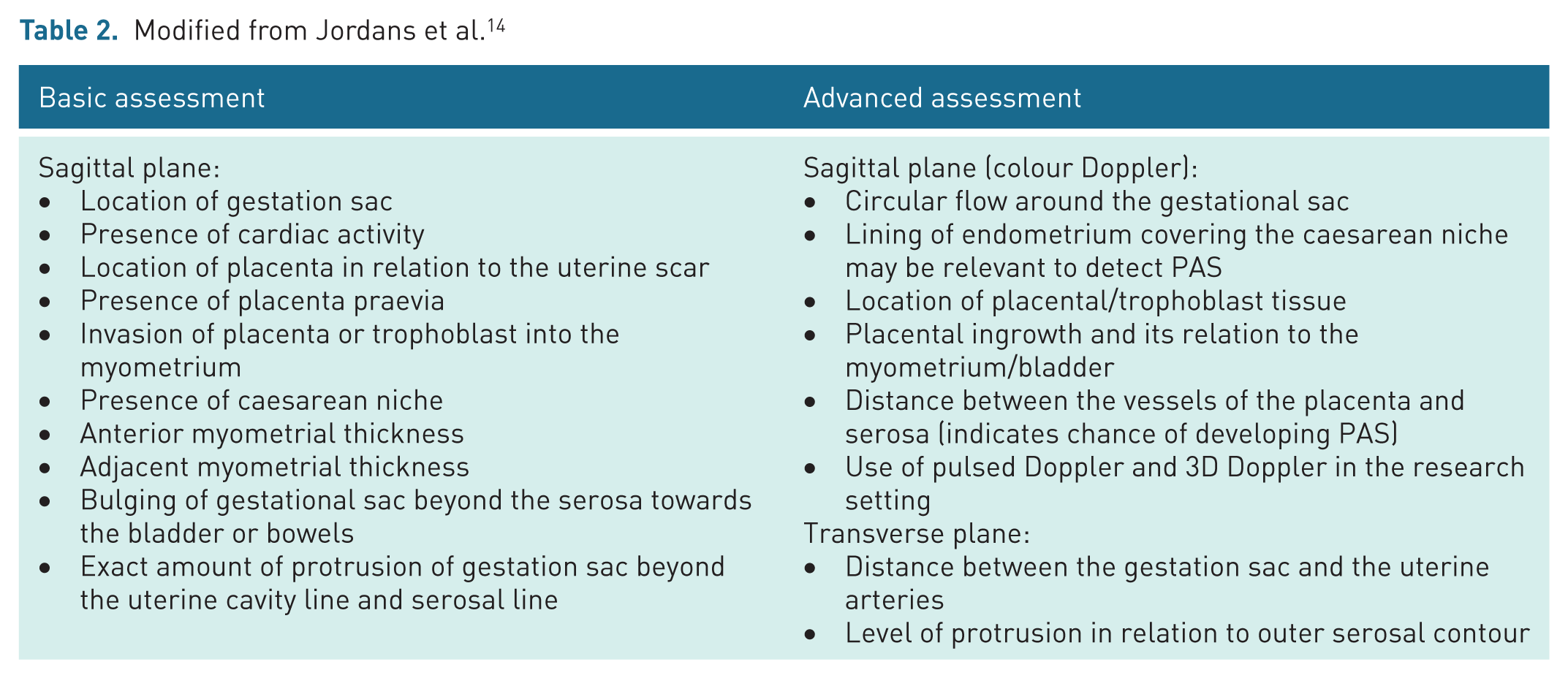
There is currently no universally accepted definition or standardised approach for recognizing and reporting the sonographic features of CSP. In 2022, Jordans et al. 14 proposed a sonographic reporting system for the diagnosis of CSP within the first 12 weeks of gestation, developed using a modified Delphi method. CSP was defined as a pregnancy implanted in, or in close contact with, the caesarean section niche. The consensus identified 10 basic and 8 advanced sonographic features to guide assessment, as outlined in Table 2.
Modified from Jordans et al. 14
To facilitate accurate description of CSP location relative to the endometrium, Jordans et al. 14 proposed a classification system categorizing CSP into three types. This system is based on two imaginary reference lines: the serosal line (SL), drawn along the outer surface of the lower anterior uterine wall, and the uterine cavity line (UCL), drawn at the endometrial–myometrial junction. The types are defined as follows (Figure 2):
This classification not only supports standardization in CSP diagnosis and reporting but also provides prognostic value, aiding in more informed counselling and management decisions.

Red line = UCL, blue line = SL. (a) Type 1 CSP–gestational sac crosses the UCL. (b) Type 2 CSP–gestational sac embedded within the myometrium and not crossing either the UCL or SL. (c) Type 3 CSP–gestational sac extends beyond the SL.
Measurement of anterior myometrial thickness, defined as the distance from the CSP to the serosal surface, should be performed upon identification of CSP. This parameter has emerged as a highly predictive first-trimester sonographic marker for PAS. The relevance and implications of this finding are explored in greater detail in the following sections of this manuscript.
Advanced imaging settings (Table 2) incorporate colour Doppler to evaluate vascular flow surrounding the gestational sac and to assess placental/trophoblastic tissue as well as the endometrium. Although no causal relationship has been established between first-trimester diagnostic ultrasound and adverse fetal outcomes, 15 the absence of evidence does not equate to evidence of absence. Accordingly, the As Low As Reasonably Achievable principle (ALARA) should be followed. 16 This entails using colour Doppler only when clinically indicated and for the shortest duration necessary. In the context of diagnosing CSP, the use of colour Doppler is justified, given the significant clinical benefit of early detection. The International Society of Ultrasound in Obstetrics and Gynaecology (ISUOG) recommends maintaining a thermal index (TI) of ⩽1.0 and limiting exposure time, ideally to under 5–10 minutes. Furthermore, fetal safety is unlikely to be compromised if the embryo lies outside the Doppler beam. 17 Therefore, when assessing placental and myometrial structures, the Doppler box should be positioned to avoid inclusion of the gestational sac or embryo.
If transvaginal ultrasound indicates abnormal placentation, it is crucial to assess the location of the gestational sac in relation to the endometrial cavity (Figure 3). This assessment is vital for guiding patient counselling, as pregnancies located outside the endometrial cavity are at a higher risk of growing outwards, leading to rupture at early gestations and therefore poorer patient outcomes.
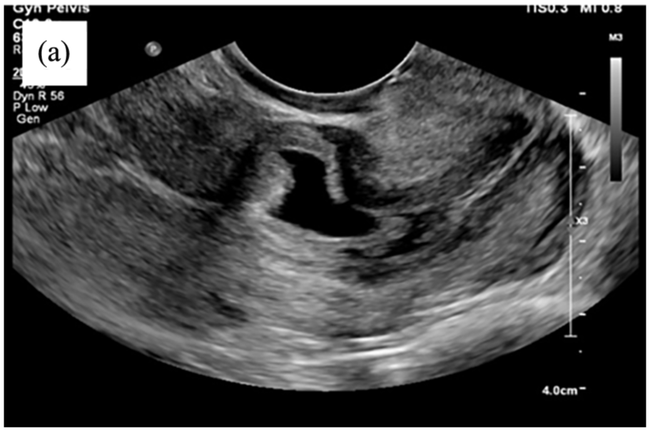
Transvaginal ultrasound demonstrating a low-lying gestational sac implanted within the caesarean niche at 5 weeks.
Timor-Tritsch et al. 18 emphasised the importance of distinguishing between a low-lying gestational sac as a CSP or a continuing intrauterine pregnancy. They proposed that this differentiation could be achieved by assessing the relative location of the centre of the gestational sac in relation to the midpoint of the uterus on a sagittal plane, measured between the external cervical os and the uterine fundus. Their findings indicated that most CSPs are located distal to this midpoint, whereas pregnancies likely to progress as intrauterine are typically located proximal to it. Figure 4 demonstrates this concept on a case of CSP (Figure 4(a)) and an intrauterine pregnancy (Figure 4(b)).
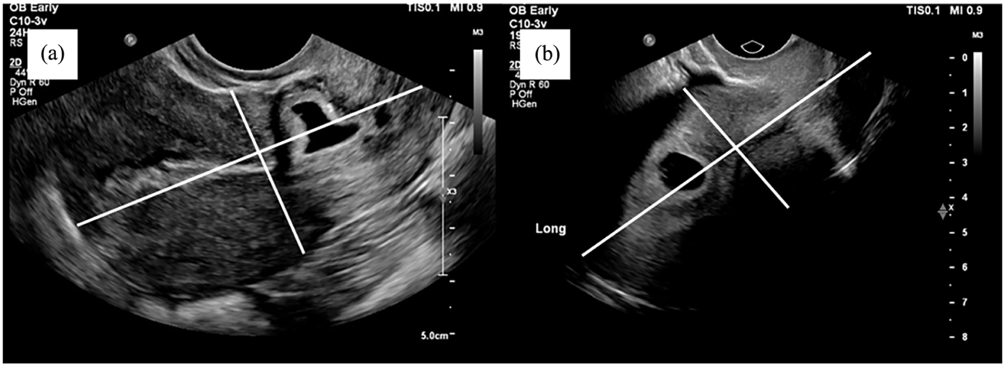
Applying the principle from the Timor-Tritsch study, in (a) the gestational sac is distal to the midpoint of the uterus, and this pregnancy was ultimately diagnosed as a CSP, whereas (b) is of an early intrauterine pregnancy, and the gestational sac is above the midpoint of the uterus.
If assessment of the entire endometrial cavity is difficult through transvaginal scanning, then a transabdominal ultrasound is often the most effective method for obtaining a clear sagittal view of the entire uterus. The importance of the transabdominal scan is demonstrated in the case shown in Figure 5. A 45-year-old woman with a history of two previous caesarean sections presented at 9 weeks of gestation to a tertiary centre for an early pregnancy scan. The initial impression from the transvaginal ultrasound was an intrauterine pregnancy that was low in the cavity, with trophoblastic tissue adjacent to the niche, increasing the possibility of PAS. However, the transvaginal ultrasound did not assess the endometrial cavity in length, and a subsequent transabdominal scan revealed that the pregnancy was in fact not within the endometrial cavity. While information from the transvaginal imaging alone, in this case, may have included counselling the patient regarding continuing the pregnancy, including delivery with a caesarean section hysterectomy, the additional information from the transabdominal imaging indicated an increased risk of rupture as the pregnancy progresses. The patient had already opted for a termination. She underwent a suction dilation and curettage, which was complicated by a 1500-ml blood loss. The significant bleeding supports the ultrasound finding of early PAS. This case highlights the importance of interpreting ultrasound findings by evaluating the entire endometrial cavity, which may require transabdominal imaging. Without this comprehensive assessment, a pregnancy located outside the endometrial cavity may go unrecognised.

Images (a) and (b) are transvaginal scans showing a low-lying gestational sac with abnormal placentation at the site of the caesarean niche. The operator measured the anterior myometrium as >5 mm; however, in retrospective review of these images, it is likely this was trophoblastic tissue around the gestational sac with no overlying myometrium. Images (c) and (d) are transabdominal scans that provide additional context, clearly demonstrating that the gestational sac is entirely within the caesarean niche and not within the endometrial cavity, thereby increasing the risk of rupture as the pregnancy advances. Highlights the endometrium, clearly seen as empty in images (c) and (d).
It is also crucial to determine whether a low-lying gestational sac will continue to develop into the endometrial cavity or grow outward, leading to uterine rupture. This determination is often challenging with a single ultrasound scan. Consequently, serial scanning is essential in these cases, as it provides the only reliable method for monitoring and detecting the progression of the pregnancy. Figures 6–8 illustrate how serial scans can be instrumental in tracking the clinical course of a low-lying gestational sac. The case presented in Figure 6 progressed to severe PAS, necessitating a hysterectomy in the second trimester. In contrast, the case in Figure 7 developed into an intrauterine pregnancy. Notably, the patient in Figure 7 did not have a history of caesarean section, placing them at a lower risk of developing PAS.

(a) Low-lying gestational sac at 13 weeks with trophoblastic tissue invading into the myometrium and extending to the cervix. (b) Placental lacunae at 13 weeks . (c) TA scan at 16 weeks showing pregnancy and placental tissue growing outside the endometrial cavity( ).
).
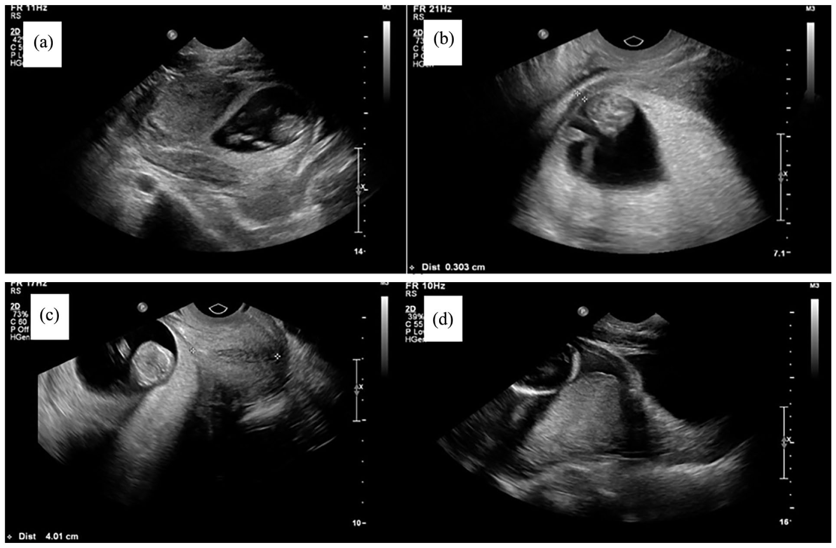
(a) Transabdominal ultrasound at 12 weeks demonstrating a low-lying gestational sac in contact with the endometrial cavity, appearing to protrude into the myometrium in a patient with no prior history of caesarean section. (b) On transvaginal imaging, the anterior myometrium is very thin. However, as can also be seen in image (c), the placenta is posterior and has no morphological features of PAS. In addition, there is no disruption of the cervix. (d) Placenta low-lying at 20 weeks and remained praevia; otherwise, the placenta remained morphologically normal throughout the pregnancy. The patient was delivered via caesarean section at the end of the third trimester, with no clinical features of PAS evident.
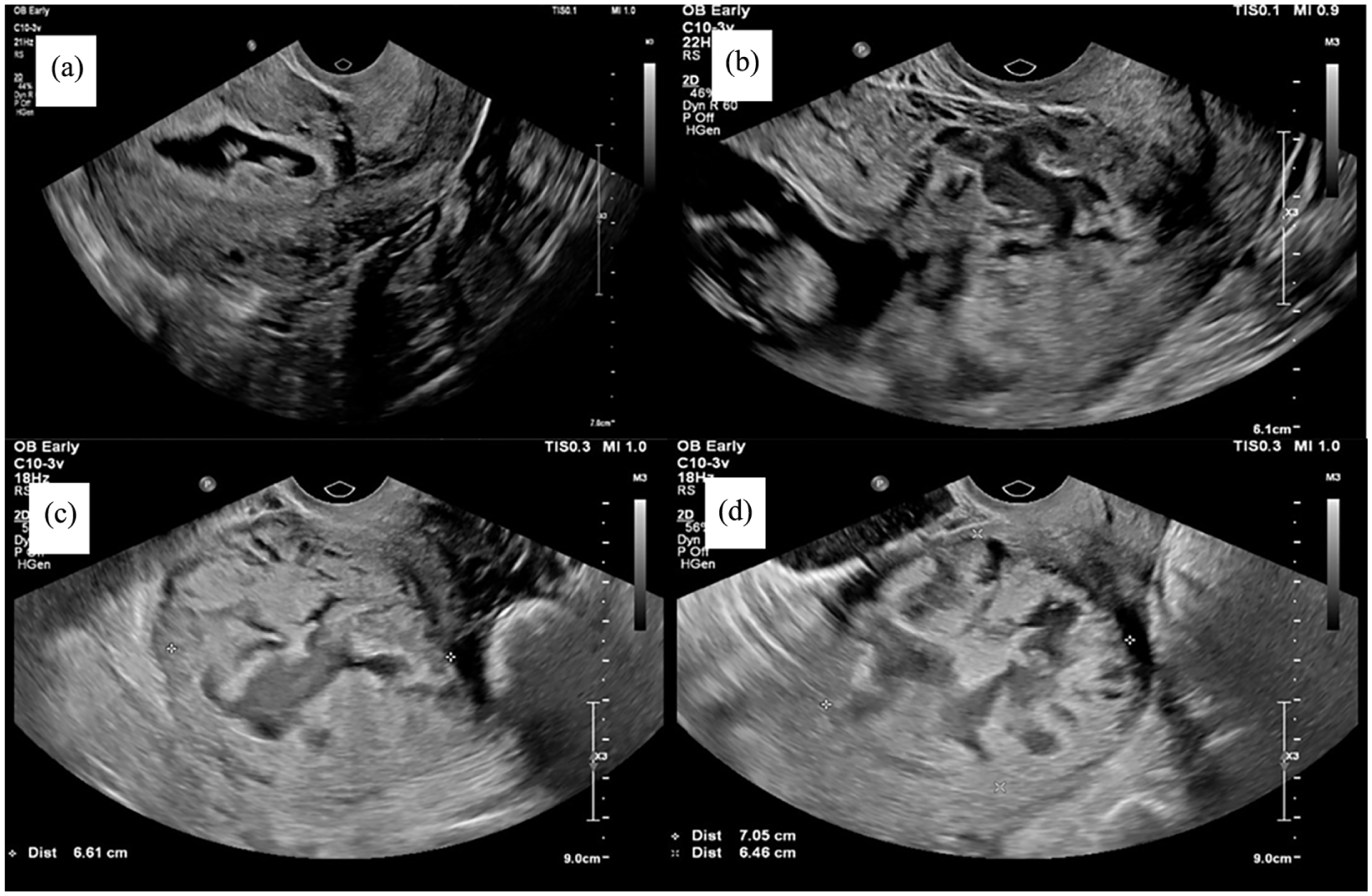
(a) The gestational sac appears to be located well within the endometrial cavity. Caesarean niche is visible below the gestational sac and does not appear to be involved in this image, with the gestational sac giving the impression that the pregnancy will continue as intrauterine. (b) Growth of trophoblastic tissue into the caesarean niche at 10 weeks. Even at this early gestation, advanced signs of PAS, such as lacunae, can be seen. (c) and (d) Abnormal trophoblast containing vascular lacunae and bulging into the surrounding myometrium at 12 weeks.
Serial scanning is particularly important in patients with risk factors for PAS, as early ultrasound signs may be subtle or absent in the first trimester, particularly before 8 weeks, and may therefore be overlooked or not recognised. Conducting serial scans allows for the identification of these cases in the late first or early second trimester, when abnormal placental invasion may become more apparent. Given that many women undergo a nuchal translucency scan at 12–13 weeks, this presents an optimal opportunity for clinicians to recognize features suggestive of PAS. It is therefore essential for clinicians to be familiar with these ultrasound findings, so they can identify PAS in at-risk patients, even if the initial scan appeared normal.
Figure 8 illustrates the case of a patient with a history of one previous caesarean section, whose pregnancy initially appeared to be a normal intrauterine pregnancy at 6 weeks. However, despite this early assessment, the patient experienced persistent vaginal bleeding. Serial ultrasounds subsequently revealed progressive invasion of trophoblastic tissue through the uterine serosa and into the cervical tissue by 10 weeks, raising suspicion of PAS, which was confirmed at 12 weeks. The patient ultimately underwent a wedge resection in the second trimester. A wedge resection is performed by first injecting a vasoconstrictor, such as vasopressin, into the myometrium around the ectopic pregnancy to minimize intraoperative bleeding. 19 An incision is then made through the myometrium to access and remove the gestational tissue, after which the affected area is excised in a wedge-shaped fashion and the bleeding points are sealed with cautery. 19 The defect is then oversewn. The procedure can be performed either laparoscopically or through open surgery, depending on patient and surgeon factors. This technique facilitates removal of the ectopic pregnancy while preserving healthy uterine tissue, thereby maintaining fertility potential. The case described in Figure 8 underscores the critical role of serial scans, particularly in symptomatic patients, in facilitating early diagnosis and appropriate management of PAS.
Anterior myometrial wall thickness
To assess whether CSPs implanted on a dehiscent scar ‘niche’ behave differently compared to those on top of a well-healed scar, Agten et al. 4 conducted a retrospective study of 17 patients diagnosed with CSP between 5 and 9 weeks of gestation who opted to continue their pregnancies. They found that all patients with CSP implanted in a caesarean niche displayed signs of PAS on ultrasound and required hysterectomy. They also demonstrated significantly lower myometrial thickness of <2 mm in the patients who developed PAS. There were no sonographic signs of PAS in the CSP group implanted on a well-healed scar, though one patient did have a hysterectomy for intractable haemorrhage, and partial accreta was subsequently histologically confirmed. The myometrial thickness for this patient was also 2 mm in the first-trimester ultrasound. It can be concluded from this study that patients with CSPs who have a myometrial thickness of 2 mm or less on first-trimester ultrasound are at higher risk of developing PAS.
In a patient with a low-lying gestational sac and a history of caesarean delivery, the smallest anterior myometrial thickness, measuring the distance from the CSP to the serosa, has emerged as a highly predictive first-trimester sonographic indicator for PAS. The reliability of this parameter lies in its consistent association with PAS across multiple studies,3,12,20 with Rac et al. 12 finding it to be the only biometric measurement that significantly improved detection of PAS.
The exact threshold below which the anterior myometrial wall thickness can improve diagnostic accuracy for PAS has been the subject of numerous investigations. As previously mentioned, Agten demonstrated that a myometrial wall thickness of <2 mm was significantly associated with PAS. Further research by Moschos et al. 20 examined various thresholds and found, in their retrospective study of 56 patients, that a threshold of <5 mm was 100% sensitive but only 74.3% specific, a threshold of <3 mm was 88.9% sensitive but achieved 100% specificity and a threshold of <4 mm was 94.9% sensitive and 97.1% specific. This highlights the challenges in selecting the most appropriate threshold to balance sensitivity and specificity in early diagnosis of PAS.
Though a consensus has not yet been reached regarding the optimal threshold for anterior myometrial wall thickness in diagnosing PAS, a measurement of <5 mm should raise concern. Patients with this finding should receive adequate counselling and careful monitoring with subsequent ultrasound for the potential development of PAS.
Ultrasound features across all gestations
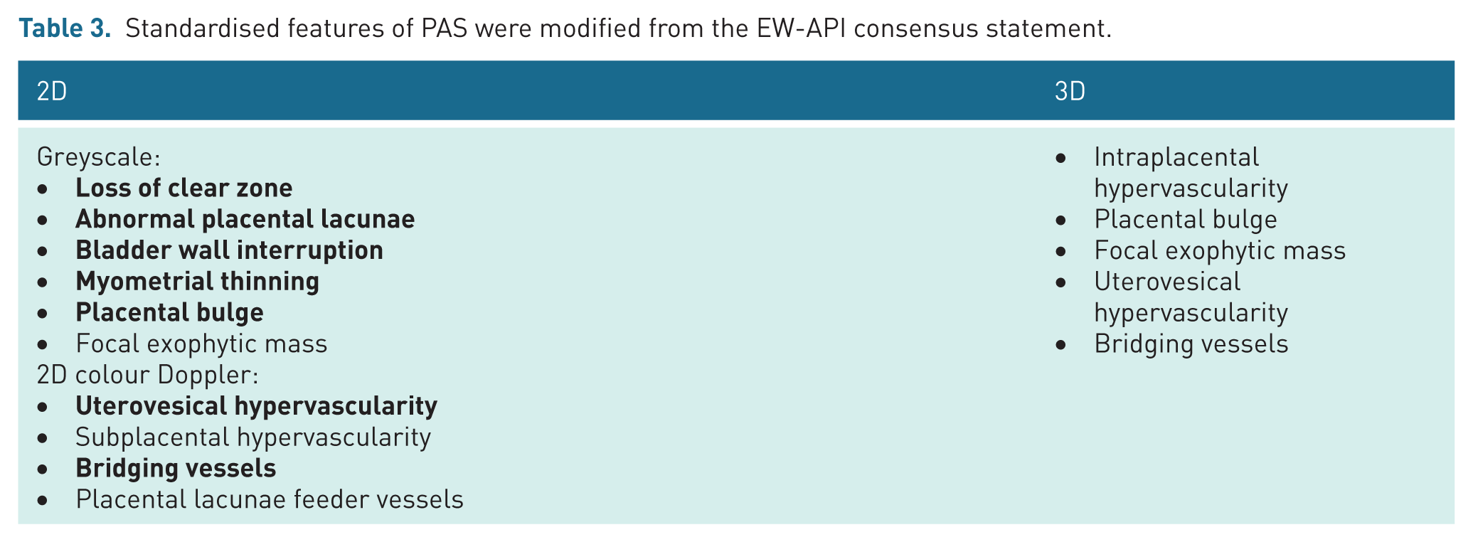
Several ultrasound indicators for PAS have been suggested, but many lack clear definitions and show varying levels of sensitivity and specificity. To improve the consistency of these ultrasound markers, the European Working Group on Abnormally Invasive Placenta (EW-AIP) developed a Delphi consensus proposal in 2015. 21 This proposal aimed to standardize the ultrasound descriptions for each sign. The EW-AIP consensus statement has not specified the sonographic signs for gestational age.
In 2023, Jauniaux et al. 22 revisited the existing criteria using a modified Delphi process. Their study reassessed the previously established signs and introduced eight additional markers identified through a comprehensive literature review. While consensus was reached on seven of the original ultrasound signs, agreement was not achieved for any of the newly proposed signs. This ongoing heterogeneity in expert opinion underscores the persistent diagnostic challenges associated with PAS.
Table 3 presents the sonographic features proposed in the EW-AIP consensus, with the seven signs that achieved consensus in the 2023 study highlighted in bold.
Standardised features of PAS were modified from the EW-API consensus statement.
Many of the ultrasound signs of PAS are subtle in the early first trimester (<9 weeks’ gestation) and may be easily overlooked. A review of our cases indicates that the most consistently observed early features include a low-lying gestational sac, a gestational sac positioned outside the endometrial cavity and a thin anterior myometrium. Other PAS-related ultrasound findings, which are discussed in detail below, may be present but are often inconsistent or only subtly detectable at this stage. Therefore, it is crucial that clinicians are not falsely reassured by the absence of these features on early pregnancy ultrasound, as PAS may still develop and become more apparent in subsequent scans.
Many of these signs described by EW-AIP can be observed in the late first trimester (9 weeks’ gestation and above) and early second trimester. They include placental lacunae, myometrial thinning, subplacental hypervascularity, placental bulge, bladder wall interruption and bridging vessels (Figures 9-12).
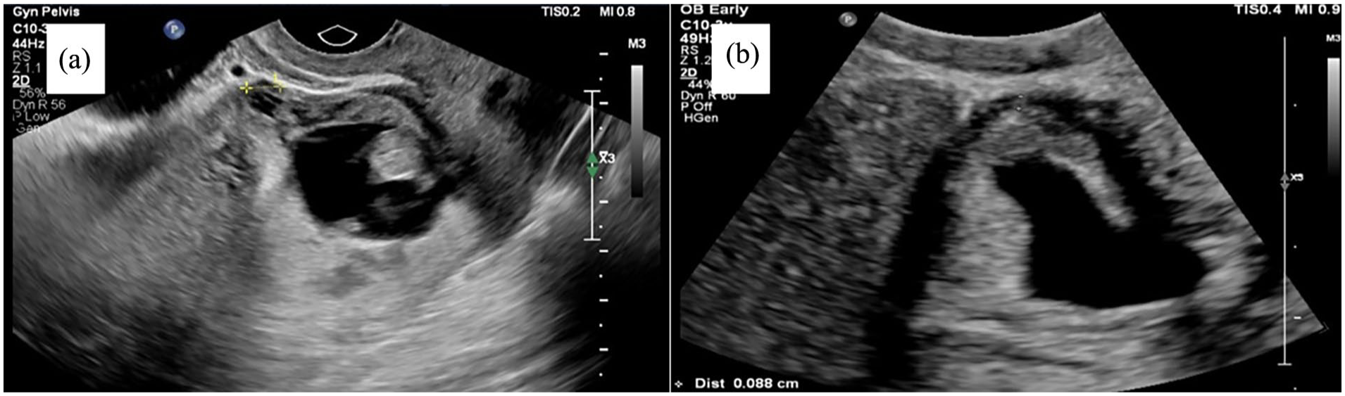
(a) No measurable overlying myometrium at 9 weeks. (b) Thin (<1 mm) anterior myometrium at 5 weeks.
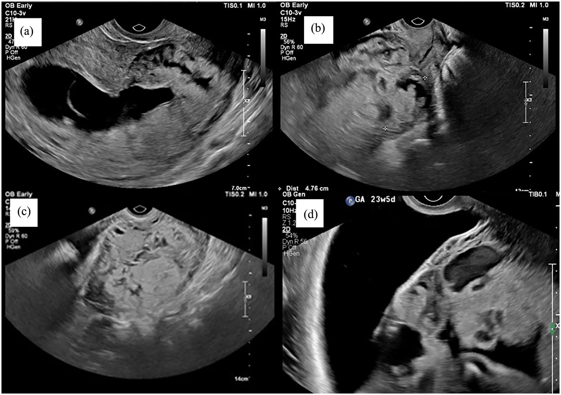
Images depicting placental lacunae at various gestational ages. (a) 10 weeks. (b) 12 weeks. (c) 16 weeks. (d) 23 weeks.
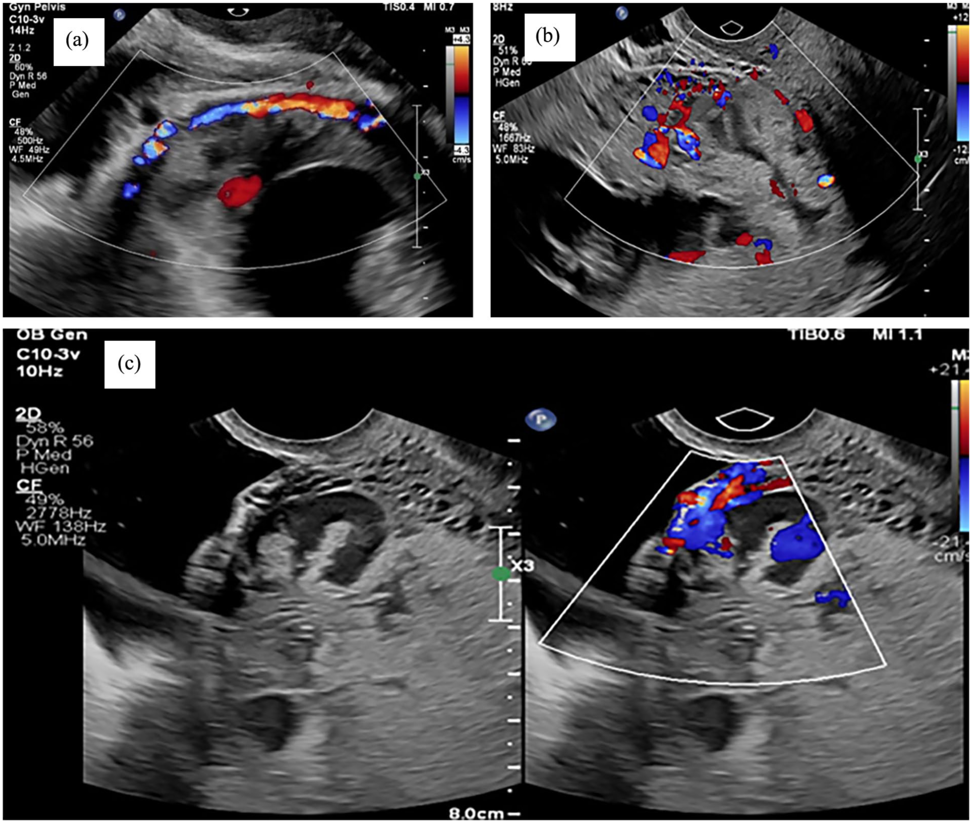
Images of subplacental hypervascularity in the first and early second trimester. (a) 9 weeks. (b) 10 weeks. (c) 16 weeks. Also demonstrates a feeder vessel.
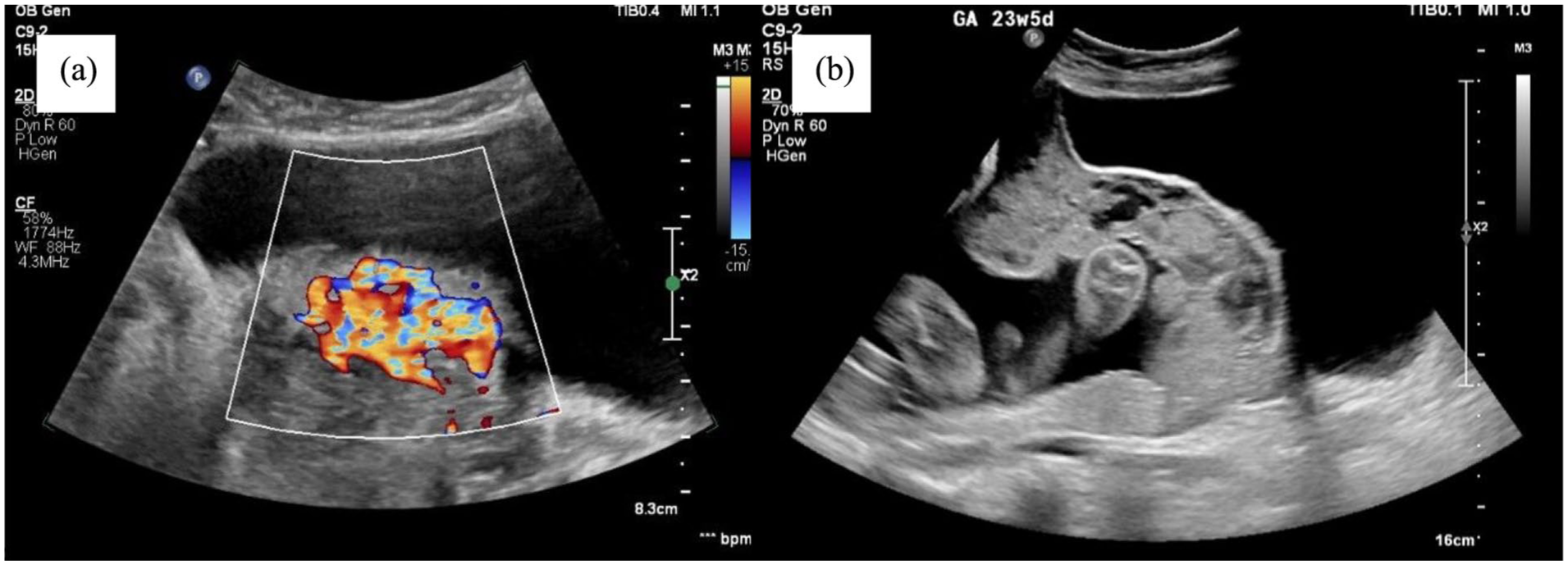
(a) USS at 16 weeks highlighting placental bulge and hypervascularity in a patient with a history of one previous caesarean section. (b) USS of the same patient at 23 weeks demonstrating a significant placental bulge and lacunae.
To evaluate the diagnostic accuracy of these signs in the late first trimester (11–14 weeks), Cali et al. conducted a retrospective analysis of prospectively collected data from 188 women at risk for PAS (defined by placenta praevia and at least one previous caesarean delivery) who underwent an ultrasound between 11 and 14 weeks. They assessed four specific signs: loss of the clear zone, placenta lacunae, bladder wall interruption and uterovesical hypervascularity. 13 The summarised findings are presented in Table 4.
Summarised from Cali et al.
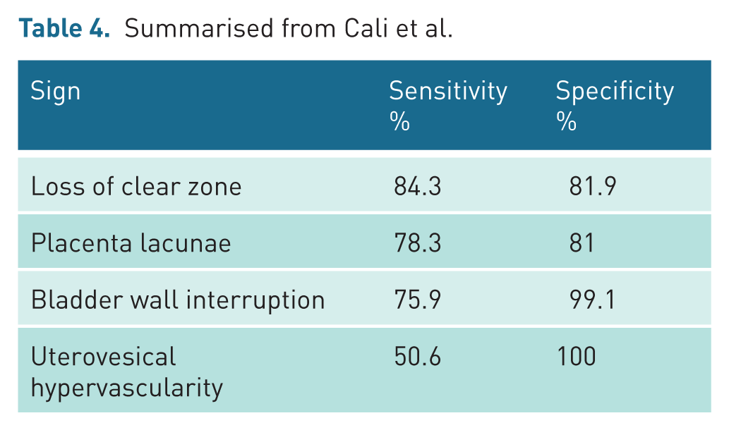
These findings underscore the overall accuracy of these four ultrasound signs in diagnosing PAS during the first trimester. Utilizing multiple signs enhances diagnostic precision. Moreover, the study achieved a 90% accuracy rate in detecting placenta percreta through the 11 to 14-week ultrasound scans, highlighting the effectiveness of early pregnancy screening in obtaining critical diagnostic insights for PAS and the positive implications this has on achieving safe patient outcomes.
Role of magnetic resonance imaging in placental accreta spectrum
Antenatal magnetic resonance imaging (MRI) serves as a valuable complement to ultrasound in the diagnosis of PAS, as it offers precise anatomical descriptions of the placenta’s invasion area. 12 In addition, MRI is a safe imaging modality during pregnancy, with no teratogenic effects on the developing fetus at any gestational age. In 2019, the Society of Abdominal Radiology (SAR) and the European Society of Urogenital Radiology (ESUR) issued a joint statement outlining recommended MRI findings for PAS to standardize the diagnostic criteria and reporting practices, enhancing consistency and accuracy in its identification. 23
A 2022 systematic review of 18 articles compared the diagnostic accuracy of ultrasound and MRI for PAS, finding no statistically significant difference between the two imaging modalities. 24 Given that MRI is more expensive and can be an uncomfortable experience for patients, ultrasound remains the preferred imaging modality in assessing and diagnosing PAS, with MRI serving best as an adjunctive tool.
Conclusion
Early diagnosis of PAS allows appropriate counselling and management, significantly improving outcomes for this potentially life-threatening condition. The role of first-trimester ultrasound in facilitating early detection is increasingly recognised. It is important to be aware that PAS signs in the early first trimester may be subtle, with classical features becoming increasingly apparent in the late first and second trimesters. Given this progression, women with risk factors, particularly those who are symptomatic, should undergo serial ultrasound examinations even if the initial early pregnancy scan appears normal.
Identifying key indicators such as CSP, anterior myometrial thinning, and placental abnormalities allows for timely intervention and tailored management strategies. This pictorial review emphasizes the importance of identifying features of PAS on ultrasound throughout the first and second trimesters of pregnancy to facilitate early diagnosis, timely intervention and tailored management strategies.
By enhancing diagnostic accuracy resulting in early detection, clinicians can provide better counselling, ensure safer surgical planning and ultimately improve maternal and neonatal outcomes.
Footnotes
Contributors
MT is the main author of the article. DN, VA and EO obtained the images and assisted in editing the article. All authors reviewed and approved the final version.
Declaration of conflicting interests
The author(s) declared no potential conflicts of interest with respect to the research, authorship, and/or publication of this article.
Funding
The author(s) received no financial support for the research, authorship, and/or publication of this article.
Acknowledgements
The authors would like to thank the sonologists and patients who have contributed the images within this educational review article.
Ethics approval
Our institution does not require ethics approval for this review, as no identifiable patient data has been included.
Permission from patient(s) or subject(s) obtained in writing for publishing their case report
YES. Verbal and written consent was obtained from patients at the Royal Women’s Hospital. Patients from Women’s Ultrasound Melbourne sign a written waiver for the use of their case for education and research purposes. All information used in this article are de-identified.
Permission obtained in writing from the patient or any person whose photo is included for publishing their photographs and images
Confirm that you are aware that permission from a previous publisher for reproducing any previously published material will be required should your article be accepted for publication,and that you will be responsible for obtaining that permission
YES.
Guarantor
DN
