






Abstract
Purpose:
To report a case of ophthalmic artery occlusion after injection of heroin into the forehead.
Methods:
Retrospective case report.
Results:
A 27-year-old patient with a history of intravenous heroin abuse presented with a left ophthalmic artery occlusion after attempting to inject heroin into a vein in her forehead. Exam was significant for diffuse retinal edema, disc edema, disc hemorrhage, and diffuse retinal hemorrhages. Findings were also noted in the widefield image, optical coherence tomography, and optical coherence tomography angiography of the retina.
Conclusions:
This patient suffered from a severe form of talc retinopathy resulting in an ophthalmic artery occlusion after injecting heroin into her forehead. To our knowledge, this is the first instance of combined ophthalmic artery occlusion and talc retinopathy resulting from intentional injection into a superficial facial vein.
Keywords
Introduction
Talc retinopathy causing multiple small emboli throughout the macula is seen in patients with a long history of intravenous drug use. Although good visual acuity is typically maintained, chronic use can lead to a variety of ischemic sequelae that result in decreased vision. Rare cases of retinal arteriolar occlusions have been documented by Tarantola et al and Lee and Sapira 1,2 ; however, there have been no reports of central retinal artery occlusions (CRAOs) or ophthalmic artery occlusions (OAOs) secondary to talc retinopathy.
There have been reports of patients who presented with CRAOs after filler injections into their foreheads, likely via retrograde flow of filler from the supraorbital or supratrochlear artery back into the ophthalmic artery. We believe that our patient suffered from an OAO with features of talc retinopathy after injecting heroin into her forehead and embolizing this arterial pathway.
Methods
This was a retrospective case report. Review by the internal review board and consent were not required because protected health information was not disclosed.
Results
Our patient was a 27-year-old white woman with a history of opioid dependence and intravenous drug abuse who presented with acute left vision loss after attempting to inject heroin into a vein in her forehead. She stated her vision abruptly went black in her left eye while injecting. The patient presented to the emergency department approximately 12 hours later. A review of systems was otherwise negative.
Her right-eye exam was unremarkable with 20/20 Snellen acuity. The left-eye exam was significant for no light-perception (NLP) vision with a relative afferent pupillary defect. Fundus exam showed a hyperemic disc with blurred edges and nasal disc hemorrhage, as well as severely constricted arterioles with box car segmentation and no cherry red spot (Figure 1A). Exam of the posterior pole showed diffuse retinal whitening with thickening in the macula. No obvious refractile particles were noted in either eye.
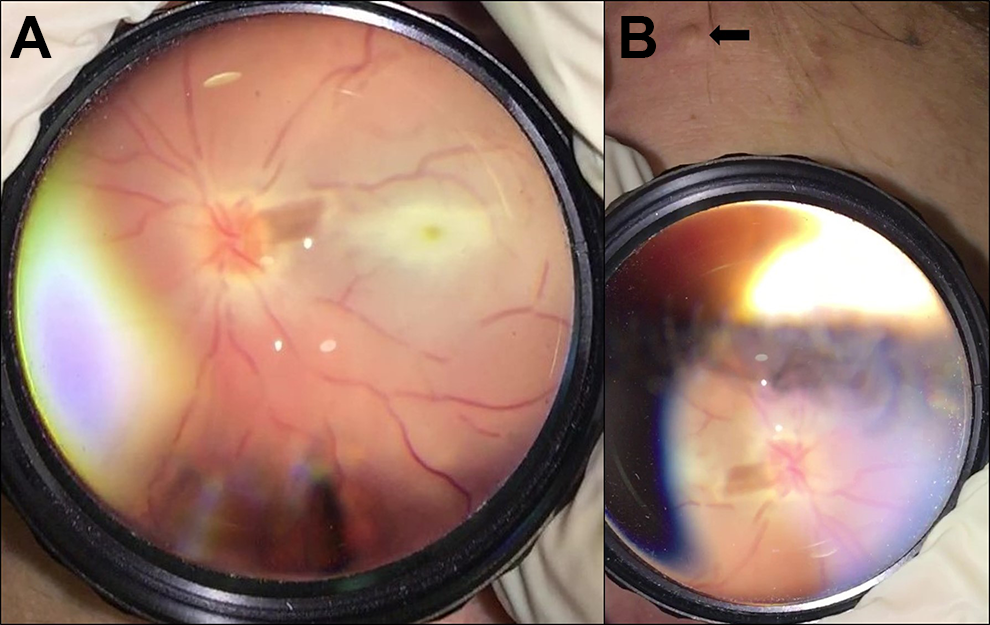
(A) Fundus photo at presentation, taken with an iPhone through a Volk Digital Clearfield lens. The photograph is shown inverted horizontally and vertically to accurately display the macula. There is disc edema, diffuse retinal whitening, severe vascular attenuation with box car segmentation, and absence of a cherry red spot. (B) The injection site (arrow) was inadvertently captured while photographing the fundus.
The patient also had ecchymosis and swelling to the left of her glabella in the area of the injection (Figure 1B). A CRAO or OAO was then presumed to be secondary to injected heroin and talc. Computed tomography angiography of the head was recommended to define the extent of the occlusion. Cardiac workup, autoimmune labs, and infectious serologies were recommended to rule out other coincidental causes of arterial occlusion. The patient left against medical advice before any of these were performed.
Upon follow-up 2 weeks later in the eye clinic, her vision remained at NLP. Exam of the anterior segment remained unremarkable. Dilated fundus exam showed new midperipheral white-centered hemorrhages (Figure 2). Optical coherence tomography (OCT) of the macula showed thickening of the inner retinal layers and subfoveal fluid, consistent with a retinal artery occlusion (Figure 3, lower). Fluorescein angiography could not be performed because of poor vascular access. OCT angiography showed severely attenuated vessels and foveal ischemia. There was poor resolution of the choroid due to retinal opacity. At the edges of the scan, where the retina was thinnest, the choroid showed poor perfusion (Figure 3, upper). The patient was sent back to the hospital for imaging, systemic evaluation, and to establish long-term follow-up. However, she again has been lost to follow-up.
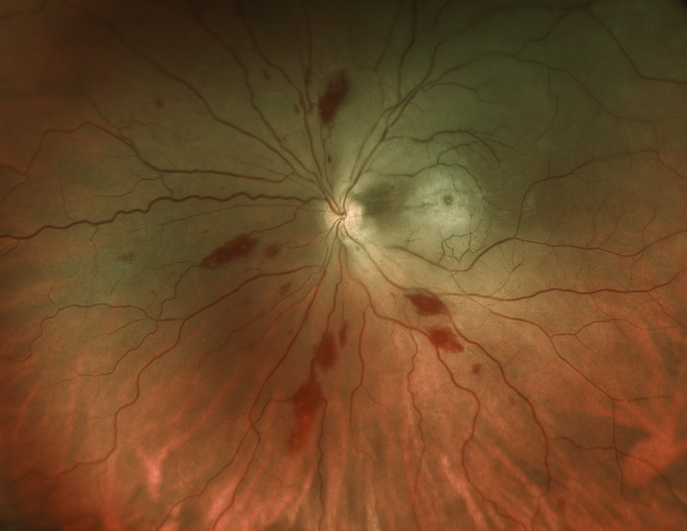
Widefield imaging 2 weeks after the initial presentation showing retinal whitening and opacity in the macula, disc hyperemia, severely constricted arterioles, and midperipheral hemorrhages in 3 quadrants.
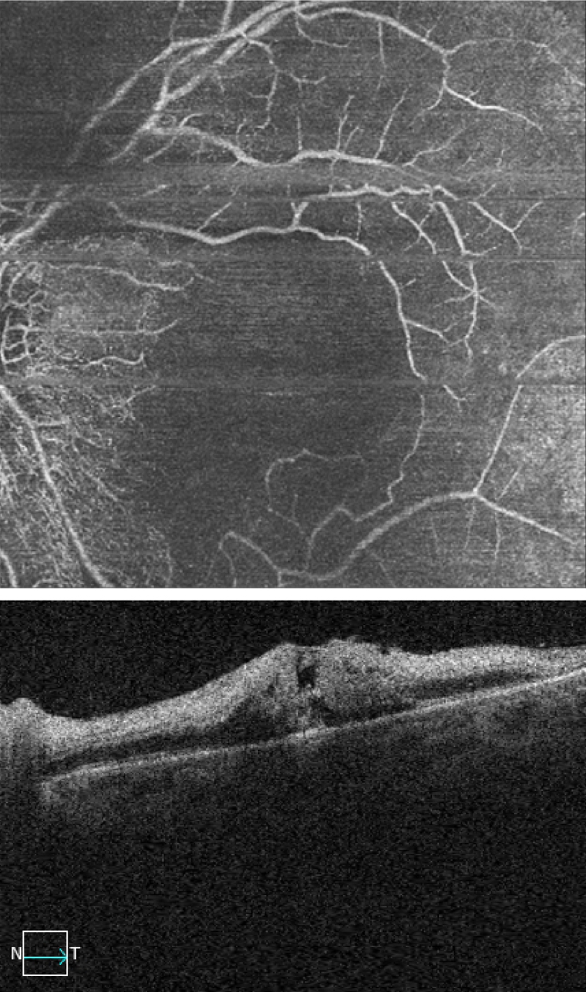
Optical coherence tomography (OCT) (lower) and OCT angiography (OCTA) (upper) of the left macula. OCT showed retinal thickening and hyperreflectivity with loss of differentiation of the inner neurosensory retina. OCTA showed diffuse foveal ischemia, vascular attenuation, and poor resolution of the choroidal circulation. The patient had difficulty positioning for the test, which caused motion artifact.
Conclusions
Various illicit drugs have been reported to cause visual complications secondary to vascular compromise. Devenyi et al reported a case of CRAO from cocaine, thought to be secondary to arterial spasm. 3 Rahman and colleagues likewise reported bilateral decreased vision due to extensive retinal and choroidal vasospasm from cocaine. 4 Heroin has also been associated with talc retinopathy. To our knowledge, this is the first reported case of an OAO due to heroin injection.
Talc retinopathy occurs when intravenous injection of crushed methylphenidate hydrochloride tablets lead to embolization of talc particles. 5 This disease typically involves scattered, inert, refractile emboli in the inner retinal layer of the macula and posterior pole. 5 Only particles smaller than 7 μm can pass through the pulmonary circulation before going to the retina. Talc particles can be as small as 5 μm. 1,6 Embolization of the pulmonary circulation in chronic intravenous drug users can lead to collateral vessels that allow larger particles to pass through. 1 Larger particles can also cause CRAOs in patients with a patent foramen ovale. 5
Although talc retinopathy usually results in minimal changes in vision, occlusive events can accumulate and eventually lead to more extensive damage. Macular or peripheral ischemia can occur, 7 leading to proliferative ischemic retinopathy. 1 Emboli may also accumulate into larger refractile particles that eventually occlude the vessel distally, as described by Zoumalan and Marmor. 6 As mentioned, only 2 cases of focal retinal arteriolar occlusions are described from talc; Lee and Sapira described a unilateral case of arteriolar occlusion secondary to talc, 2 whereas Tarantola et al described bilateral focal arteriolar occlusions secondary to injected talc in a patient with a patent foramen ovale. 1,5 No cases of CRAO or OAO were reported.
There have been multiple reports of facial cosmetic fillers injected into the forehead and upper cheek causing this complication. Vision loss is thought to be secondary to injection of the filler into a branch of the ophthalmic artery, such as the supraorbital or supratrochlear artery. The force of the injection causes retrograde flow of the filler into the ophthalmic artery, then embolization once pressure is released from the syringe. 8
Other distal branches of the ophthalmic artery that may lead to this include the angular, zygomaticotemporal, zygomaticofacial, and dorsal nasal arteries. 8 Another mechanism was attributed to possible vessel compression from filler injected adjacent to a vessel. 8,9 Beleznay and colleagues studied 98 patients with visual changes after filler injections in their 2015 review; the glabella was noted to be the most common culprit site, which was the case for our patient. 10 Other complications from filler injections that were also noted in our patient included ecchymosis and swelling, which was secondary to blood extravasation or rupture of vessels during injection. 8,9
Although extensive imaging was not available, we considered this to be an OAO because of the patient’s rapid onset of persistent NLP vision. 11 Since there was evidence of retinal emboli 2 weeks after the patient presented with vision loss, we expect that this resulted from simultaneous occlusion of multiple branches of the ophthalmic artery, including the central retinal artery and ciliary arterioles to the optic nerve. The most likely cause was an additive in the patient’s heroin introduced into the ophthalmic artery by retrograde injection of either the supraorbital or supratrochlear artery. Therefore, we expect that this is a site-specific complication arising directly from the patient’s choice of a forehead vein.
Footnotes
Acknowledgments
We acknowledge Chuck Terranova, director, Lions Diagnostic Imaging Center, for his expertise in acquiring and preparing the images for publication, and the Lions Vision Beyond Sight Foundation for continual support of our imaging department.
Ethical Approval
This case report was conducted in accordance with the Declaration of Helsinki. The collection and evaluation of all protected patient health information was performed in a Health Insurance Portability and Accountability Act (HIPAA)-compliant manner.
Statement of Informed Consent
Informed consent was obtained prior to administering care to the patient, including permission to use clinically acquired data and images for publication.
Declaration of Conflicting Interests
The author(s) declared no potential conflicts of interest with respect to the research, authorship, and/or publication of this article.
Funding
The author(s) received no financial support for the research, authorship, and/or publication of this article.