
Abstract
A surface deposition treatment like electroless Ni–B deposition, which is a new candidate to use in a wide range of engineering applications owing to many advantages, including low cost and good wear resistance, may improve the antibacterial activity and physical properties of stainless steel biomedical devices. In the present study, the structural and antibacterial properties of electroless Ni–B coatings deposited on AISI 304 stainless steels under different deposition conditions were investigated. Escherichia coli, the most important causative organism for infection, were used as the testing bacteria for in vitro test, including incubation at 37°C and 24 h. X-ray diffraction for crystallographic examination and scanning electron microscopy for morphological analysis were also used. The characterisation results showed that the antibacterial activity of the steel substrates deposited with coatings having especially high NaBH4 concentrations (1·2 g L−1), thus being amorphous, was strongly improved. Furthermore, the bactericidal performance difference of the coatings exhibiting cauliflower-like surface morphology was more obvious than that of the others. Electroless Ni–B surface treatment may be utilised for increasing the lifetime of stainless biomedical devices.
Introduction
Today, surgical and implant industries that use austenitic stainless steels are being pushed to develop new materials that improve the quality of medical care and the way treatments are delivered. The resulting escalating costs call attention to the need for process understanding, rapid innovation and solution efficiency. Antimicrobial coatings reduce infectious complications to prolong the useful life of devices and implants. A number of methods have been reported for synthesising the antibacterial coatings, such as calcium phosphate, 1 1,2 hydroxyapatite,3–5 diamond-like carbon,6–11 TiO2,12–14 TiN, Al2O3,15 etc., as biofilm on a variety of metallic substrates. Electroless Ni–B coating is a new candidate to use in a broad range of industry applications due to its unique properties, such as cost effectiveness, thickness uniformity, good wear resistance, lubricity, good ductility and corrosion resistance and excellent solderability and electrical properties. The aim of the present study is to investigate the effect of deposition parameters on the antibacterial properties of electroless Ni–B coatings, which has not been reported yet.
Experimental
AISI 304 stainless steels (rectangular coupons of size 15×15×5 mm) were used as substrate materials for the deposition of Ni–B coating. The surface of the coupons was ground to a roughness value of Ra⩽0·1 μm using a SiC emery paper with 1200 mesh grit and then polished with α-alumina having 0·05 μm grain size. Before deposition, they were degreased with acetone, rinsed with distilled water and picked in 15HCl for 15 s and then washed thoroughly with distilled water and dried in air with a fan.
The deposition parameters and their levels, to be able to affect the structural and tribological properties of electroless Ni–B coating, were arranged according to the Taguchi L9 (34) method. Controllable parameters and their levels were selected, as described in Table 1. The surface and cross-section morphologies of the treated coating were examined by SEM (JEOL 6400). Atomic force microscopy (Nanomagnetics Instrument) was used to view more clearly the surface topography and to characterise the surface roughness. The X-ray diffraction (XRD) pattern of the coating was determined by XRD (Rigaku D/Max 2000) using Cu Kα radiation.
Used experimental plan*
*Constant parameters: concentration of ethylenediamine of 90 g L−1 and concentration of sodium hydroxide of 90 g L−1, lead nitrate of 0·0145 g L−1 and pH≈13·5.
The antibacterial activity of electroless Ni–B coating on a stainless steel substrate was determined using Escherichia coli O157∶H7 (E. coli, Gram negative) by a disc diffusion method on nutrient agar medium. Uncoated stainless steel was used as a control. Bacterial suspensions (108 colony forming units/mL final cell concentrations) were poured into Petri dishes (9 cm) from flasks containing 25 mL sterile nutrient agar. Then, the bacteria were spread out evenly on top of the medium, covering the whole area of the testing Petri plate by a sterile swab. In this way, the large part of the bacteria was on the culture medium. Samples of the testing Petri plates were kept in the incubator at 37°C for E. coli for 24 h. Measuring the diameter of the circular shape around the samples (inhibition halo) provided information about the sensitivity or resistance of the cultivated microorganism to the biocide coating. All of the tests were carried out in triplicates.
Results and discussion
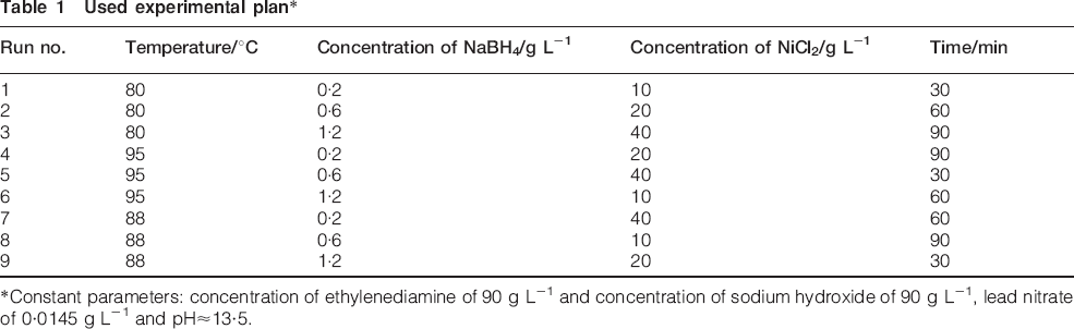
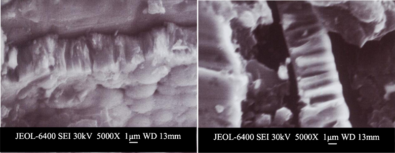
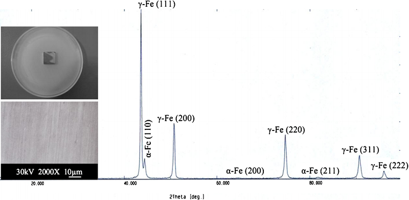
Figure 1 displays a typical example of the morphologies of the cross-section of electroless Ni–B coatings, which were deposited under run 6 conditions. When the SEM photos are examined, they draw attention to a colonial and uniform Ni–B coating structure having a cauliflower-like morphology and a thickness of ∼4 μm. This result confirms the typical Ni–B coating mentioned in the literature.16–18 The XRD spectra, the surface morphology and the antibacterial behaviour of the stainless steel substrate in Fig. 2 and the samples treated by electroless Ni–B deposition process under nine deposition conditions are given all together in Figs. 3–5. The XRD result for the steel substrate shows the crystal planes of γ-Fe (111) at 2θ≈43·7°, α-Fe (110) at 2θ≈44·6°, γ-Fe (200) at 2θ≈50·8°, α-Fe (200) at 2θ≈64·7°, γ-Fe (220) at 2θ≈74·7°, α-Fe (211) at 2θ≈82·1°, γ-Fe (311) at 2θ≈90·7° and γ-Fe (222) at 2θ≈96·1° (Fig. 2). Depending on the deposition parameters, different XRD spectra and morphological structures were revealed. Accordingly, no deposition on some samples, i.e. runs 1, 4 and 7, is observed, which reflect crystalline austenitic iron phases, whereas the diffraction patterns of some of them declare an amorphous nature because the major crystalline peaks are submerged in the NixBy phases nucleated on the substrate. Six different types of structures were observed in electroless Ni–B coating samples produced at nine different deposition conditions with respect to morphology. The first type of structure includes pea-like spherical grains, as shown in Fig. 3b (at run 2). The second type resembles a maize plant, but with partially porous structure, as shown in Fig. 5b (at run 8). The third type indicates a structure where the grains have formed nodules in doubles or triplets, which are named ‘primary nodular’, as shown in Fig. 4b (run 5). The fourth type is the structure that emerges by the formation of nodules similar to blackberries or a bunch of grapes by small grains, as shown in Fig. 3c. The fifth type is a broccoli structure that is composed of much smaller granules, as shown in Fig. 5c (at run 9). Last, the sixth type is the typical cauliflower morphology, where the nodules are formed by accretion of coarse grains, as shown in Fig. 4c (run 6).
Cross-sectional SEM images of electroless Ni–B coating
X-ray diffraction spectrum, surface morphology and antimicrobial effect of AISI 304 stainless steel substrate against E. coli
X-ray diffraction spectra, surface morphologies and antibacterial properties of electroless Ni–B coatings deposited under a run 1, b run 2 and c run 3 deposition conditions
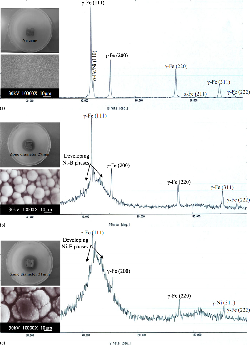
X-ray diffraction spectra, surface morphologies and antibacterial properties of electroless Ni–B coatings deposited under a run 4, b run 5 and c run 6 deposition conditions
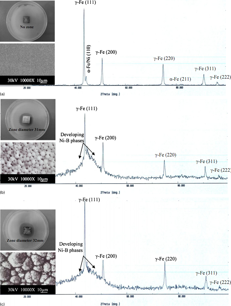
X-ray diffraction spectra, surface morphologies and antibacterial properties of electroless Ni–B coatings deposited under a run 7, b run 8 and c run 9 deposition conditions
When Figs. 2, 3a, 4a and 5a were examined, an evident inhibition halo is not observed for the uncoated substrate steel and for samples 1, 4 and 7, where the NaBH4 amount is at a low value (0·2 g L−1); thus, there is no significant film growth. However, samples 3, 6 and 9, in which the NaBH4 amount has the highest value (1·2 g L−1) and amorphous growth is observed together with a cauliflower-like surface morphology, exhibit the best antibacterial activity. Samples 2, 5 and 8 also have a granular morphology and less amorphous quality in terms of crystallography; it is attributed to the lower NaBH4 concentration. Consequently, the antibacterial performance of the coatings was also developed parallel to the morphological and crystallographic development based on the NaBH4 amount. Table 2 summarises the deposition parameter–inhibition halo relations of the samples treated under nine electroless deposition conditions. When the effect of NiCl2 amount, bath temperature and deposition duration on the structural and antibacterial activity is analysed, it can be stated that there is no linear correlation between these parameters and the structural and morphological development and antibacterial activity. Consequently, these parameters alone are not definitive parameters, but the NaBH4 amount is predominant in all deposition parameters. When the results obtained from the interaction effects are examined, the dominant effect of NaBH4 amount will be seen more clearly.
Deposition parameter(s)–inhibition halo relations of samples treated under nine electroless deposition conditions

Conclusions
The electroless Ni–B deposition process may be a significant promise for increasing the lifetime of stainless biomedical devices due to its ability to provide a hard, wear, antibacterial and corrosion resistant surface. The present study shows that the antibacterial properties of electroless Ni–B coatings change significantly depending on the composition of electroless plating baths and deposition parameters. The microstructural peculiarities, such as grain size, surface morphology and texture, which are closely related to the electroless deposition parameters, will also be determined based on the antibacterial performance. Some differences in the antibacterial behaviours of electroless Ni–B coatings exhibiting the amorphous quality in different levels and the cauliflower-like surface morphology developing in different types can be related to the contact area stemming from the surface morphology of the coating. As a result, it is seen that the most effective parameter on the antibacterial activity of electroless Ni–B coatings as well as the structural properties is the NaBH4 concentration. Better determination of the relations between the morphology of the coating and its antibacterial properties can be accomplished only with collaboration of interdisciplinary efforts.
Footnotes
Acknowledgements
The present research is a part of the BOREN (Institute of National Boron Research of Turkey) project supported by grant no. BOREN 2008.Ç-0178. The author would like to thank BOREN for funding the project.