
Abstract
This paper investigates the influence of nanostructured ceria (CeO2) coating on high temperature oxidation behaviour of 2·25 Cr-1 Mo steel. The isothermal corrosion study of the micrometre size and nanostructured CeO2 coated specimens was conducted in air oxidation environment at 973 K for 8 h. The corrosion rate and reaction kinetics were studied, and the post-corroded scales were characterised by field emission scanning electron microscopy and X-ray diffraction. The results clearly indicate that the nanostructured CeO2 coated specimen improves the oxidation resistance in comparison to the micrometre size CeO2 coated specimen. The improvement of oxidation resistance in the case of nanostructured CeO2 coating can be attributed to the formation of semimolten zone, which consists of nanostructured CeO2 particle. On the other hand, in the absence of semimolten nanostructured zone, the micrometre size CeO2 coated specimen shows higher corrosion rate in comparison to the nanostructured CeO2 powder coated specimen.
Introduction
Nanomaterials have attracted immense interest with numerous technological developments in the past two decades. Nanomaterials exhibit different physical and chemical properties than their bulk counterparts. The radical change in the physicochemical properties of nanocrystalline materials with respect to their bulk counterparts is due to high surface energy. Cerium oxide (CeO2) coating materials are extensively used for high temperature oxidation protection of various Cr based alloys. The ceria in nanocrystalline form has significant physical and chemical properties, which can tremendously enhance the oxidation/corrosion of the alloys.
Most high temperature alloys rely on the formation of protective Al2O3 and Cr2O3 scale on their surface to resist high temperature oxidation.1–3 However, the cracking and spallation of the oxide scale restrict the high temperature application of such alloys. The cracking and spallations of the oxide may be induced by the stresses arising from the oxide growth process or from the difference in coefficient of thermal expansion between the oxide and the alloys. 1 The additions of Ce, Y, Zr, La (rare earth elements) or their oxides improve the high temperature oxidation resistance in chromia and alumina forming alloys due to a decrease in scale growth rate and improved resistance to scale spallation by improving the scale–alloy adhesion.4–8
The improvement of oxidation resistance of nanocrystalline CeO2 coated alloys over the microsize CeO2 coated alloys has been reported in the past by several researchers.9,10 2·25 Cr-1 Mo steels are the popular candidate materials in steam generators/boilers for thermal power plant.11,12 Apart from that, the Cr–Mo steels are also used in petroleum processing industries, refining and pressure vessel for coal liquefaction. 13 High temperature oxidation/corrosion leads to drastic material degradation of the Cr–Mo steels and finally result in premature failure of the components.14–16 The earlier studies have indicated that the coating of micrometer size CeO2 is not so effective at higher temperature for prolonged exposure of time. Nanoparticles of CeO2 are expected to improve the oxidation resistance of chromia and alumina forming alloys because of their easy dissolution in the grain boundaries, which blocks the short circuit diffusion paths more effectively compared to micrometer size CeO2 particles. Preliminary investigation on the improvement of high temperature oxidation resistance of Ni, Cr and Ni–Cr superalloys with the application of nanocrystalline ceria coating was carried out by several researchers.17–19 However, detailed investigation of nano-CeO2 coated Cr–Mo steels to improve the high temperature oxidation resistance is not reported yet.
The present investigation therefore attempts to bring forward understanding related to the effect of nano-CeO2 coating over 2·25 Cr-1 Mo steel on high temperature corrosion oxidation behaviour under air oxidation environment at 700°C. At the same time, the studies are also carried out for micrometer size CeO2 coated specimen to compare the oxidation rate. The estimation of the corrosion rate and reaction kinetics are necessary supplement in this investigation along with post-corroded scale characterisation.
Experimental
Initial material
The initial material for the present investigation was normalised (1223 K/30 m in/air cooled) and tempered (1003 K/60 min/air cooled) 6·0 mm thick 2·25 Cr-1 Mo steel plate (Ilsenburger Grobblech, Germany). The chemical composition of the material is 0·11C, 0·48Mn, 0·22Si, 0·008S, 0·012P, 2·03Cr, 0·99 Mo and balance Fe.
Preparation of nanopowder
The initial powder for the investigation was micrometer size CeO2 powder (Shenzhen Xinyuan Abrasive Company Limited, Guandong, China) of 20 μm size. The powders were examined by scanning electron microscopy (SEM) to reveal the morphological characteristics. The powders were further subjected to mechanical milling by planetary ball mill (Res PM 200, Retsch Ltd, Germany) by wet milling method. The milling was performed by mixing toluene with the powder for 10 h. Finally, the powders were dried to produce the nanosize. The size of the milled powders was then measured in particle size analyser (Zeta Nano Zs, Malvern Ltd, UK). The powders were also examined in SEM (S-3000N, Hitachi limited, Japan) to reveal the morphological characteristics. The size and shape of the micronsize and nanosize powders are shown in Fig 1.
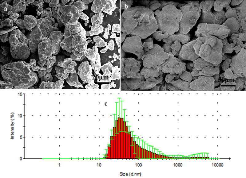
Particle morphology of a micrometre size CeO2 powder, b nanosize CeO2 powder and c particle size distribution of nanosize CeO2 powder (Colour version available online)
Preparation of coating
The substrate materials (2·25 Cr-1 Mo steel) were first subjected to the grit blasting by alumina powder (Al2O3) in pressure air blasting machine. Air pressure during blasting was maintained at 5 kg cm−2. The micrometer size CeO2 powder was directly plasma sprayed to the substrate material using atmospheric plasma spray coating unit (SG-100, Paraxair USA) at Metallizing Equipment Co Pvt Ltd, Jodhpur, India. A robotic arm (Kuka Robots, Germany) was used for uniform coating on all sides of the specimen over the substrate. The nanosize powder, because of its extremely fine size, was first subjected to agglomeration through spray drying before plasma spray coating. The agglomerated powders were then sprayed through plasma coating process to form the adherent coating. This type of coating is designated as ‘nanostructured coatings’. The same weight of micrometre size and nanosize powder was used for the coating purpose. The plasma spraying parameters for the coating are given in Table 1.
Spray parameters as employed during atmospheric plasma spray coating
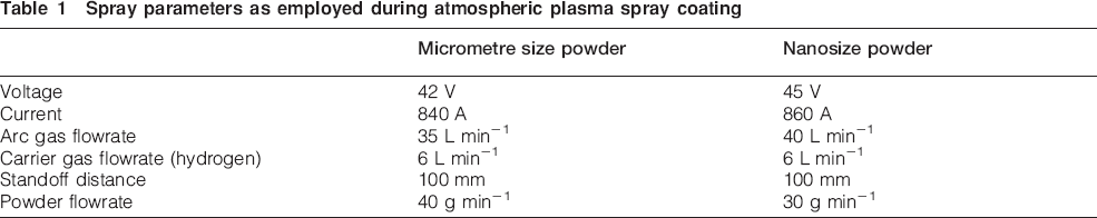
Figure 2a depicts the SEM surface morphology of the micrometre size powder coated specimen. The figure indicates the presence of a few porosities along with some unmelted ceria (CeO2) particles on the top surface. At the same time, the surface morphology of nanostructured coated specimen shows adherent coating without porosities and cracks (Fig. 2b). The nanostructured coating at higher magnification confirms the presence of nanoparticles possibly of CeO2 on the coating surface (Fig. 2c).
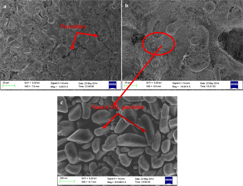
Surface morphology of as received a micrometre size ceria (CeO2) coated specimen, b nanosize ceria (CeO2) coated specimen and c nanosize ceria (CeO2) coated specimen at higher magnification (nanoscale) (Colour version available online)
High temperature corrosion test
High temperature corrosion test was carried out in a vertical tubular furnace attached with digital weighing balance. Specimens for corrosion tests were placed in the central heating zone of the furnace before heating the furnace in the desired temperature. The furnace was first heated to 300°C at 3°C min−1, followed by heating at a rate of 5°C min−1 up to 700°C. The furnace temperature was continued up to 8 h with the accuracy of the temperature level ±3°C. The isothermal corrosion test was carried out in air oxidation environment at 700°C using 8 h, and the weight change of the specimen during the corrosion study was measured at a digital weighing balance (Metler Toledo) at different intervals of time up to an accuracy of ±.01 mg. After 8 h duration, the specimen inside the furnace was subjected to cooling at a rate of ±4°C.
Results and discussion
Corrosion rate and reaction kinetics
The corrosion rate and reaction kinetics of micrometre size and nanostructured CeO2 coated specimen in isothermal corrosion study are presented in Fig. 3. The figure suggests that the reaction rate of nanostructured CeO2 coated specimen is much lower than that of the micrometre size CeO2 coated specimen. The figure also shows the drastic improvement of oxidation rate in case of nanosize ceria coated specimen. The reaction kinetics of both the coated specimen follows the parabolic growth rate (Fig. 3), which indicates that the oxidation process is diffusion controlled and governed by the outer cation migration and inner anion migration.
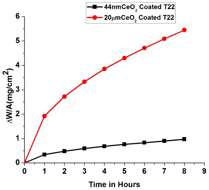
Kinetic behaviour and oxidation rate of micrometre size CeO2 coated and nanostructured CeO2 coated specimen (Colour version available online)
The parabolic rate constant (Kp) are calculated by the following equation
The parabolic rate constants of micrometre size CeO2 coated and nanostructured CeO2 coated specimens are calculated and found to be 10·314×10−4 mg2 cm−4 s−1 and 3·26×10−5 mg2 cm−4 s−1 respectively. The result indicates that there is a significant reduction of parabolic rate constant in the case of nanostructured CeO2 coated specimen.
Characterisations of post-corroded specimens
Coating microstructure
The coating microstructure after the oxidation process is characterised using field emission SEM. The microstructure of micrometre size CeO2 specimen consists of molten and some unmelted CeO2 particle. No semimolten zone is found in this type of coating. The figures also show some cracks in between the molten and unmelted zone (Fig. 4a). The formation of cracks is important for the migration of cation during the oxidation process. It provides the short circuit diffusion path for outward cation migration. On the other hand, the coating microstructure of the nanostructured CeO2 coating consists of semimolten zone along with fully molten zone (Fig. 4b). The existence of semimolten zone is obvious in plasma spray coating of nanostructured powders because of incomplete melting of the particle. The semimolten zone at higher magnification shows the presence of nanosize CeO2 powder (Fig. 4c). The presence of nanosize particle within semimolten zones in different plasma spray has been described in literature.21–23 As this coating is formed by a mixture of particles that are fully molten and semimolten in the spray jet, some authors have described this microstructure as ‘bimodal microstructure’.20–23
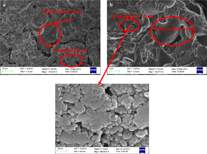
Coating microstructure and SEM surface morphology of a micrometre size CeO2 coated specimen, b nanostructured CeO2 coated specimen and c CeO2 nanoparticle within semimolten zone (Colour version available online)
The plasma spray coating of ceramic powders is intrinsically associated with melting of the particles. In case of nanosize CeO2 powder plasma spray coating, some of the particles are fully molten in the thermal spray jet, while some of the powders are in semimolten condition. In the case of particle in fully molten condition, the nanostructural characteristics of the powder particle completely disappeared, while in the region of semimolten zone, the nanostructural features of the powder particle are retained as shown in Fig. 4b and c. The presence of nanostructural features is important in connection with the oxidation resistance of CeO2 coated alloy. The nanostructured particles, because of their easy dissolution in the grain boundaries, subsequently restrict the short circuit diffusion path more effectively and improved the corrosion resistance to a significant extent. In contrast, the post-corroded micrometre size CeO2 coated specimen shows the fully molten zone and the unmelted zone. The coating also shows the porosities and cracks (Fig. 4a). The presence of microcracks and porosities/voids is also reported in different literatures.24–26 The low oxidation resistance may be attributed to inhomogeneously distributed microcracks, porosities and finally less adhesion CeO2. The porosities and cracks act as short circuit diffusion path for inner migration of oxygen and increase the oxidation rate.
X-ray mapping analysis
The X-ray mapping analysis of the micrometre size and nanostructured CeO2 coated specimen is shown in Figs. 5 and 6. The oxygen mapping of Fig. 5 suggests the inner migration of oxygen from the environment towards the substrate through the coating and diffusion layer. At the same time, the outward migration of Fe and Cr towards the coating and diffusion layer is observed. This indicates that the transport mechanism of micrometre size CeO2 coated sample is governed mainly by inner migration of oxygen through cracks and porosities, which result in more oxidation products. In contrast, the X-ray mapping analysis of post-corroded nanostructured CeO2 coated specimen (Fig. 6) shows that inward migration of oxygen towards diffusion layer and substrate is less in comparison to the micrometre size powder CeO2 coated specimen. At the same time, the outward cation migration of Fe and Cr is blocked completely due to the absence of porosities and cracks on the coating and also the presence of nanosized CeO2 particle in the coating.
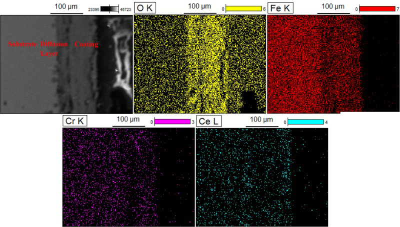
X-ray mapping analysis of post-corroded micrometre size coated CeO2 coated specimen (Colour version available online)
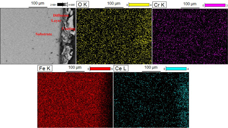
X-ray mapping analysis of post-corroded nanostructured CeO2 coated specimen (Colour version available online)
X-ray diffraction analysis
The X-ray diffraction (XRD) analysis of micrometre size powder coated sample (Fig. 7a) confirms the presence of CeO2, CeFeO2 and Fe2O3 phase. The results also show that CeO2 exists as a stronger phase, while CeFeO2 exists as a weaker phase. The presence of CeO2 and CeFeO2 in the surface indicates that the Ce ions are found to be segregated along the grain boundaries of the scale. On the other hand, the XRD analysis of the nanostructured CeO2 coated specimen shows the presence of CeFeO2 as a stronger phase and CeO2 and Fe2O3 as weaker phases. The nanostructured CeO2 particle in the coating has more tendency to form complex CeFeO2 compound during the oxidation process. The strong presence of CeFeO2 compound in the nanostructured coating effectively restricts the outward cation migration (Fe and Cr) and significantly improves the oxidation resistance.
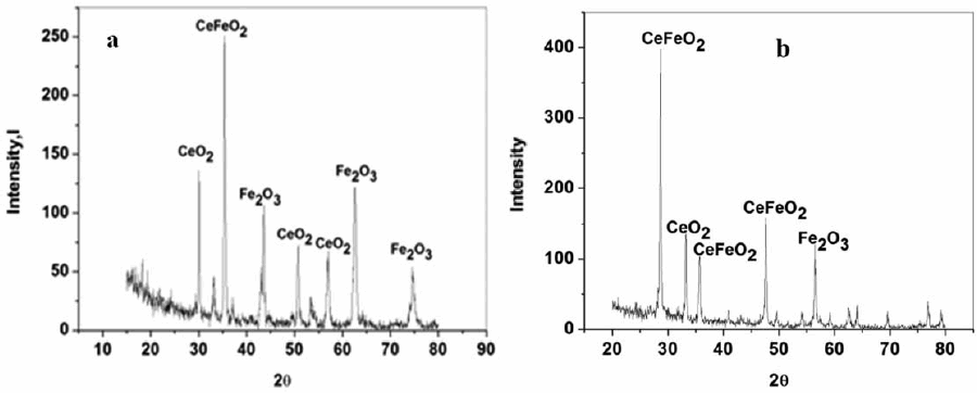
XRD analysis of a post-corroded micrometre size CeO2 coated specimen and b post-corroded nanostructured CeO2 coated specimen
Conclusion
The plasma spray coated nanostructured CeO2 coated specimen significantly improves the corrosion rate in comparison to the micrometre size coated CeO2 coated specimen in air oxidation environment at 700°C up to 8 h. Both the micrometre size CeO2 and nanostructured CeO2 coated specimen follows parabolic rate kinetics, which indicates that the corrosion is governed by diffusion controlled growth (i.e. outer cation and inner anion migration) during oxidation process. The parabolic rate constant (Kp) of the nanostructured specimen is much lower than the micrometre size coated specimen.
The surface morphology of the post-corroded micrometre size CeO2 consists of unmelted zone, fully molten zone without formation of any semimolten zone. The presence of CeO2 segregated in this zone improves the oxidation rate, but at the same time, the formation cracks on the coating acts as a short circuit diffusion path for outward cation migration and increases the oxidation rate. In contrast, the surface morphology of the nanostructured CeO2 coated specimen consists of molten and semimolten zones. The nanosize CeO2 particle exists in the semimolten zone. The nano-CeO2 particles, by easy dissolution at the grain boundaries, effectively block the short circuit diffusion path for outward cation migration and improve the oxidation rate to a significant extent.
The X-ray mapping analysis of the micrometre size coated sample indicates that the transport mechanism of the oxidation process is governed by the inner migration of an oxygen (anion) instead of outward migration of cation. In contrast, the X-ray mapping analysis of nanostructured CeO2 coated specimen shows that the inner migration of oxygen is much less than the micrometre size coated specimen, which further indicates that the oxide scale formation of nanostructured coating is much thinner in comparison to the micrometre size coated specimen. The XRD analysis demonstrates that the micrometre size CeO2 coated and nanostructured CeO2 coated specimen shows the presence of CeO2, CeFeO2 and Fe2O3 phases. The existence of stronger CeFeO2 phase in the case of nanostructured CeO2 coating effectively blocks the outer cation migration and significantly increases the oxidation resistance.