






Abstract
Background: Diabetic motor neuropathy is expressed as the loss of function and the contracture of the intrinsic muscles of the foot, leading to the classic claw toe deformity. This deformity predisposes the foot to ulcerations on the dorsum or tip of the toes or an interdigital ulcer over a condyle between the toes. We present our results of a modified resection arthroplasty for the treatment of this difficult problem. Materials and Methods: In this study, 72 toes (57 feet) with a deformity in the second to fifth toe accompanied by chronically infected ulcers were involved. All patients underwent modified resection arthroplasty of the PIP or DIP joint depending on the ulcer location. The second toe was involved in 27 cases (38%), the third toe in 11 cases (15%), the fourth toe in 19 cases (26%), and the fifth toe in 15 cases (21%). With the exception of 4 patients, all had a positive culture, including 7 cases of MRSA. The mean followup was 28.7 ± 8.1 months. Results: The mean wound healing time was 25.6 ± 6.2 days. Three cases eventually required toe amputation but there was no proximal spread of infection. No recurrence of a claw toe or ulcer occurred in the remaining toes. Conclusion: We believe that modified resection arthroplasty for toe deformities with chronic infected ulcers in diabetic patients is a good treatment alternative to toe amputation.
Introduction
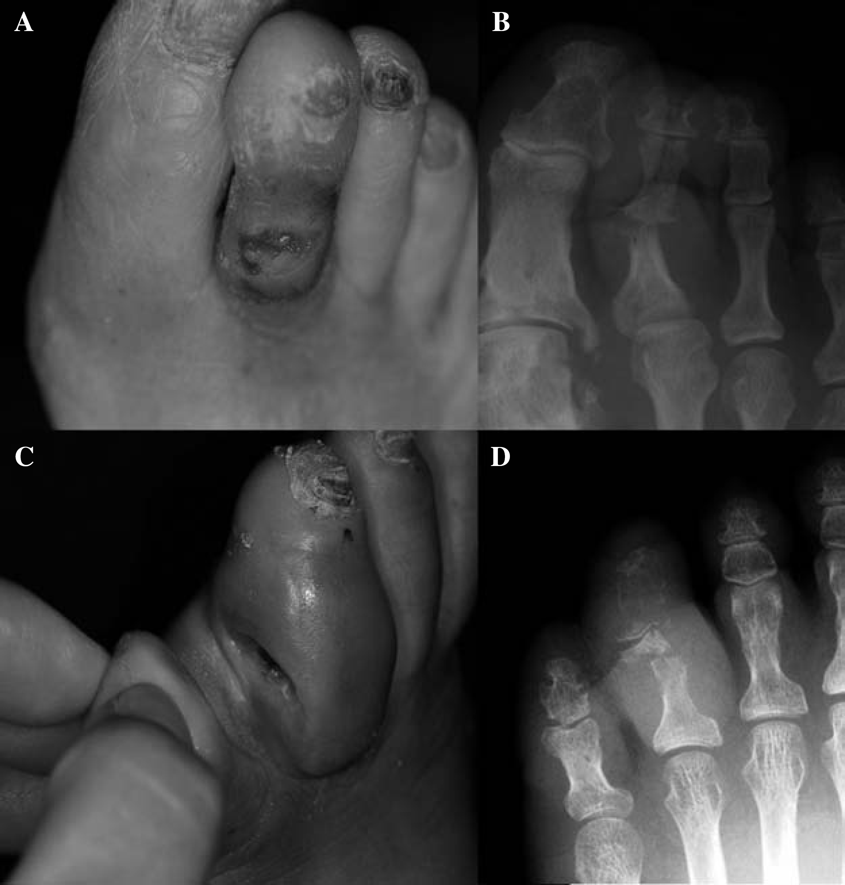
Recently, as the number of diabetic patients and their life spans has increased, the incidence of diabetic foot problems has also increased.6,7 Up to 20% of hospitalized diabetic patients have medical issues associated with their feet. 4 Neuropathy contributes to ulcerations and deformities on the foot. 14 Decreased intrinsic muscle function in the feet due to neuropathy can lead to a characteristic ‘neuropathic posture’ because of the unopposed activity of the extrinsic muscles. In this case, the arch is raised, toes are clawed, and pressure is concentrated in several areas, including the heel, the metatarsal heads, the proximal interphalangeal joints, and the tips of the toes. Force generated during walking stimulates callus formation or ulceration at pressure points. In the claw toe deformities of diabetic patients, wounds usually occur on the dorsum of the proximal interphalangeal (PIP) joint 3 (Figure 1, A and B). Biomechanical changes in periarticular tissue, particularly within the small joints, have been recognized as producing thickened skin, diminished range of motion, and joint contractures in diabetic patients which also predispose to the formation of toe ulcers. 13 Loss of sensation and a tight shoe can produce an interdigital ulcer over a condyle of the phalanx between the toes 3 (Figure 1, C and D).
With the exception of toe amputation, treatment protocols for the management of deformities in the second to fifth toes with chronic infected ulcers have not been established. 3 Once an entire toe has been amputated, even the pressure of a properly fitted prescription foot will cause the two adjacent toes to move toward the defect from either side. As these toes drift into the varus and valgus, respectively, they touch as if filling a void, thus promoting another defect in which other toes may also drift in “domino” fashion. 3 For these reasons, we attempted to find an alternative method for managing toe deformities accompanied by chronic ulcers with or without osteomyelitis that did not include toe amputation.
Classic resection arthroplasty has been used to manage fixed forms of claw toe or hammer toe deformity. 5 In the original resection arthroplasty, the distal condyles of the proximal phalanx are resected and the extensor tendon and the dorsal skin of the PIP joint area are sutured with one stitch. In this study, we modified steps to completely remove the infected phalangeal bone and the suturing of the dorsal wound and extensor tendon. The aim of this study was to evaluate the results of modified resection arthroplasty in toe deformities accompanied by non-healing ulcers with or without osteomyelitis in the second, third, fourth, or fifth toe of diabetic patients.
Materials and Methods
Among patients with diabetic foot ulcers who visited this hospital from May 2003 to March 2006, those with a toe deformity and ulcers that had been present for more than 6 weeks with or without evidence of soft tissue or bone infection were eligible. All patients had a second to fifth toe deformity accompanied by chronic ulcers treated with modified resection arthroplasty of the PIP or DIP joint. Among the selected subjects, the patients who had an untreated vasculopathy documented by Doppler ultrasound and/or three-dimensional CT-angiography were excluded. Patients with vasculopathy who underwent angioplasty or bypass surgery, and then modified resection arthroplasty were included. Fifty-two patients (57 feet and 72 toes), composed of 28 men and 24 women with a mean age of 62.3 ± 11.2 years, were enrolled. The right:left:both sides ratio was 26:21:5. In 11 feet, two or more toes were involved simultaneously (7 feet had 2-toe involvement, and 4 feet had 3-toe involvement). Routine laboratory blood tests were performed and blood tests representing diabetic control (HbA1c, fasting and 2-hour postprandial blood sugar levels) were checked. Additionally, simple radiography, 3-phase Tc-99m bone scanning, Gram staining, and bacterial cultures at the wound site were performed on the first day of hospitalization and on the day of surgery. After surgery, we recorded the total wound healing time, complications, additional toe amputations, and recurrence rates. The mean duration of followup was 28.7 ± 8.1 months.
Operative Technique and Postoperative Treatment
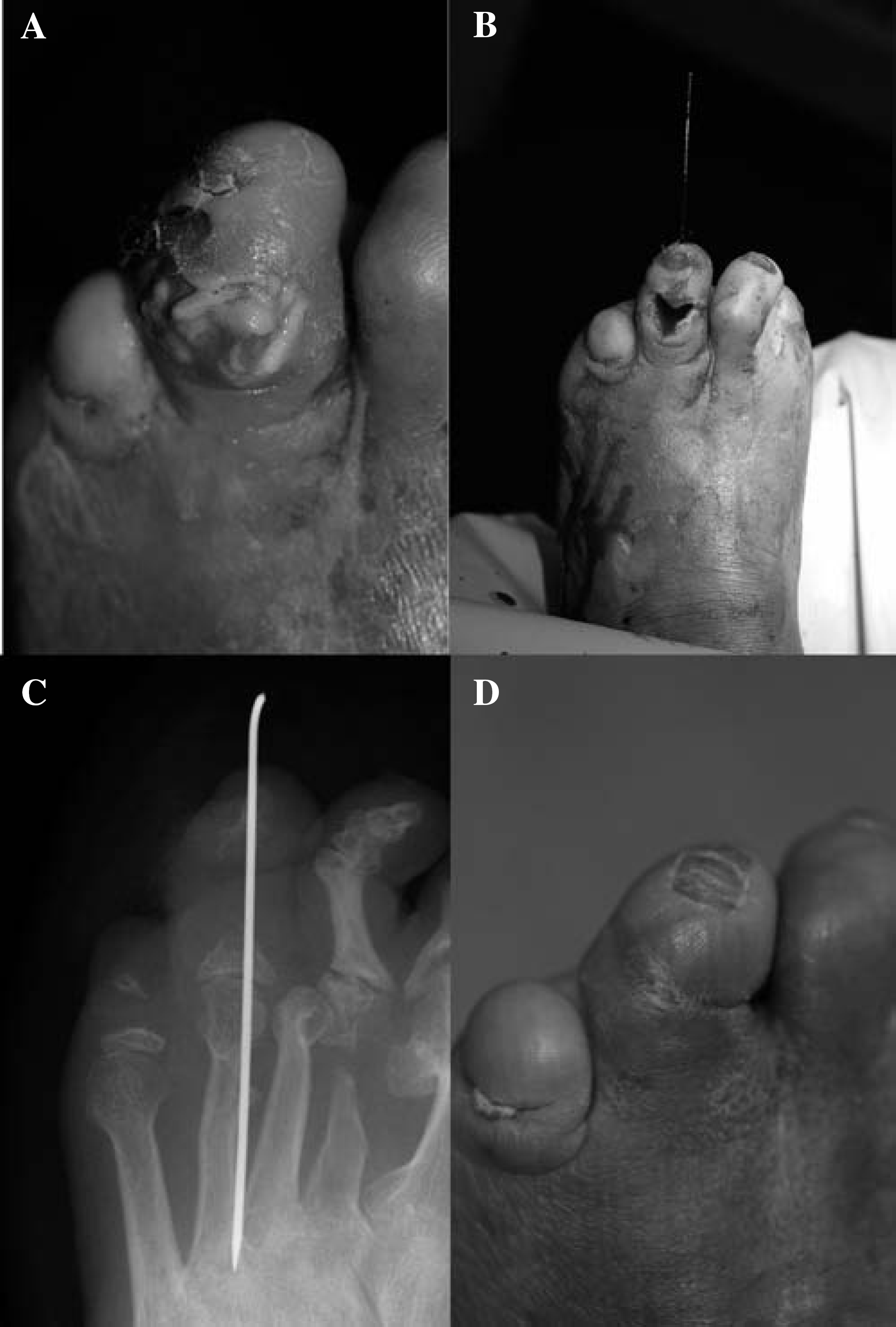
A toe block was selectively performed. In cases where two or more toes were involved simultaneously or debridement of the foot was necessary to control the propagation of infection, an ankle block, or popliteal block, was performed. Tourniquets were not necessary. Almost all wounds associated with claw toes were located on the dorsum of the PIP joint (Figure 2A). Through the dorsal wound or the addition of a longitudinal skin incision on the original wound, the PIP joint was exposed. In cases of an interdigital ulcer from compression of the condyle of the phalanx between the toes, the ulcer formed on the lateral side of toe which was included in the exposure. All infected tissue, including bone and tendon (particularly extensor tendons, capsules, and ligaments), were thoroughly debrided (Figure 2B). At this time, care was taken to avoid damage to the plantar digital artery that runs adjacent to the flexor digitorum longus tendon. All extensor tendons along the dorsum of the PIP joint were gone due to infection or the debridement. The extensor tendon, which might have a role in the proximal propagation of infection, was cut more proximally than the wound. Preoperative radiographs provided guidance regarding which bone should be removed. The finger was the most useful tool in detecting the remaining bone debris. If no osteomyelitis is identified, then the distal condyles of the proximal phalanx are resected transversely in the supracondylar region with a bone-cutter. Copious irrigation was performed. A Kirschner wire 1.2 mm in diameter was introduced at the proximal interphalangeal joint and driven distally until it exited the tip of the toe. Then, with the digit aligned in the correct position, the Kirschner wire is driven proximally to stabilize the arthroplasty site (Figure 2C). The pin was bent at the tip of the toe to prevent proximal migration. If the PIP joint was infected, primary closure was not performed, but if the PIP joint was free of symptomatic infection, primary wound closure was performed. In cases where primary closure was impossible due to infection, the dorsal wound closed via secondary intention.
Following closure, a loose, well-padded dressing was applied. A hard sole postoperative shoe and weightbearing as tolerated was allowed after surgery. After the wounds healed, the Kirschner wire was removed, and regular and consistent movement of the foot with custom-made shoes and molded insoles was encouraged following education on foot care. Followup evaluations were performed every 2 to 3 months for at least 1 year for each patient.
Results
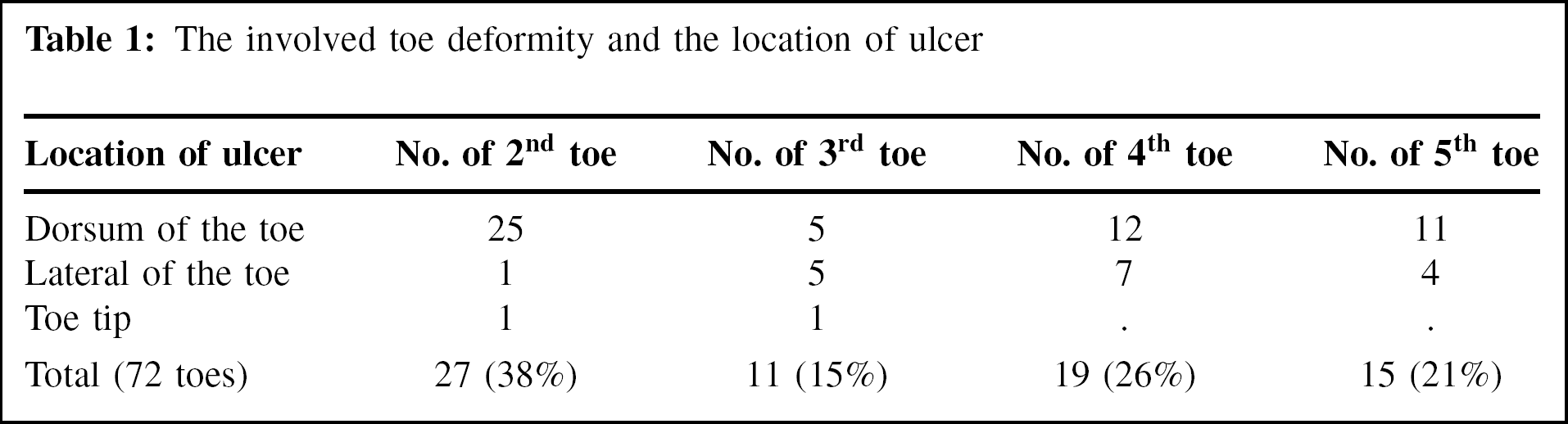
The mean duration of diabetes was 15.4 ± 5.1 years. The mean duration of diabetic foot ulcer presence before hospitalization was 11.8 ± 9.1 weeks. Thirty-eight patients (73%) had diabetes exceeding 10 years'duration and 44 patients (85%) could not detect a 5.07 (10 g) Semmes-Weinstein monofilament. Twenty patients (38%) had an increased BUN or creatinine, and four were dialysis patients. Among diabetic indicators, the median HbA1c was 8.6 ± 2.2%, indicating uncontrolled diabetes in most patients. The majority of patients had acute infection symptoms on the day of admission, and 41 toes (57%) had a swollen “sausage toe” 15 appearance. There were 17 toes with a lateral toe ulcer related to intercondylar compression between the toes. 55 toes had a chronic ulcer located on the dorsum of the PIP joint or DIP joint or toe tip. Involved toes included the second toe in 27 cases (38%), the third toe in 11 cases (15%), the fourth toe in 19 cases (26%), and the fifth toe in 15 cases (21%) (Table 1). Among 15 feet with fifth toe involvement, seven had a co-existing ulcer on the fifth MTP joint area.
All but four patients demonstrated positive bacterial growth on culture studies. Staphylococcus aureus (including seven cases of MRSA) was the most common pathogenic organism in 23 patients (44%), and two or more organisms were identified in 13 patients (25%). The positive results of three phase bone scanning were recognized in all involved digits, and bony destruction on a radiograph appeared in 41 toes (57%).
All patients underwent modified resection arthroplasty. The mean wound-healing time was 25.6 ± 6.2 days. Following surgery, there was no proximal propagation of infection. Three cases of toe amputation were eventually performed. Two cases of toe amputation were necessary when excessive removal of necrotic or infected tissue resulted in toe necrosis after surgery. In one additional case of toe amputation, after the modified resection arthroplasty surgery on the fifth toe, toe necrosis resulted and propagated to the next toe. Eventually, the second to fifth toes were removed.
The involved toe deformity and the location of ulcer
Prior to the modified resection arthroplasty, eight cases of vasculopathy were managed with endovascular balloon dilatation and stenting (6 cases) or bypass surgery (2 cases). Three or more weeks after the vascular intervention, the modified resection arthroplasty was performed. There were 12 toes that underwent re-operation for the removal of infected bone debris that caused non-healing wounds and continuous drainage. No recurrence of claw toe or ulcer was identified during followup. On physical examination, various degrees of toe stiffness in the PIP and MTP joints was identified in all cases, but this stiffness did not visibly impact gait. There were five cases of calluses on the plantar surface of the PIP joint caused by severe limitations in dorsiflexion of the MTP joint. Charcot neuroarthropathy or other deformities were not observed (Figure 2D).
Discussion
The presence of foot ulcers in diabetic patients is extremely debilitating and dramatically increases the risk of lower extremity amputation.6,7 Additionally, costs associated with the treatment of foot ulcers in diabetic patients is a major expense in the overall care of this patient group, even though a recent report indicated that diabetic foot osteomyelitis could be effectively managed with oral antimicrobial therapy, with or without limited debridement. 11 In this study, we focused on claw toe deformities that promoted recurrent ulcer formation. It is widely accepted that osteomyelitis in the phalanges is rarely cured by antibiotics alone and usually requires partial or complete toe amputation. 9 Additionally, patients exhibiting toe deformities with chronic non-healing ulcers are more likely to necessitate toe amputation. In this study, 57% of the toes exhibited osteomyelitis of the phalangeal bone with swelling known as ‘sausage toe’, another possible indicator for toe amputation. 15 We were able to salvage most of the toes and avoid amputation through massive debridement as well as modification of the resection arthroplasty.
Sensory neuropathy and the development of claw toes due to small muscle atrophy has an important role in the development of foot ulcers.2,8,10,12,14 The period of time from the onset of diabetes to the formation of claw toe deformity in diabetic patients is not well-known. In this study, however, 73% of patients had diabetes exceeding 10 years'duration and 85% of patients could not detect a 5.07 (10 g) Semmes-Weinstein monofilament.
Classic claw toe deformities lead to ulceration on the dorsum of the digits. The claw toe deformity promotes a second region for ulceration beneath the metatarsal head as the pressure of the base of the hyper-extended proximal phalanx pushes down on the metatarsal head through its intact but contracted tendons. 3 In this study, ulceration beneath the MTP joint area was so rare that only 7 cases of ulcers over the fifth MTP joint were noted. Some patients with claw toe deformity with toe tip ulcers can be managed without surgery. In this study, the number of patients who underwent surgery due to toe tip ulcers was just two.
In our experience, when sufficient amounts of infected bone were removed, the resultant shortening of the toe length did not cause a floppy toe. Shortening of the toe was seen in patients where excessive removal of phalangeal bone took place to control infection. On followup, toes with prolonged healing time or continuous drainage were those cases in which the bone debris of osteomyelitis had not been clearly removed. Simple x-rays provide a great deal of information to determine the amount of bone to be resected. Moreover, the surgeons'fingers were useful in locating remnants of bone debris in patients'toes. The main limitation of this study was we could not assess the objective functional results such as lesser toe scale due to a high rate of neuropathy and previous fore-foot surgery associated with other toe problems.
Conclusion
A foot ulcer is one of the most common complications associated with diabetic patients and can lead to a decreased quality of life.1,16 The rapid healing of chronic ulcers is paramount in improving the quality of life for diabetic patients. The modified resection arthroplasty is an excellent treatment option for managing toe deformities with non-healing ulcers.