
Abstract
This paper examines the corrosion behaviours of carbon steel immersed in sterile natural sea water with and without strain Bacillus cereus. Electrochemical studies, including Tafel plots and electrochemical impedance spectroscopy (EIS) were performed to evaluate the variation of the corrosion behaviour of carbon steel in medium containing B. cereus as compared to the sterile control samples. The results of Tafel plot measurements showed significant reduction in the corrosion rate in the presence of bacterial biofilm produced by strain B. cereus. The EIS data showed that the charge transfer resistance is greater in a medium containing B. cereus and increases with immersion time.
Introduction
Biofilms are densely packed multicellular communities of microorganisms attached to a surface or interface. 1 Bacteria seem to trigger biofilm formation in response to specific environmental cues such as nutrient and oxygen availability. 2 The growth of biofilm is considered to be a result of complex processes involving transport of organic and inorganic molecules and microbial cells on the surface, adsorption of molecules on the surface (formation of the conditioning layer) and initial attachment of microbial cells followed by their irreversible adhesion facilitated by production of extracellular polymeric substances (EPS).3,4,5–6 A large number of studies have reported accelerated corrosion of metallic materials in the presence of different microorganisms. Extensive research for example has shown increased corrosion rates in the presence of sulphate reducing bacteria (SRB).7,8–9 In addition to the SRB, there are several other microorganisms that have been found to affect the corrosion processes. Rajasekar et al. studied corrosion behaviour of steel API 5LX in diesel–water system containing Bacillus cereus. They revealed an increase in the corrosion rate. 10 Yuan and Pehkonen also observed an increase in corrosion of stainless steel in nutrient rich artificial sea water medium in the presence of Pseudomonas. 11 The influence of activity of bacterial isolates (Serratia marcescens ACE2, B. cereus ACE4) on degradation of corrosion inhibitor and the dissolution of API 5 LX steel has been evaluated by Muthukumar et al. 12 They showed that the two bacterial strains have the ability to degrade the aromatic and aliphatic hydrocarbon present in inhibitor of corrosion and increases the rate of dissolution of metal.
While the majority of microbiologically influenced corrosion studies have focused on the accelerated corrosion rates, corrosion inhibition due to the presence and the activity of microorganisms within biofilms has also been reported in the literature.13,14–15 In order to explain the inhibition mechanism, some researchers reported that protective layers might be passive oxide products formed and entrapped in a biofilm matrix or biofilm matrix itself. 15
Biofilm formation on metals immersed in a marine environment can occur in a few hours. It is the result of deposition of inorganic and organic materials such as phosphates, phosphonates,16,17,18–19 EPS20,21 like carbohydrates, proteins and lipids. The adsorbancy of exopolymeric substance is known to modify the surface properties of the substrate by modifying the surface charge.
Aerobic bacteria were found to decrease the rate of metal corrosion due to biofilm formation.6,20 Perez et al. 22 revealed that the strong adhesion of biofilm could inhibit the damage to the steel for a time. However, at longer times, the same chemical environment, surrounding the biofilm due to microbial metabolism, can lead to a quite corrosive environment for the steel. The microorganisms may induce corrosion inhibition according to two general mechanisms 23 : (i) a neutralisation of the action of corrosive substances present in the environment; (ii) forming protective films or stabilising pre-existing protective films on metal.
It was reported24–25 that biofilm forming aerobic bacterium Pseudomonas and facultative anaerobic Escherichia coli were able to protect carbon steel from corrosion. These authors suggested that the biofilm provide corrosion protection by reducing the oxygen concentration on the metal surface. They also illustrate that increased biofilm depth caused greater corrosion reduction. 26 It was also revealed that in presence of Pseudomonas flava, a significant reduction in the corrosion rate of mild steel was noticed. 27 They suggested that this bacterium stain has the effect of improving the corrosion inhibitive properties of phosphates film. According to Wu et al., 28 the results of mass loss showed that the corrosion rate of the specimens in the single Pseudomonas system was faster than that in sterile sea water but slower than in natural sea water. It has been shown that micropitting corrosion takes place underneath the corrosion product. Pitting corrosion of 304 stainless steel due to presence of Pseudomonas aeruginosa in biofilm state was also reported when investigating the microbiologically influenced corrosion. 29 Relationships between pit characteristics and corrosion properties were investigated by Chaves and Melchers. 30
The corrosion inhibition caused by iron reducing bacteria, Shewanella algae or Shewanella ana, was also observed for aluminium, mild steel and brass exposed to artificial sea water containing growth medium. 31 Pitting attack of aluminium in Luria Bertani medium was significantly reduced by the secretion of anionic peptides by Bacillus biofilms.21,32 Chongdar et al. 33 have studied the behaviour of mild steel immersed in phosphate buffered basal salt solution containing two aerobic bacteria, Pseudomonas alcaligenes and Pseudomonas cichorii. They show that in the medium containing P. cichorii, a significant decrease in the corrosion rate was observed. The corrosion behaviour of the aluminium and copper in modified baar's medium containing aerobic bacteria, Bacillus brevis or Pseudomonas fragi, was also studied. It was noticed that the biofilms secreted by both bacteria reduce the corrosion rate of these two metals. It was suggested that protection occurs because of the elimination of oxygen. 20 Ornek et al. 21 studied the corrosion behaviour of the aluminium in artificial sea water containing Bacillus subtilis. They demonstrated that in presence of a bacterial biofilm produced by these bacteria, pitting was observed during the first 2 days; however, for the remainder of the exposure time, the aluminium was passive. The study of corrosion activity of abiotic sample with that of the sample colonised with Bacillus mycoides indicates inhibition of aluminium. 34
Bacillus cereus is a gram positive, aero-anaerobic respiratory type, rod shaped bacterium that is widely distributed in several biotopes. It belongs to the Bacillus kind. Among the numerous Bacillus species, 35 B. cereus can be of marine environment origin.
While many reviews describe the growth and the effect of SRB on the environmental parameters and corrosion behaviours of carbon steel,36,37–38 to our knowledge, no work has dealt with the effect of introducing strain B. cereus on the corrosion performance of carbon steel in sea water. The present study aims to better understanding the influence of strain B. cereus bacterium on the corrosion behaviour of carbon steel in natural sea water as a function of immersion time, using scanning electron microscopy (SEM), energy dispersive spectroscopy (EDS), open circuit potential (E OCP), polarisation resistance, Tafel plots and electrochemical impedance spectroscopy (EIS).
Materials and methods
Materials
The coupons of A60 steel composed of the following elements with a mass ratio of 0.4%C, 0.045%P, 0.045%S and 0.009%N and remaining Fe were used as a working electrode. Before each experiment, the exposed surfaces (0.5 cm2) were sequentially abraded with 600, 800, 1000 and 1200 silicon carbide (SiC) paper, followed by ultrasonic bath in acetone for 5 min, sterilised by immersion in ethanol, washed with sterile distilled water and dried aseptically in air.
Medium
The B. cereus strains used in this study were first isolated from marine environment (in the region of Boumerdes located in Algeria), and then immersed in sterile sea water. The sea water analysis gave the following composition in grams per litre: chlorides sodium 27.2, sodium magnesium 3.6, magnesium sulphate 1.45, calcium sulphate 1.31 and other components in low concentrations. The pH was 8.1.
The microorganisms were incubated 24 h on a beef extract agar at 28°C. All tests were carried out in natural sea water. This medium was sterilised by autoclaving for 20 min at 120°C under 1.3 bar pressure. The sterile medium was used as a control for all the experiments. The contaminated medium contains 1 mL of inoculum of B. cereus.
Isolation of B. cereus
Two sea water samples were put in sterile flasks and were immediately taken to the laboratory to look for aerobic and anaerobic bacteria. For the anaerobes, a 1 mL sample was taken from the sterile tube and in which 2 mL of tryptone glucose extract agar (TGEA) was added. Once the agar was solidified, 1 mL of sterile liquid paraffin was added to ensure anaerobiosis. Then, the whole was incubated at 37°C between 24 and 48 h in the oven.
To seek for aerobic bacteria, decimal dilutions of the sample were first prepared (10− 1 and 10− 2) in sterile physiological water. In order to develop these aerobic bacteria, TGEA was used as a culture medium.
The biological analysis shows the presence of some yeasts as well as clamydospores. The dominant flora consist of bacteria forming round, grey and large colony, with irregular contours. Anaerobic microorganisms were not detected. The enrichment of these colonies required the use of 10 mL of brain heart infusion broth, in which some colonies were transplanted from the analysis of sea water. The incubation was carried out at 37°C between 18 and 24 h.
Our analysis revealed the presence of bacteria. The observation of these bacteria showed long bacilli Gram positive of regular shape isolated or in short chains. In addition, we have noticed the presence of endospores. Biochemical tests, revealed also the presence of catalase. The identification was confirmed with the API 20 E identification system. The bacteria observed positive interaction with Voges Prauskauer, citrate, nitrate and gelatin. The analysis demonstrated that these isolate bacteria belong to the B. cereus group, which is spore forming, aerobic, mostly mesophilic and grow at a pH up or equal to 7. These bacteria can stand high salt concentrations.
Electrochemical techniques
The variation in time of the open circuit potential E OCP, the polarisation resistance R p, Tafel plots and impedance measurements were performed in a conventional three-electrode cell. The working electrode was an A60 steel (0.5 cm2), and the auxiliary and reference electrodes were a platinum wire and a saturated calomel electrode respectively. The electrochemical measurements were performed using a measuring device consisting of an Autolab PGSTAT-30 driven by GPES and FRA 4.9 Software (Eco Chemie, the Netherlands) controlled by a PC.
Experiments of potential sweep were performed at a scan rate of 0.5 mV s− 1 from the E OCP. For electrochemical impedance measurements, an ac amplitude voltage of 5 mV and an applied frequency ranging from 1 kHz to 0.01 Hz has been used. For EIS data modelling and curves fitting method, the Equivalent Circuit Software (Equivcrt) is used. This programme is based on non-linear least squares fitting, which allows non-ideal electrochemical behaviour to be modelled.
All potential values mentioned in electrochemical measurements were measured with respect to saturated calomel electrode.
Surface analysis
Environmental scanning electron microscopy (XL30 ESEM) coupled with EDS was used to examine the surface and the characteristic corrosion of the A60 steel samples after 6 days of exposure to sterile medium with and without B. cereus. In addition, we highlight the presence of biofilm and its evolution over time on the surface of the electrode immersed in the contaminated medium using a microbiological analysis carried out over 6 days. The technique of transferring the biofilm from the A60 steel surface to the culture medium was carried out according to the protocol, which consisted of imprinting the surface on agar TGEA Petri dish under sterilised atmosphere. After a day of immersion, we put the surface of the disc, which contains biofilm, on agar TGEA where it begins to form biofilm. The same procedure is repeated on the third and the sixth day. The Petri dishes are then incubated in a drying oven at 37°C for 48 h.
Results and discussions
Scanning electron microscopy analysis
Figs. 1 and 2 show micrographs of the electrode surface immersed in sterile sea water for 6 days with the presence and absence of B. cereus. In sterile medium (Fig. 1), a duplex thick layer is formed on the steel surface. It is composed of a compact inner layer and a porous outer layer. As can be seen, the porous layer presents poor adhesion and instability. There are many cracks over its surface, and they might be induced by intrinsic physical growth stresses. 39 The addition of B. cereus in the solution leads to the formation of a supplementary layer, with different structural characteristics (Fig. 2). Two areas are distinguished: a dark area (the inner layer), in which iron is the widespread species, and compounds containing phosphorus can also be found. The clear area (outer layer) is porous and is due to the heterogeneous biofilm formation. As can be seen (Fig. 2a), B. cereus cells are dense and aggregate to form bacterial clusters or irregular biofilm. They are rod shaped, with generally the same size and morphology, measuring length of ∼3.55 and 0.96 μm in width.
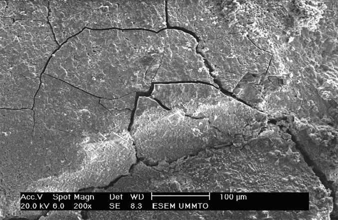
Images (SEM) of A60 steel after 6 days of exposure in sterile seawater
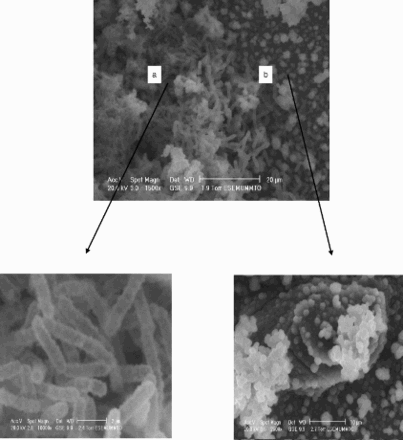
a bacterial community; b biofilm
The EDS analysis of the layer formed on the carbon steel immersed in sterile sea water with the presence and absence of B. cereus is given in Table 1. As can be seen, in the sterile sea water without B. cereus, the sodium chloride salt is present on the surface (Table 1), while the presence of phosphorus indicates corrosion products containing phosphates.
Energy dispersive spectroscopy analysis corresponding to abiotic and biotic system
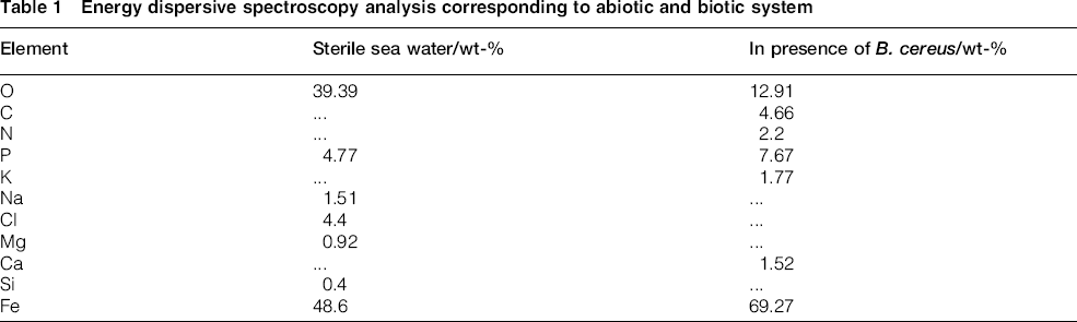
In the presence of a bacteria, EDS spectrum indicated the presence of of Ca, S, P and Fe, and traces of C and N are also identified (Table 1), suggesting the presence of biofilm and EPS. C and N may form a C–N bond, which is well known to be a functional group of proteins. Thus, one can suggest that A60 steel immersed in a medium containing B. cereus may be constituted by an oxide layer of iron phosphate covered by bacteria and EPS.
Fig. 3 presents photographs showing the evolution of the biofilm on the surface sample according to the immersion time. After the first day of immersion, on can notice the presence of few bacterial cells aggregated over the surface. The extension and accumulation of bacteria in visible colonies are well revealed after 3 days of exposure. With increasing immersion time (after 6 days of immersion), the biofilm seems smooth and become more and more dense and compact.
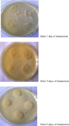
Development of biofilm on A60 steel with exposure time in media containing B. cereus sterile sea water
Open circuit potential EOCP
The variations of E OCP with time exposure of AC60 steel in aerated sterile sea water with and without B. cereus is shown in Fig. 4. In sterile sea water, E OCP abruptly decreases during the first 25 h of immersion, and then remained relatively constant throughout the period of exposure, reaching − 0.65 V after 20 days. In the presence of bacteria, E OCP versus time data show an initial shift in potential towards the negative direction reaching − 0.68 V after 5 days. E OCP continuously increased and reached − 0.66 V after 9 days of exposition, and then decreased. After that, E OCP remained constant, reaching − 0.67 V after 20 days.
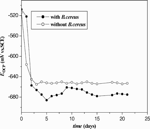
E OCP as a function of time of A60 steel in sterile sea water with and without B. cereus
The shift in E OCP to negative values during carbon steel OCP exposure, in the presence of bacteria, may be explained by a rate reduction in cathodic process caused by a significant decrease in oxygen concentration at the carbon steel/electrolyte interface. It is well known that the respiration of bacteria in a biofilm generates areas with different characteristics. In aerated medium, the bacteria within the top layer of the biofilm will use the oxygen for energy production. At a certain depth within the biofilm, all the oxygen will have disappeared. There will be less and less bacteria because the bacteria above are using oxygen faster than it can diffuse from the water into the film. 40
Polarisation resistance Rp
The main advantage of the polarisation resistance measurement technique is the possibility to monitor continuously the instantaneous corrosion rate of a metal exposed to a corrosive environment. Therefore, this technique is very fit for the detection of changes in corrosion rates due to the existence of bacteria, inhibitors, sunlight, biocides, etc.
Fig. 5 shows the variation of R p with exposure time of A60 steel in the sterile medium in the presence and the absence of B. cereus. In the infected medium, it was interesting to note the feature that shows that at the beginning of the immersion, R p increases with time and stabilises at a value close to 10 KΩ. This behaviour suggests that the surface of the infected electrode is quickly changing in the first 5 days of immersion. In the sterile medium, R p increases during the first 2 days of exposure and continues until a maximum value close to 1.31 kΩ before decreasing. Note that the values of estimated R p are higher in the presence of bacteria compared to those obtained in the sterile environment. In other words, the B. cereus has an inhibitory effect on the corrosion of A60 steel.
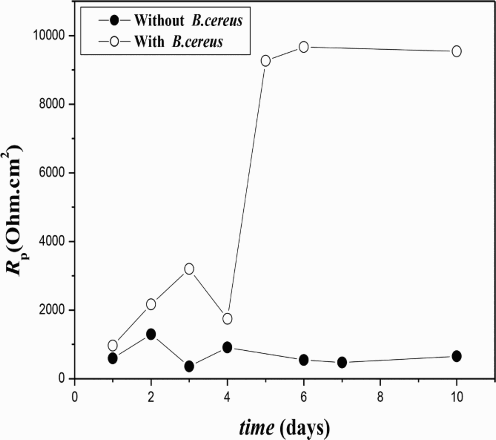
Estimated values of polarisation resistance as function of exposure time (with and without B. cereus)
The growth of biofilms is a combination of adhesion of organic and/or inorganic macromolecules, EPS production and microbial growth. The major components of EPS are proteins and polysaccharides. A function frequently attributed to EPS is their general protective effect on biofilm microorganisms against external environment. It was postulated that the exopolysacharides produced by the bacterial strains reduced the corrosive activity as it prevented the interaction of the metal surface with the external environment. 41
Bacillus cereus could produce EPS with good effect on corrosion inhibition. According to Bragadeeswaran et al., 42 the biofilm formed microorganisms produce EPS, which may serve as corrosion inhibitor for stainless steel. The EPS produced by B. cereus is a heteropolysaccharide, which influences the corrosion. A direct relationship between the rate of corrosion and the concentration of EPS was also revealed.
Potentiodynamic polarisation curves
Fig. 6 shows potentiodynamic polarisation curves of the A60 steel exposed to sea water in the absence and in the presence of B. cereus as a function of exposure time. A marked difference between the polarisation curves of the carbon steel with and without B. cereus after 1 day of exposure can also be observed. In free B. cereus containing medium, the anodic current density drastically increases, and higher current density values were obtained. Whereas, in the presence of B. cereus, a narrow passive plateau is observed with low passive current densities, confirming the inhibitive role of incorporating B. cereus in sea water in the corrosion process.
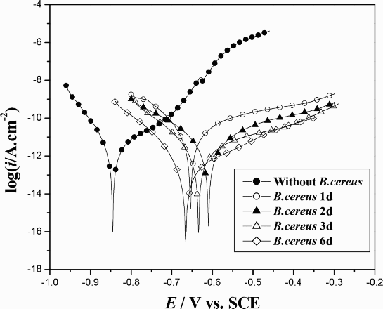
Tafel plots of A60 steel in sterile sea water with and without B. cereus, scan rate 0.5 mV
According to the reported literature,43,44 if the displacement in corrosion potential is more than ± 85 mV with respect to the corrosion potential of the blank, the inhibitor can be considered a distinctive cathodic or anodic type. In the present study, one can notice that there is a displacement in the corrosion potential >100 mV with respect to the corrosion potential of the blank; therefore, the B. cereus can be classified as anodic inhibitor.
In B. cereus containing medium, one can notice that the shape of the curves does not vary with time, which indicates that the mechanism of anodic and cathodic process that takes place on the surface of the electrode does not change. It is also clearly observed that corrosion current densities decreased with the exposure time, and significantly reduced from 18.43 μA cm− 2 after 1 day of exposure to 3.14 μA cm− 2 after 6 days of exposure (Fig. 7). The decrease in the corrosion rate of the electrode with immersion time is due to the reinforcement of the protective film on its surface. Until 6 days of exposure, B. cereus could produce more and more EPS with good effect on corrosion inhibition.
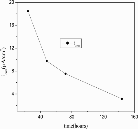
Corrosion current density and corrosion potential of A60 steel in sterile sea water with B. cereus as function of exposure time
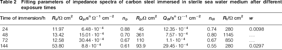
Regarding the marine environment, corrosion process generally occurs under diffusion control. However, the non-appearance in Tafel plots of the limiting diffusion current of dissolved oxygen is often limited by other factors. One such factor is the gradual build up of the corrosion product layer, which progressively retards the diffusion of oxygen from the external environment. It was also reported that the O2 reduction process on iron carbon steel is complex since it depends on the state of the oxide film including the presence of metallic ions [Fe (II), Fe (III)]. 45
In addition, in open air sea water, the predominant cathodic half cell reaction is

Electrochemical impedance spectroscopy measurements
Electrochemical impedance spectroscopy was increasingly used for the characterisation of biofilms. It is considered as a powerful tool to the study of bacteria adhesion to a specimen surface. In this work, EIS measurements were performed to evaluate and to understand how the biofilm influenced the corrosion rate of carbon steel in sterile sea water.
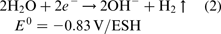
Impedance spectra of A60 steel, recorded at the open circuit potential, in sterile sea water, with and without B. cereus, as a function of exposure time are illustrated in Fig. 8.
Impedance spectra of A60 steel immersed in sterile sea water (a,b without B. cereus, c,d with B. cereus) for various exposure times (a,c Nyquist plots; b,d magnitude Bode plots)
Because of the fact that important high frequency features are difficult to distinguish in a complex plane plot, Bode modulus plots were added for suitable representation of the impedance spectra.
Fig. 8a and b shows respectively typical Nyquist and Bode modulus plots of A60 steel immersed from 1 to 6 days in sterile sea water without B. cereus.
As can clearly be seen, one can differentiate between two frequency time constants. The characteristic change in the slope of the impedance magnitude with frequency variation indicates the presence of more than one time constant. This allows the determination of two capacitive contributions involved in the measured impedance. As shown by the changes in the capacitive loops diameter and magnitude over time, we observed different corrosion behaviours in the concerned environment.
During the first 2 days, the capacitive loop diameters and magnitudes increased rapidly, after which they fluctuated and decreased. On the basis of surface analysis, which showed the presence of phosphorus, the observed fluctuation according to immersion time is possibly due to the response of the iron–phosphate layer based phosphorous compounds that is formed. The decrease in diameter of Nyquist plot for immersion time >2 days suggests localised breakdown of phosphate inhibitor film.
As reported by Castaneda et al.,
46
the presence of KH2PO4 and K2HPO4 in the medium may result in the precipitation of the phosphorus based compound on the surface of the sample. According to previous works,46,47 various species such as chloride, phosphates and organic compounds existing in sea water may precipitate on the surface of the sample. The precipitation of iron phosphate/iron phosphonates occurs at the interface according to the following reactions48,49


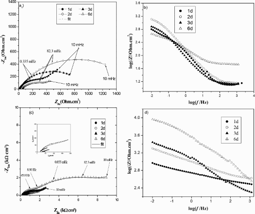
For the interpretation and the exploitation of the impedance diagrams, the different spectra were fitted by an equivalent electrical circuit. Several equivalent circuits have been suggested in the literature to model the response of the biofilms. The equivalent circuit can be developed based on a general knowledge of the physical events that occur at the interface. Several models of circuits to fit the experimental data were tested. The best agreement between experiment and fitting was obtained with the equivalent circuits shown in Fig. 9. The values of χ2 were all < 10− 3 and showed good fits.
Equivalent circuit proposed for impedance measurements
To take into account the non-ideal behaviour of the capacitive elements, constant phase elements (CPE) were introduced. The CPE behaviour can be attributed to different reasons. Spreading effects that are caused by the irregularity of the surface or biofilm irregularity on the sample surface can explain it. 50 Some insoluble material can also influence the dielectric properties of the biofilm capacitor.
The impedance of a CPE is defined as follows

The EIS data of the A60 steel in sterile sea water in the absence and presence of biofilm can be interpreted in terms of equivalent circuit of two time constant model shown in Fig. 9. R s is the electrolyte resistance, R t and CPEdl are respectively the charge transfer resistance and the pseudo double layer capacitance. R p and CPEp are respectively the resistance and pseudocapacitance of the external porous film, and W represents the diffusion impedance through the porous film. W was included to achieve an optimal fit between the measured impedance and simulation model.
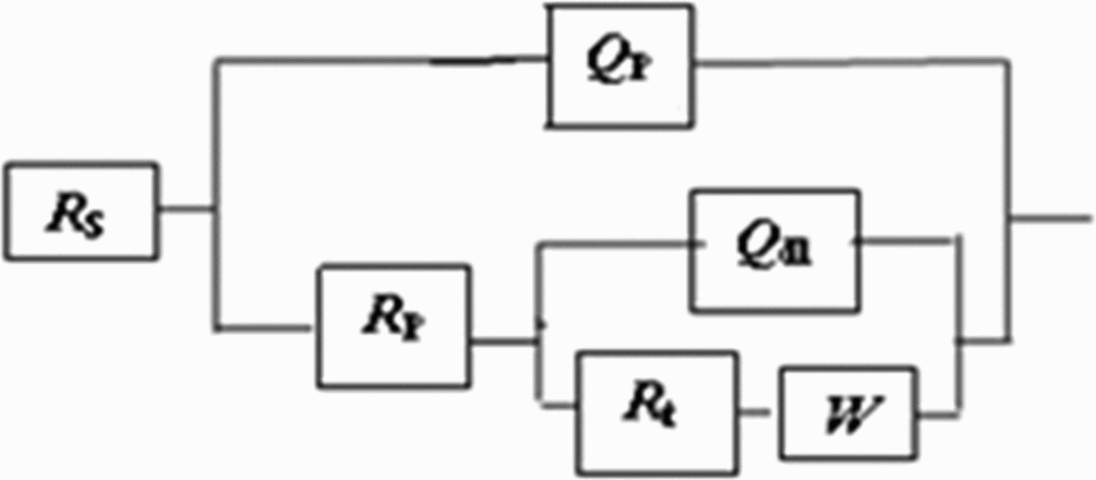
Calculated values of the electrical circuit component for the sterile medium with and without B. cereus obtained from the best fit with the experimental impedance diagrams are summarised in Tables 2 and 3.
Fitting parameters of impedance spectra of carbon steel immersed in sterile sea water medium after different exposure times
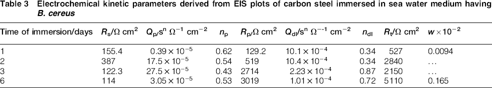
Electrochemical kinetic parameters derived from EIS plots of carbon steel immersed in sea water medium having B. cereus
In the absence of B. cereus, R ct and R p increased during the first 2 days of exposure and then decreased. The high value of R ct (close to 1 KΩ cm2) observed after 2 days of exposure may be interpreted by the resistive character of the iron phosphate layer in relatively short exposure time. Whereas, after 2 days of immersion, R tc and R p decreased, reflecting a change in the surface state of the samples. Two suggestions can be put forward to explain such behaviour: (i) after a certain period of immersion, the outer porous layer of corrosion products, containing phosphates, becomes less stable and less adherent and so lose it protectiveness role. The higher n p values indicate that the porous outer layer is almost homogeneous until 2 days of exposure. The lower n p value after 6 days of immersion indicates surface inhomogeneity. This finding corroborates well with SEM observations. As reported previously, many cracks are shown over its surface after 6 days of exposure.
(ii) After 2 days of immersion, the dissolution of the metallic inner layer is more marked due to the more pronounced pitting corrosion at this stage. The CPE component n dl can be used as the measure of surface inhomogeneity, its decrease being connected with certain increase in surface metal roughening. As can be seen from Table 2, values of n dl after 2 days of immersion are lower than that obtained at the beginning of exposure, revealing the high heterogeneity of the inner layer.
Concerning the A60 steel immersed in B. cereus containing sterile sea water, values of the electrical circuit components are reported in Table 3. It is interesting to note that values of R t are higher than that in the culture of isolated bacteria, confirming the inhibitory effect of B. cereus on the corrosion of A60 steel compared to that found without B. cereus. We can mention the increase in R t over time, indicating the presence of corrosion inhibitor. The biofilm acts as a limited barrier to ion transport, which could attenuate the aggressiveness of the corrosion attack.
We can also notice that as far as the immersion time increases, values of R p increase. It is well documented that EPS production is required for biofilm formation (initial attachment) and development. 51 So, the increase in R p might be explained by the formation of a homogeneous layer produced by the organo metal (FeEPS) complex formed after the interaction of the dissolved iron (Fe2+) with EPS secreted by the bacteria during its growth. This layer reinforces the porous film formed by phosphates. According to Jin et al., 52 EPS extracted from different culturing stages contained different proportions of protein and polysaccharide but with similar functional groups. All types of EPS could inhibit iron corrosion and the EPS from the stationary stage had the highest inhibition efficiency.
Values of n ∼0.3 were cited in the literature in the case of porous electrodes. 53
Conclusions
Bacillus cereus can adhere onto carbon steel surface to form biofilm in sea water under aerobic conditions. The SEM results show the formation of heterogeneous biofilm layer on the carbon steel substrate due to presence of a bacterium B. cereus.
The E corr value of the medium inoculated with bacteria shifted more negative value than the E corr values of the medium, suggesting that the increase in the corrosion resistance of A60 steel was due to the reduction in the oxygen concentration at the electrode surface by biofilm.
Potentiodynamic polarisation curves showed a significant reduction in the corrosion current densities in the presence of B. cereus. The decrease in the corrosion rate of the electrode with immersion time is due to the formation of protective film on its surface.
The impedance spectra measurements also have revealed the inhibitory effect of the biofilm formed by B. cereus.
Acknowledgement
The authors wish to thank the team of Laboratoire physics and chemistry of materials for their support in the production of this work