Abstract
 Open accessAbstractFirst published 2026

Select search scope: search across all journals or within the current journal


 Open accessAbstractFirst published 2026Open accessResearch articleFirst published 2026
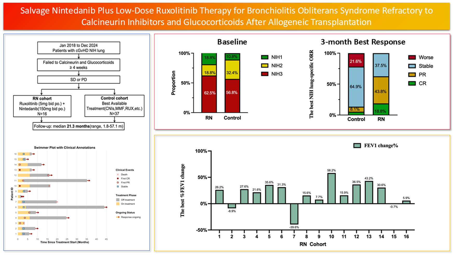
Bronchiolitis obliterans syndrome (BOS) is a severe pulmonary complication following allogeneic hematopoietic stem cell transplantation (allo-HSCT) with limited therapeutic options once refractory to standard immunosuppression. We conducted a pilot study from January 2018 to December 2024, enrolling consecutive patients with BOS defined by NIH criteria who failed glucocorticoids and calcineurin inhibitors for ≥4 weeks. Sixteen patients received salvage therapy with ruxolitinib 5 mg twice daily and nintedanib 150 mg twice daily (RN cohort) in continuous 28-day cycles for up to six cycles, while 37 contemporary patients served as controls. At baseline, NIH lung scores in the RN cohort were 18.8% NIH 1, 18.8% NIH 2, and 62.5% NIH 3. The median number of treatment cycles was 3.5 (range, 1–6). At 3 months, 11 patients (68.8%) achieved ≥10% improvement in %FEV1 from baseline (median = 26.5%, range = 15.6%–58.2%). By NIH lung response criteria, the overall response rate (ORR) was 62.5% (12.5% complete response, 50.0% partial response) in the RN cohort versus 13.5% (5.4% complete, 8.1% partial) in controls. Notably, hematologic toxicities were less frequent with RN therapy than in controls. These findings suggest that low-dose ruxolitinib combined with nintedanib is an effective and well-tolerated salvage regimen for BOS after allo-HSCT and warrant confirmation in a prospective phase II study.
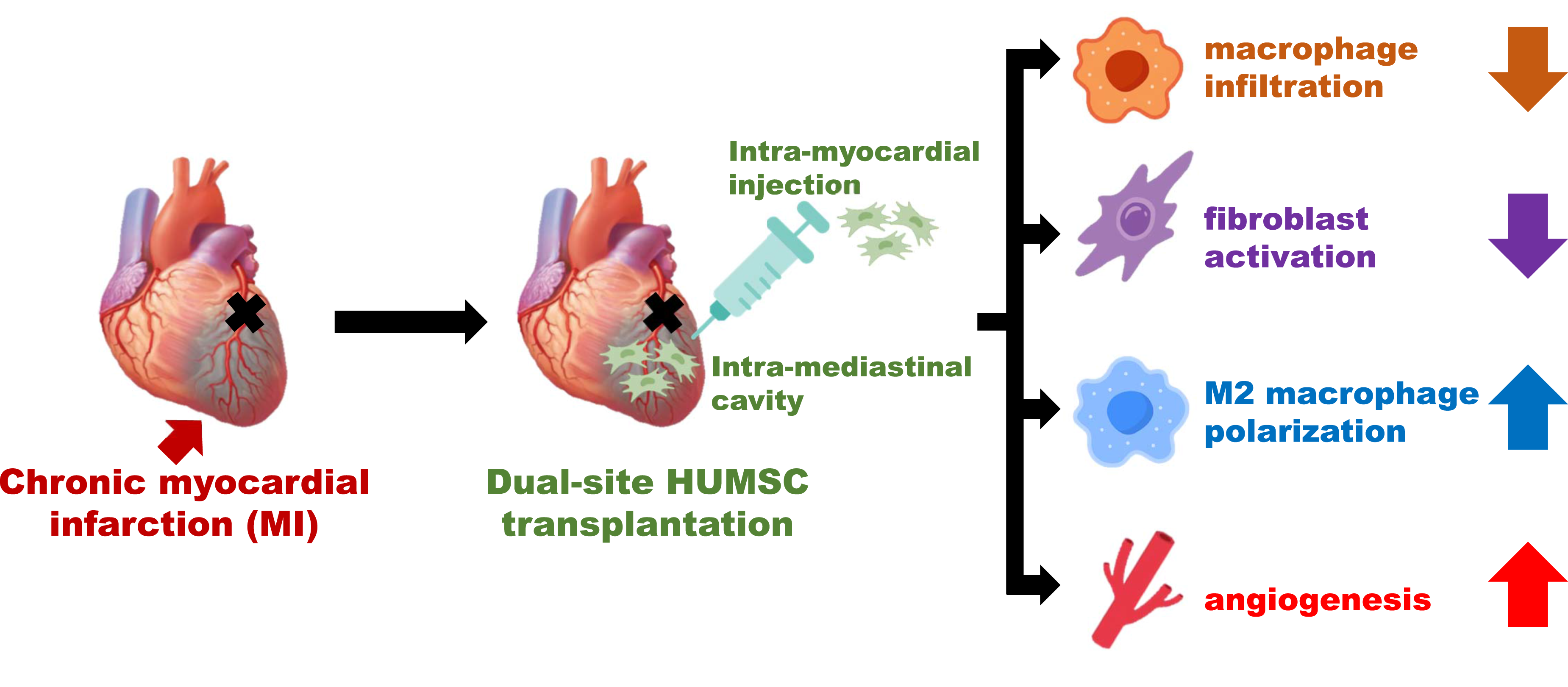
Open accessResearch articleFirst published 2026Myocardial infarction (MI), mainly caused by coronary artery occlusion, remains a leading cause of death worldwide. Although many patients survive after emergency treatment, chronic MI often develops, underscoring the need for effective therapies. This study evaluated the therapeutic potential of human umbilical mesenchymal stromal cells (HUMSCs) in a rat model with chronic MI. MI was induced by permanent ligation of the left anterior descending artery. Seven days post-ligation, 4×10⁶ HUMSCs were transplanted into the peri-infarct myocardium, while an additional 2.5×10⁷ HUMSCs were introduced into the mediastinal space around the ligation site. Successful model establishment was confirmed by elevated cardiac biomarkers and characteristic electrocardiographic changes. Echocardiography and magnetic resonance imaging demonstrated significant impairments in myocardial strain dynamics, reduced ejection fraction, and diminished fractional shortening, all of which improved following HUMSC transplantation. The transplantation also reduced macrophage infiltration, increased M2 macrophage polarization, suppressed fibroblast activation, attenuated fibrosis, and promoted angiogenesis, ultimately preserving cardiomyocytes and improving cardiac function. The transplanted HUMSCs were detected in rat’s myocardium without differentiating into cardiomyocytes or endothelial cells. These findings suggest that adequate HUMSC transplantation offers a promising therapy to attenuate progression of chronic MI or heart failure.
Open accessResearch articleFirst published 2026 , Valeria Iansante, Sharon C. Lehec , [...]
Liver transplantation is the only curative option for end-stage liver disease, but its applicability is limited by donor shortages. Hepatocyte transplantation offers a promising alternative, particularly for pediatric acute liver failure, yet its success relies on the availability of viable cells. Cryopreservation enables off-the-shelf use, though long-term storage may impair cell quality. This study investigates whether cryostorage duration, donor characteristics, and organ retrieval parameters affect the viability and functionality of cryopreserved primary human hepatocytes (PHHs). We conducted a retrospective analysis of 144 thawing events across 81 GMP-grade hepatocyte batches, cryopreserved for up to 14 years. Donor age ranged from 3 days to 70 years. Viability and functionality were assessed using MTT assays, albumin and urea secretion, and CYP450 activity. Linear regression was used to analyze correlations (
This is a visual representation of the abstract.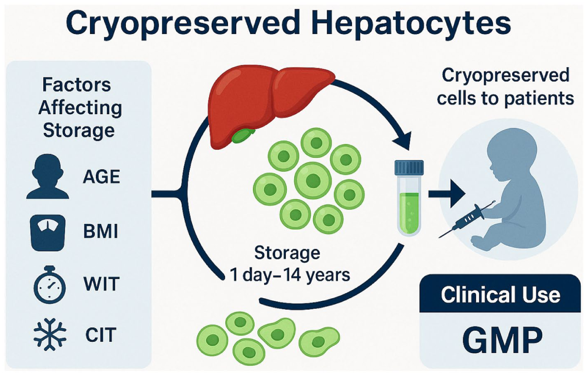
Open accessReview articleFirst published 2026, Xiaojian Zhu, Yi XiaoAutoimmune diseases (AIDs) are a class of diseases caused by autoimmune intolerance, which can be divided into systemic and organ-specific diseases. AIDs affect approximately 10% of the global population and rank among the leading causes of disability and mortality. At present, immunosuppressive agents are the first choice for the treatment of AIDs. B-cell-targeted therapies—particularly CD20 monoclonal antibodies—have brought new hope for systemic AIDs, yet a subset of patients still respond poorly. As a rapidly developing cellular immunotherapy technology, Chimeric antigen receptor T cell (CAR-T) plays an important role in the treatment of hematological malignancies. CAR-T targeting B-cell-specific antigens can rapidly deplete circulating B cells, thereby reducing the formation of autoantibodies, which has become the basis for research on CAR-T in the treatment of autoimmune diseases. Currently, many studies are underway, and CAR-T and its derivative therapies bring new hope for the treatment of autoimmune diseases.
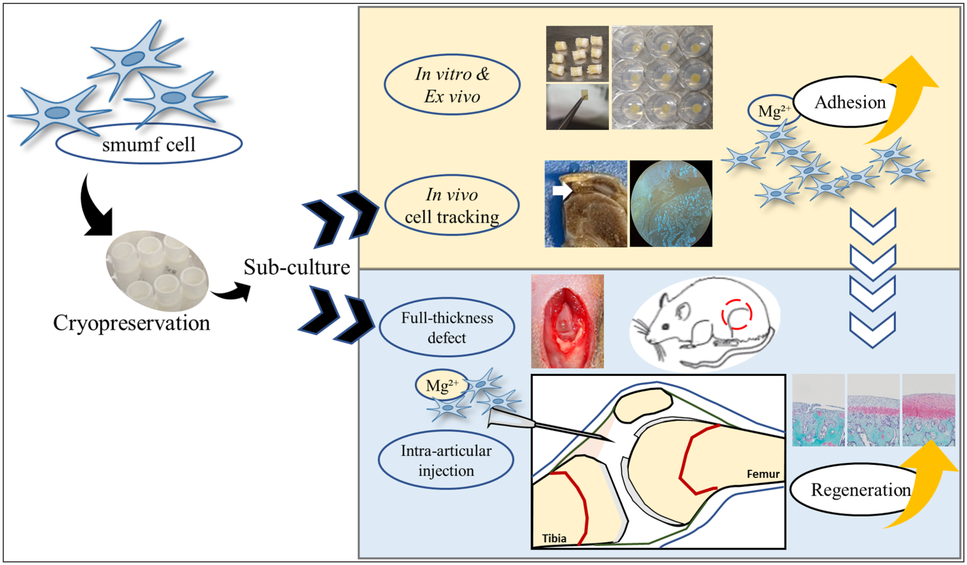
Open accessResearch articleFirst published 2026The effective application of allogeneic mesenchymal stem cells (MSCs) has the potential to enhance cartilage regeneration. This study aimed to evaluate the therapeutic efficacy of intra-articular (IA) injections of small umbilical cord–derived fast proliferating cells (smumf cells) combined with magnesium (Mg2+) in a rat model of full-thickness cartilage defects (FTDs). Adhesion of smumf cells was assessed on type I collagen-coated surfaces
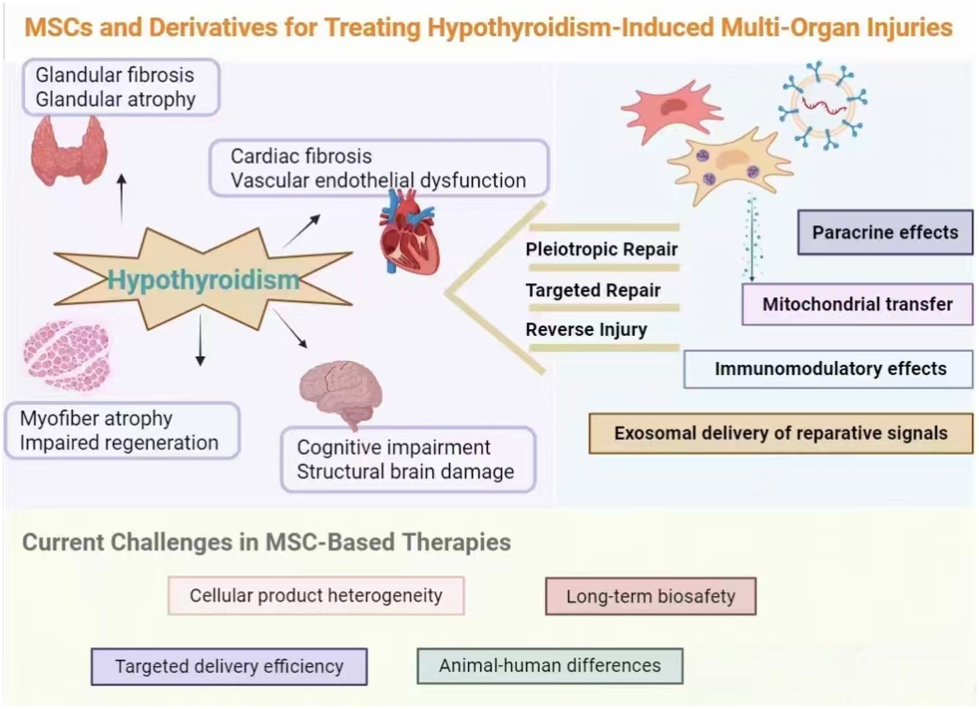
Open accessReview articleFirst published 2026Hypothyroidism, a prevalent endocrine disorder characterized by insidious onset and prolonged progression, leads to metabolic slowdown and multiorgan dysfunction, significantly compromising patients’ quality of life. While hormone replacement therapy improves most patients’ serum thyroid function, it faces limitations, including individual response variations, lifelong medication requirements, and potential risks, with no reversal of existing tissue damage. In recent years, the mesenchymal stem cells (MSCs) and their derivatives have gained prominence in immunomodulation and regenerative medicine because of their abundant sources, low immunogenicity, potent paracrine effects, and multidirectional differentiation capabilities for tissue repair. Their unique advantages in modulating the immune microenvironment, promoting angiogenesis, reducing fibrosis, and stimulating endogenous cell regeneration offer novel strategies to overcome treatment bottlenecks in hypothyroidism and its complications, effectively repairing thyroid and multiorgan damage in animal models. This review synthesizes literature from the PubMed and Web of Science databases, incorporating relevant basic research and clinical trials. This study examined the systemic impacts of hypothyroidism, evaluated the experimental and clinical roles of the MSCs in thyroid tissue reconstruction and organ protection, and analyzed molecular mechanisms, including immune regulation, the antioxidant stress response, anti-apoptosis, and cellular function restoration. Clinical application potential is assessed alongside critical challenges such as standardized preparation, long-term safety,
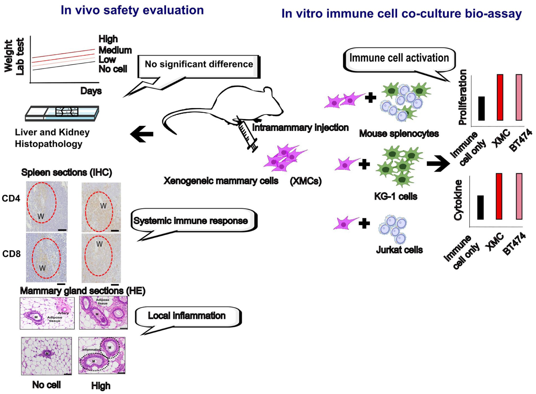
Open accessResearch articleFirst published 2026, Ting-Yu Lin , [...]Xenogeneic cells isolated from safe animal tissues and expanded
Open accessEditorialFirst published 2026Diabetes mellitus (DM) is a metabolic disorder with chronic hyperglycemia due to insulin deficiency and/or impaired insulin action. DM is a common disease, but it often threatens a patient’s quality of life. Cellular replacement therapy using insulin-producing cells is a promising therapy for severe DM because of the proper provision of internal insulin according to a change in blood glucose concentrations. This Special Collection, “Cellular Replacement Therapy for Diabetes,” was planned to appeal the current status and present novel translational trials of this therapy. Ten specialists presented their research, which attempted to overcome three limitations of the current cellular replacement therapy, including the regulation of immunity, limited donor supplies, and establishment of a preferable transplant site for cellular replacement therapy. Regarding regulation of immunity, potential of mesenchymal stem cells and immunomodulatory splenocytes is clarified. Regarding alternative donors, the current status of porcine islet xenotransplantation and therapy using multipotent stem cell–derived cells is introduced. And regarding transplant site, possibility of liver surface and subcutaneous tissue is elucidated.
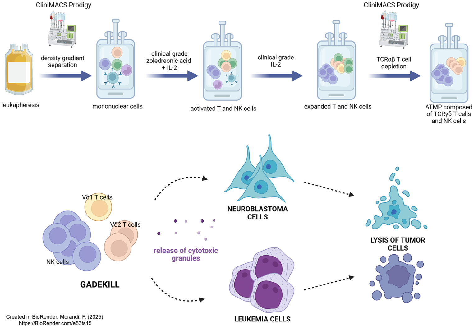
Open accessResearch articleFirst published 2026, Martina Della Lastra, Gino Tripodi , [...]γδ T lymphocytes and NK cells are effective to kill tumors or viral-infected cells avoiding graft versus host disease (GvHD), thus they have attracted high interest as potential tool for adoptive cell therapy. We generated an advanced therapy medicinal product (ATMP) composed of mature γδ T and NK cells to provide an innovative tool to protect patients against tumor relapse and life-threatening infection after haploidentical hematopoietic stem cell transplantation. The ATMP was manufactured and validated in a GMP facility and was obtained from leukapheresis stimulated with zoledronic acid and IL-2, afterward depleted of αβ T lymphocytes using the CliniMACS Prodigy. The ATMP is characterized by high homogeneity, cell viability, cytotoxic abilities, stability after cryogenic preservation, and it was virtually free of αβ T and B lymphocytes. Both NK and γδ T cells were activated and characterized by high expression of cytotoxic and activating receptors including NKG2D, CD16, NKp30, NKp44, and NKp46. Furthermore, γδ T lymphocytes and NK cells were cytotoxic against myeloid leukemia or neuroblastoma cells. In conclusion, we implemented a novel ATMP to be shortly translated into clinical practice, which may be used in the post-transplant phase as efficacious immunotherapy in neuroblastoma and leukemic pediatric patients.
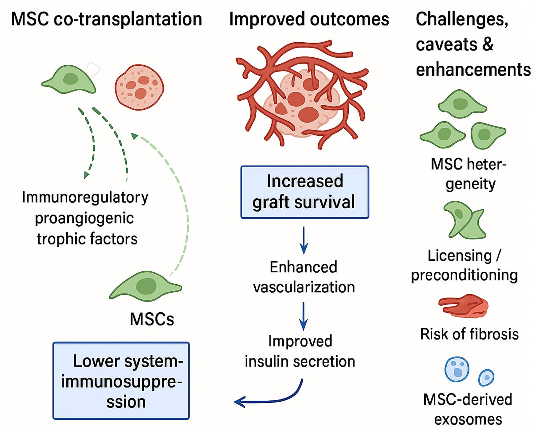
Open accessReview articleFirst published 2026, Carl Randall Harrell, Crissy Fellabaum , [...]Islet cell transplantation holds great promise for restoring glycemic control in patients with type 1 diabetes. However, its long-term efficacy remains limited due to poor islet survival, immune rejection, and insufficient vascularization. Mesenchymal stem cells (MSCs) have emerged as potent biological adjuvants capable of addressing these challenges through a range of molecular mechanisms. MSCs secrete a variety of growth factors, immunoregulatory and pro-angiogenic molecules that enhance viability of islet cells, modulate the immune response, promote neo-angiogenesis and enhance islet engraftment. In addition, MSC-derived exosomes (MSC-Exos) have been identified as key mediators, delivering regulatory microRNAs and proteins that replicate many of the beneficial effects of MSCs in a cell-free format. MSC-Exos act as small RNA carriers and immunomodulators, promoting islet survival and functional integration. Understanding the molecular interplay between MSCs, their exosomes, and the islet microenvironment provides crucial insights for the development of advanced co-transplantation strategies. Accordingly, in this review article, we summarized current knowledge about molecular mechanisms that are responsible for MSC-dependent improvement of islet cell transplantation and we highlighted the translational potential of MSC and MSC-Exos-based approaches in improving islet graft outcomes for type 1 diabetes.
Open accessArticle commentaryFirst published 2026Enhancer–promoter (E-P) interactions are central to cell-type-specific transcriptional programs, yet the molecular machinery that establishes and maintains these loops has remained poorly defined. A recent study by Jiang et al, published in
Open accessResearch articleFirst published 2026Enterocutaneous fistula (ECF) traditionally relies on surgical treatment and lacks effective therapies to promote tissue regeneration. This study constructed a composite system based on hypoxia-preconditioned human umbilical cord mesenchymal stem cell–derived exosomes loaded onto a gelatin sponge (GS Hypo-Exos), aiming to explore its potential application in ECF treatment. First, systematic characterization of exosomes was performed: transmission electron microscopy, nanoparticle tracking analysis, western blot, and flow cytometry confirmed successful exosome extraction. Material evaluation indicated that the gelatin sponge exhibited high porosity and good biocompatibility. Scanning electron microscopy confirmed successful loading of exosomes onto the sponge surface, and
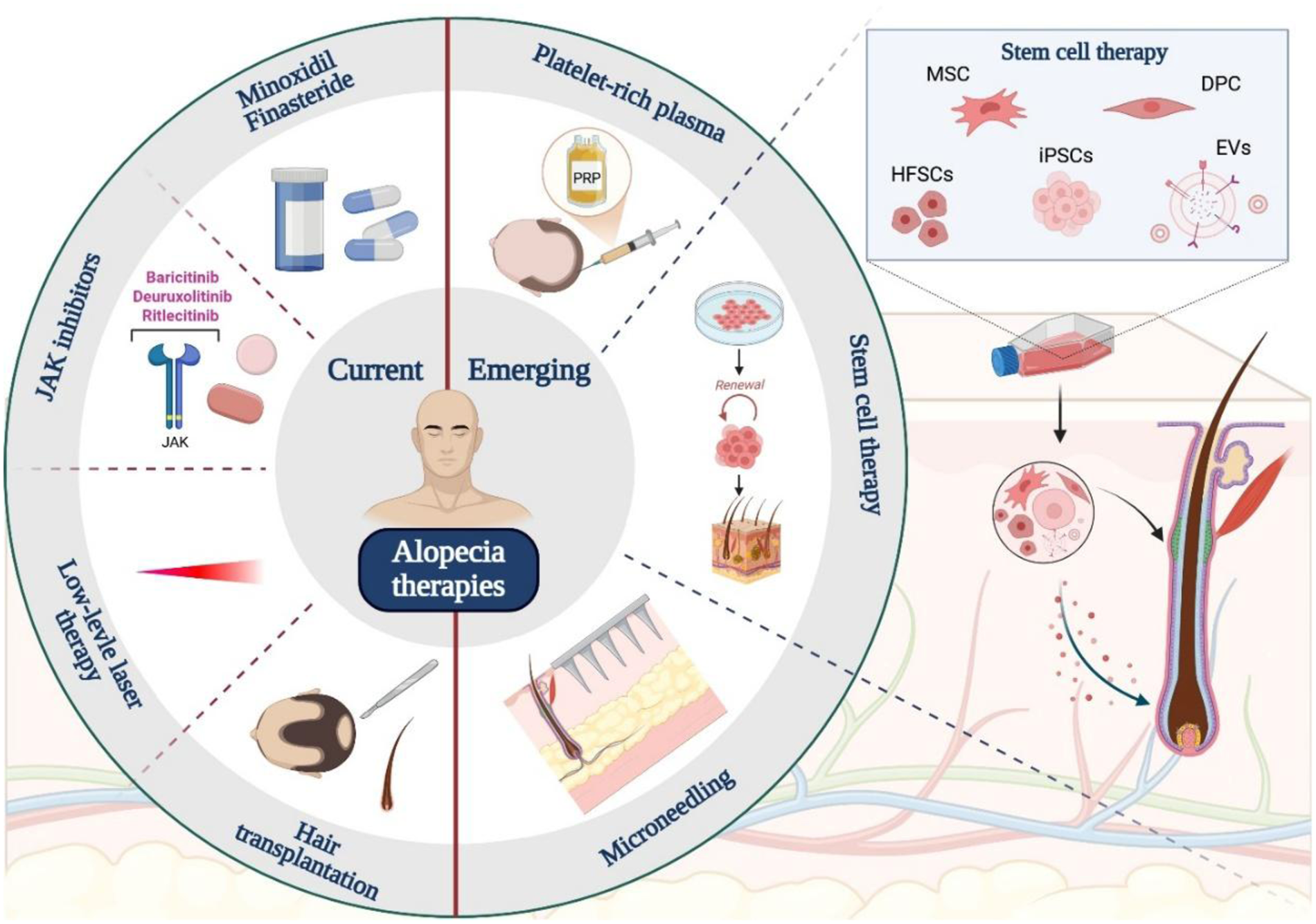
Open accessReview articleFirst published 2026The natural cycle of hair growth and shedding is continuous. However, an abnormal rate of hair regrowth can signal alopecia, a condition characterized by excessive hair loss and thinning. Alopecia is a common dermatological concern globally, affecting individuals across all ages and sexes, often resulting in psychological distress and diminished quality of life. Despite the availability of various current therapy options such as minoxidil, finasteride, JAK inhibitors, low-level laser, and hair transplantation, these approaches are often limited by variable efficacy or side effects. Consequently, there is growing research interest in regenerative therapies using stem cells as a promising avenue to overcome these shortcomings. Stem cells, characterized by their ability to self-renew and differentiate into multiple cell types, offer the potential to promote hair follicle regeneration. Among the different stem cell types under investigation, hair follicle stem cells (HFSCs), mesenchymal stem cells (MSCs), and induced pluripotent stem cells (iPSCs) have attracted attention due to their regenerative capabilities and immunomodulatory properties. This narrative review explores the underlying causes of alopecia, current and emerging therapies, particularly emphasizing the therapeutic potential of stem cells and their specialized roles in promoting a supportive environment for the hair follicle and scalp. It also examines ongoing clinical and preclinical studies involving stem cell–based therapies for hair loss, aiming to assess their feasibility as future clinical solutions for alopecia.
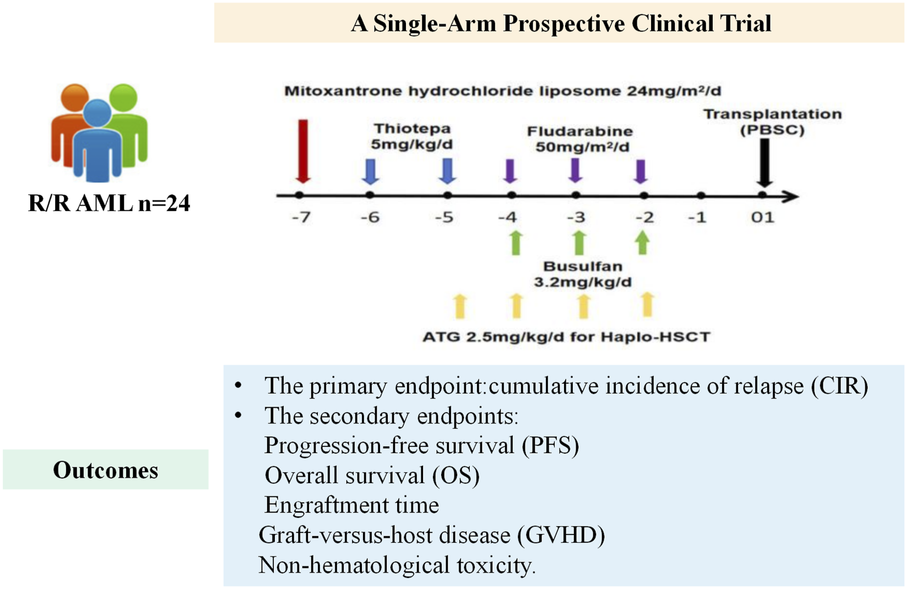
Open accessResearch articleFirst published 2026To investigate the efficacy and safety of MTBF as conditioning regimen for salvage allogeneic hematopoietic stem cell transplantation (allo-HSCT) in patients with relapsed or refractory acute myeloid leukemia (R/R AML). We conducted a single-arm prospective clinical trial (NCT06385808). The main outcome was cumulative incidence of relapse (CIR), and the secondary outcomes were progression-free survival (PFS), overall survival (OS), engraftment time, graft-versus-host disease (GVHD), and non-hematological adverse effects. Twenty-four R/R AML patients from the First Affiliated Hospital of Xi’an Jiaotong University were enrolled, and all achieved engraftment. Mucositis was the predominant toxicity and was managed appropriately. The two-year CIR was 10%. The two-year OS and PFS were 78.9 ± 11.1% and 51.2 ± 19.3%, respectively. Two patients experienced relapse, and three patients died of infection. Subgroup analyses demonstrated that maintenance treatment post-transplantation may improve OS. The MTBF regimen for salvage allo-HSCT in R/R AML exhibited notable anti-leukemia activity and tolerable toxicity. (NCT06385808, Efficacy and Safety of MTBF Conditioning Regimen for Salvageable Allo-HSCT in the Treatment of R/R AML).
Open accessArticle commentaryFirst published 2026, Erlie JiangSteroid-refractory acute graft-versus-host disease (SR-aGVHD) remains a major therapeutic challenge after allogeneic hematopoietic stem cell transplantation. Although ruxolitinib and basiliximab are widely used, their limitations highlight the need for safer, more effective, and scalable biologic strategies. Xenopax, a humanized anti-CD25 monoclonal antibody, has demonstrated encouraging results across real-world and clinical studies. The multicenter RELAX study reported a 28-day overall response rate of 64.5% and a 2-year overall survival of 68.0%, with reduced corticosteroid exposure and infection rates compared with historical basiliximab cohorts. Importantly, RELAX incorporated an economic assessment, showing a lower incremental cost per responder and reinforcing xenopax as a cost-efficient option for SR-aGVHD. Beyond salvage therapy, xenopax is gaining momentum in prophylactic settings, including as a potential replacement for methotrexate in haploidentical transplantation. Looking ahead, integration with autonomous decision-support systems—most notably daGOAT, the first AI platform validated to prescribe risk-adapted GVHD prophylaxis—may further strengthen CD25-targeted approaches. Together, these developments position xenopax as a versatile biologic with the potential to reshape both treatment and prevention paradigms in GVHD management.
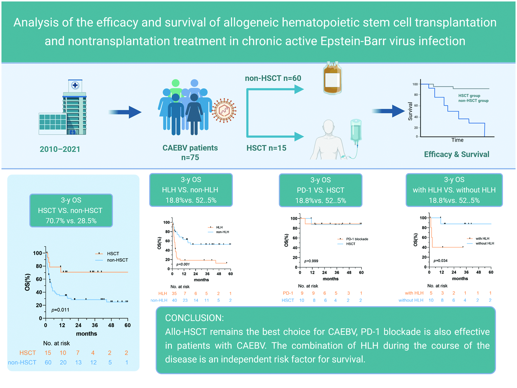
Open accessResearch articleFirst published 2026This retrospective study evaluated 75 patients with chronic active Epstein–Barr virus infection (CAEBV) to compare the efficacy and survival outcomes of allogeneic hematopoietic stem cell transplantation (HSCT) and programmed death-1 (PD-1) blockade therapy. Patients were classified into HSCT and non-HSCT groups. The primary endpoints were overall response rate (ORR), overall survival (OS), and event-free survival (EFS). HSCT significantly improved ORR, 3-year OS, and 3-year EFS compared to non-HSCT treatment. Subgroup analysis showed that PD-1 blockade achieved outcomes comparable to HSCT in a subset of patients; however, HSCT remained superior, overall, particularly in patients without hemophagocytic lymphohistiocytosis (HLH). The presence of HLH was identified as an independent risk factor for inferior survival. In conclusion, allogeneic HSCT remains the preferred curative strategy for CAEBV, whereas PD-1 blockade represents a promising alternative for carefully selected patients. Early recognition and management of HLH are crucial for improving prognosis.
Open accessReview articleFirst published 2026Intramedullary cellular transplantation within the human spinal cord has historically been regarded as a highly experimental intervention associated with substantial theoretical risk. However, cumulative clinical evidence progressively challenges this perception. Multiple early-phase clinical studies have demonstrated the technical feasibility and procedural tolerability of intramedullary and intralesional cellular delivery in spinal cord injury (SCI), employing heterogeneous cellular products, including bone marrow-derived mononuclear cells, mesenchymal stromal cells (MSCs), and neural stem cells. Despite considerable heterogeneity in cellular composition, dosing strategies, injection volumes, and delivery paradigms, severe injection-related complications remain uncommon. Reported adverse events are predominantly mild and transient, while procedure-induced neurological deterioration has not emerged as a reproducible safety signal. These observations suggest that controlled spinal cellular administration may represent a biologically tolerated intervention rather than an intrinsically destabilizing procedure. Beyond procedural considerations, long-term biological safety remains a central dimension of translational evaluation. While theoretical concerns persist — particularly regarding tumorigenesis, clonal selection, and potential genomic instability associated with

Open accessArticle commentaryFirst published 2026The global shortage of human donor livers poses a formidable barrier to treating end-stage liver diseases and unresectable hepatic malignancies, leaving millions of patients without life-saving options each year. While genetically engineered pig-to-human xenotransplantation has achieved significant breakthroughs in cardiac and renal fields, liver xenotransplantation has long lagged due to the liver’s intricate metabolic, synthetic, and immunological functions. The recent report by Zhang et al. in the
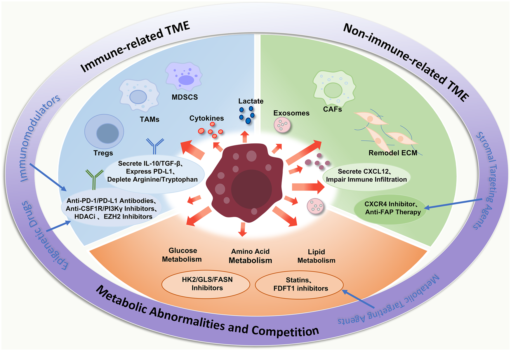
Open accessReview articleFirst published 2026The tumor microenvironment (TME) plays a significant role in the occurrence, development, and prognosis of lymphomas. The mechanisms of each TME component in lymphoma and related therapeutic strategies are reviewed in this article. In the immune-related TME, regulatory T-cells (Tregs), tumor-associated macrophages (TAMs), and other immune cells mediate immune suppression and evasion. In the non-immune-related TME, cancer-associated fibroblasts (CAFs) and extracellular matrix (ECM) are involved in tumor progression, drug resistance, and immune evasion. In addition, aberrant glucose, amino acid, and lipid metabolism in tumor cells not only sustain their rapid proliferation and survival but also reshape the TME to favor immune escape, thereby worsening patient outcomes. Various treatment strategies targeting the components of the TME and metabolic abnormalities have brought new hope for the treatment of lymphoma. Further in-depth investigation into the intricate crosstalk between the TME and lymphoma cells will be instrumental in developing more effective, personalized precision therapies to improve patient survival.
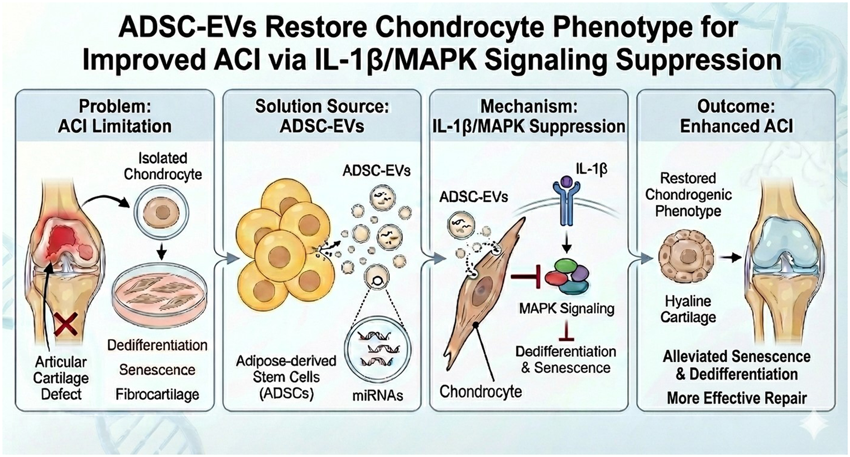
Open accessResearch articleFirst published 2026, Ling-Hua Chang, Che-Wei Wu , [...]Autologous chondrocyte implantation (ACI) has long been regarded as the gold-standard chondrocyte-based therapy for articular cartilage repair. The main challenge in ACI is that chondrocytes lose their chondrogenic phenotype after monolayer expansion
Open accessResearch articleFirst published 2026Metabolic dysfunction–associated steatotic liver disease (MASLD) is the most common chronic liver disorder and can progress to steatohepatitis and fibrosis; although approved pharmacotherapies for metabolic dysfunction–associated steatohepatitis (MASH) with fibrosis remain limited. Autologous chemically induced liver progenitor (CLiP) cells, generated from mature hepatocytes without genetic modification, have shown therapeutic promise in rodents, but their efficacy has not been tested in large animals. Six female Clawn miniature pigs (15–42 kg) were fed a high-fat, high-cholesterol diet to induce MASLD with biopsy-proven fibrosis (Brunt stage ≥1). Animals were assigned to CLiP transplantation (
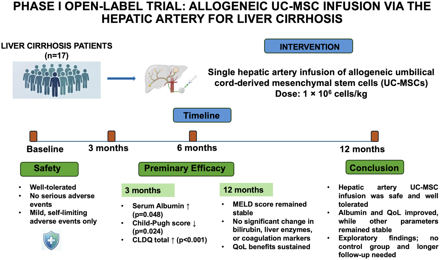
Open accessResearch articleFirst published 2026Liver cirrhosis causes substantial morbidity, and options beyond transplantation are limited. Umbilical cord–derived mesenchymal stem cells (UC-MSCs) may support liver repair, but their delivery via the hepatic artery has not been evaluated. This study assessed the safety and exploratory efficacy of allogeneic UC-MSCs infusion in cirrhosis. Twenty patients with liver cirrhosis entered a single-center open-label pilot trial in Hanoi, Vietnam, between 2020 and 2023; 17 completed follow-up and were included in exploratory analyses. All received a single hepatic artery infusion of UC-MSCs at 1 × 10⁶ cells/kg and were assessed at baseline, 3, 6, and 12 months for liver function, Model for End-stage Liver Disease (MELD) and Child–Pugh scores, Chronic Liver Disease Questionnaire (CLDQ), and adverse events. No serious adverse events occurred; mild events were self-limited. Albumin increased at 3 and 6 months (
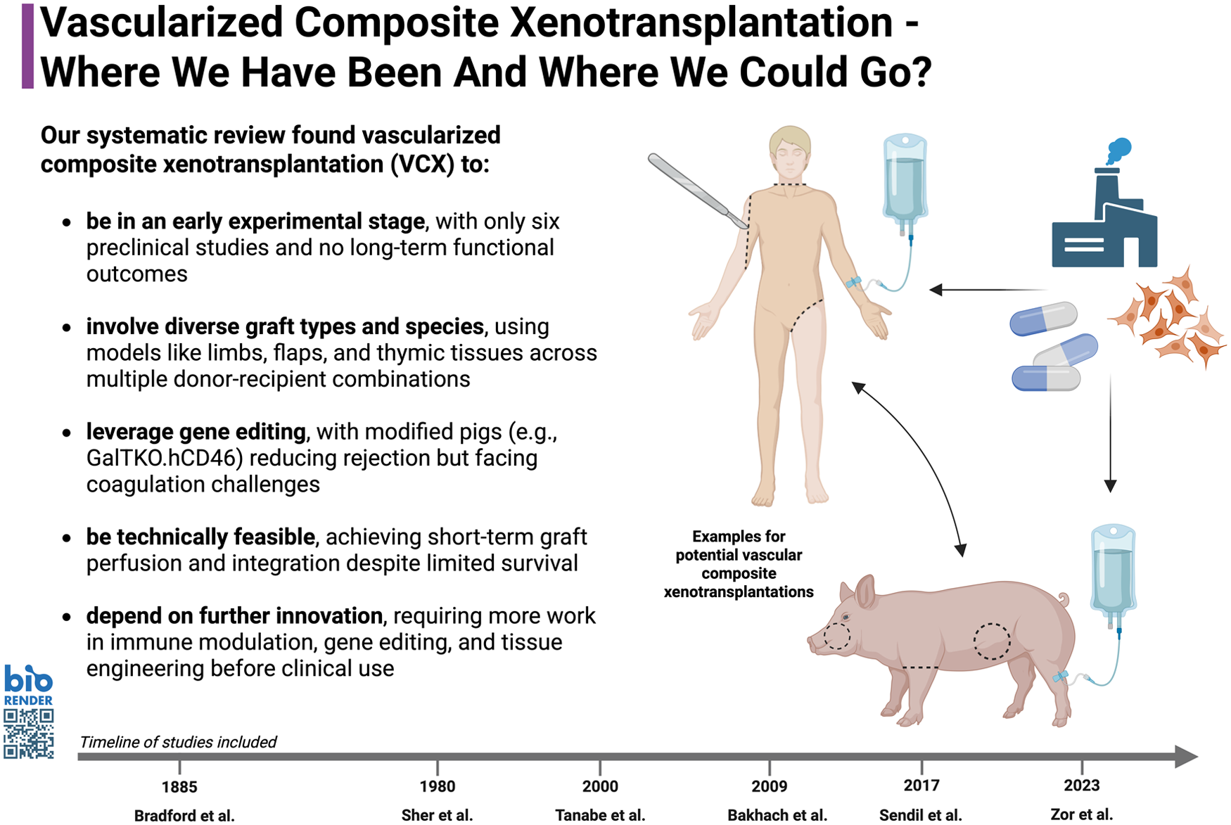
Open accessReview articleFirst published 2026Vascularized composite allotransplantation (VCA) enables functional and aesthetic reconstruction after complex tissue loss but remains limited by donor scarcity and immunosuppression. Xenotransplantation, using tissues from other species, offers a potential solution. While progress in solid organ xenografts has accelerated, vascularized composite xenotransplantation (VCX) remains largely experimental. This study presents the first systematic review of VCX, tracing its development over the past century and preclinical efforts to overcome immunologic and technical barriers. A systematic review following Preferred Reporting Items for Systematic Reviews and Meta-Analyses (PRISMA) 2020 guidelines searched PubMed, EMBASE, Cochrane, Web of Science, and Google Scholar for English-language VCX studies. Methodological quality was assessed using the Newcastle-Ottawa Scale, SYRCLE Risk of Bias tool, and Oxford Levels of Evidence. Of 257 records, six preclinical studies (1885–2023) met inclusion criteria. Donor species included pigs, rabbits, dogs, and humans; recipients were rodents or nonhuman primates. Most used heterotopic grafts (e.g., limbs, groin flaps, thymosternal blocks) combining skin, muscle, bone, and nerves. Techniques included cryopreservation, microsurgical anastomosis, and genetically modified pig grafts (e.g., GalTKO.hCD46). All demonstrated short-term survival without long-term function. Despite technical feasibility, VCX remains experimental. Advances in gene editing, immune modulation, and tissue engineering are essential for clinical translation and may ultimately redefine reconstructive transplantation.
Open accessResearch articleFirst published 2026 , [...]Cell transplantation using cell sheet technology is a promising regenerative approach that enables the delivery of a large number of viable cells while preserving cell–cell interactions and extracellular matrix. However, the clinical application of autologous cell sheets is limited by donor-site invasiveness, prolonged preparation time, and high manufacturing costs. Allogeneic cell sheets may overcome these limitations, but their therapeutic effects and immunological profiles require further clarification. In this study, we investigated the tissue-repair effects and immune responses associated with allogeneic skeletal muscle-derived cell (SMDC) sheet transplantation. In vitro analyses showed that human SMDCs suppressed activated T-cell proliferation in a cell number-dependent manner, lacked co-stimulatory molecules, and expressed immune checkpoint ligands, suggesting a potentially low-immunogenic and immunomodulatory phenotype. For in vivo evaluation, L8-derived syngeneic comparator sheets and allogeneic SMDC sheets derived from different rat strains were transplanted onto the serosal surface in a rat gastric ulcer model. SMDC sheet transplantation promoted early ulcer repair without increasing systemic inflammatory responses, as assessed by serum C-reactive protein levels. Histological analyses revealed limited macrophage and T-cell infiltration at the transplantation sites, although the extent of local immune responses varied depending on donor–recipient strain combinations. Transcriptomic analysis of ulcer tissues showed that the L8-derived syngeneic comparator and selected allogeneic groups shared downregulation of inflammation-related pathways, whereas another allogeneic donor strain induced a distinct transcriptional profile. These findings suggest that allogeneic SMDC sheets can promote early tissue repair without inducing overt systemic inflammatory activation. However, donor–recipient strain compatibility may influence local immunological and transcriptional responses after transplantation. The observed effects are consistent with early paracrine and immunomodulatory mechanisms, although direct cell tracking and more detailed immunological analyses are required. Allogeneic SMDC sheets may represent a potential ready-to-use strategy for gastrointestinal tissue repair, provided that appropriate donor selection and further preclinical validation are performed.

Open accessReview articleFirst published 2026, Jian Chen, Xijie Tang , [...]Extracellular vesicles (Evs) act as a natural intercellular message transmitter, Evs can carry proteins, ribonucleic acid (RNA) and other bioactive substances, and have rich biological regulatory functions. Because of its low immunogenicity and high biocompatibility, it has become a popular research object in drug delivery. These remarkable properties also create new opportunities for modern therapy. However, due to the complex preparation process, there are challenges in terms of targeting accuracy, load release controllability, and pharmacokinetic optimization, and many problems may be encountered in reality. Based on real-life biomedical experiments, this paper summarizes the methods and types of Evs loading drugs, membrane modification methods, and the use of biological materials to improve the release efficiency, so as to provide reference for future research on engineered Evs.

Open accessResearch articleFirst published 2026B-cell maturation antigen (BCMA) chimeric antigen receptor (CAR) T cell therapy has demonstrated promising efficacy in relapsed/refractory multiple myeloma (MM). However, evidence supporting its use in earlier treatment settings remains limited. This study presents a preliminary case series evaluating the feasibility of BCMA CAR-T cell therapy as post-induction consolidation in newly diagnosed multiple myeloma (NDMM) patients who did not proceed to autologous stem cell transplantation (ASCT). Four patients were included in this analysis. After induction therapy, all patients achieved very good partial response or better and did not proceed to ASCT due to ineligibility or personal preference. They subsequently received BCMA CAR-T cell therapy as consolidation treatment. All patients tolerated the BCMA CAR-T cell infusion well. Cytokine release syndrome occurred in all cases, but was limited to grades 1–2, and no immune effector cell–associated neurotoxicity syndrome was observed. Following therapy, patients maintained or further deepened responses and ultimately reached stringent complete response (sCR) and minimal residual disease (MRD) negativity. During a median follow-up period of 17.8 months, all patients remained in sCR status and MRD negativity without the need for additional anti-tumor therapy, and none experienced relapse or disease progression. These preliminary results suggest that BCMA CAR-T cell therapy as post-induction consolidation is feasible and associated with manageable toxicity in NDMM patients not proceeding to ASCT, warranting further evaluation in larger prospective studies.
Open accessExpression of concernFirst published 2026 Open accessResearch articleFirst published 2026
Islet transplantation is a promising treatment for diabetes, but the shortage of donor islets limits its broad application. Induced pluripotent stem cells (iPSCs) provide an alternative source for generating insulin-producing cells; however, whether the somatic cell origin of human iPSCs influences pancreatic endocrine differentiation remains incompletely defined. In this study, we generated iPSCs from human pancreatic duct cells (HD-iPSCs) and compared their differentiation propensity and functional characteristics with human fibroblast-derived iPSCs (HF-iPSCs) under identical differentiation conditions. HD-iPSC-derived cells showed higher expression of pancreatic endocrine and β-cell-associated markers, including insulin, PDX1, and FOXA2, compared with HF-iPSC-derived cells. Flow cytometric analysis further confirmed a higher proportion of insulin-positive cells in differentiated HD-iPSC-derived cells. Functionally, HD-iPSC-derived cells exhibited greater glucose-stimulated C-peptide secretion than HF-iPSC-derived cells, although their secretory capacity remained lower than that of native human islets. Following transplantation into streptozotocin-induced diabetic mice, HD-iPSC-derived cells reduced blood glucose levels more effectively than HF-iPSC-derived cells, and insulin-positive grafts were detected in vivo. These findings suggest that human pancreatic duct cell-derived iPSCs have enhanced pancreatic endocrine differentiation potential compared with fibroblast-derived iPSCs. Although further maturation and optimization are required, pancreatic duct cells may represent a favorable somatic cell source for generating iPSC-derived insulin-producing cells for diabetes cell therapy.
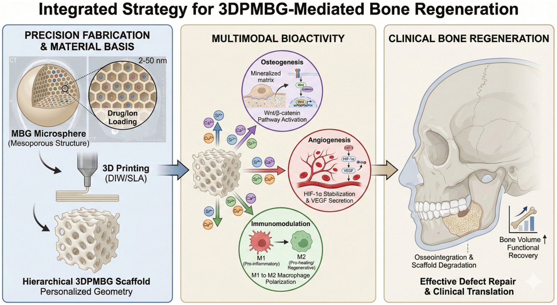
Open accessReview articleFirst published 2026Craniomaxillofacial (CMF) bone defects pose significant regenerative challenges due to complex anatomy and physiological demands. While autologous bone grafting remains the gold standard, it is limited by donor-site morbidity and supply constraints. Mesoporous bioactive glass (MBG), characterized by its ordered nanoporous structure and superior bioactivity, offers a promising alternative. This review systematically analyzes the integration of MBG with 3D printing technologies, including direct ink writing, stereolithography, selective laser sintering, and fused deposition modeling. We critically evaluate physicochemical challenges such as rheological optimization and thermal devitrification while elucidating the “osteo-immune-vascular” axis orchestrated by these scaffolds. Specifically, we discuss how ionic dissolution products modulate macrophage polarization, stabilize hypoxia-inducible factor-1α (HIF-1α) to induce CD31ʰⁱEmcnʰⁱ vessel formation, and activate Wnt/β-catenin signaling. Despite promising preclinical data, clinical translation faces hurdles regarding regulatory approval and manufacturing standardization. Future developments in 4D printing, AI-driven inverse topology design, and organ-on-a-chip validation represent a paradigm shift from passive substitution to active regeneration, paving the way for the tissue-engineered reconstruction of complex CMF defects.
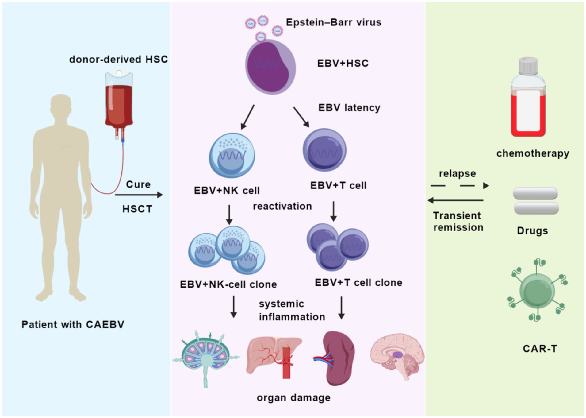
Open accessReview articleFirst published 2026Chronic active Epstein–Barr virus (CAEBV) infection is a rare and highly lethal lymphoproliferative disorder. The pathological basis of this condition involves Epstein–Barr virus (EBV) persisting in hematopoietic stem cells, driving clonal expansion of T cells or natural killer (NK) cells, and subsequently triggering systemic inflammatory responses and multi-organ failure. Current treatment modalities, encompassing antiviral medications, immunosuppressants, and cytotoxic chemotherapy, offer only transient remissions, with the majority of patients ultimately experiencing relapse. Recent single-cell sequencing and chimera studies have confirmed that EBV-infected hematopoietic stem cells constitute the “seed” cell population for CAEBV initiation and maintenance. This finding indicates that allogeneic hematopoietic stem cell transplantation (allo-HSCT) is the only treatment known to date that can fully eradicate viral reservoirs and restore normal immunity, suggesting that it may represent a curative strategy. Nevertheless, transplantation timing, donor matching, conditioning intensity, and transplant-related complications have been shown to have a significant impact on long-term prognosis. The clinical decision-making process necessitates a high degree of individualization, incorporating molecular risk factors, disease activity, and comorbidities. Advancing research into the latent-lytic cycle regulation mechanisms of EBV, in addition to the clinical translation of small-molecule inhibitors targeting viral proteins and EBV-specific adoptive cell therapies, holds great promise for the future. One such potential avenue for future research is the development of an integrated “pre-transplant viral load reduction-post-transplant relapse prevention” strategy. This approach shows great potential in reducing transplant-related mortality and continuously improving survival outcomes for CAEBV patients.
Open accessCorrectionFirst published 2026 Open accessResearch articleFirst published 2026, Bo Shao, Kui Ye , [...]
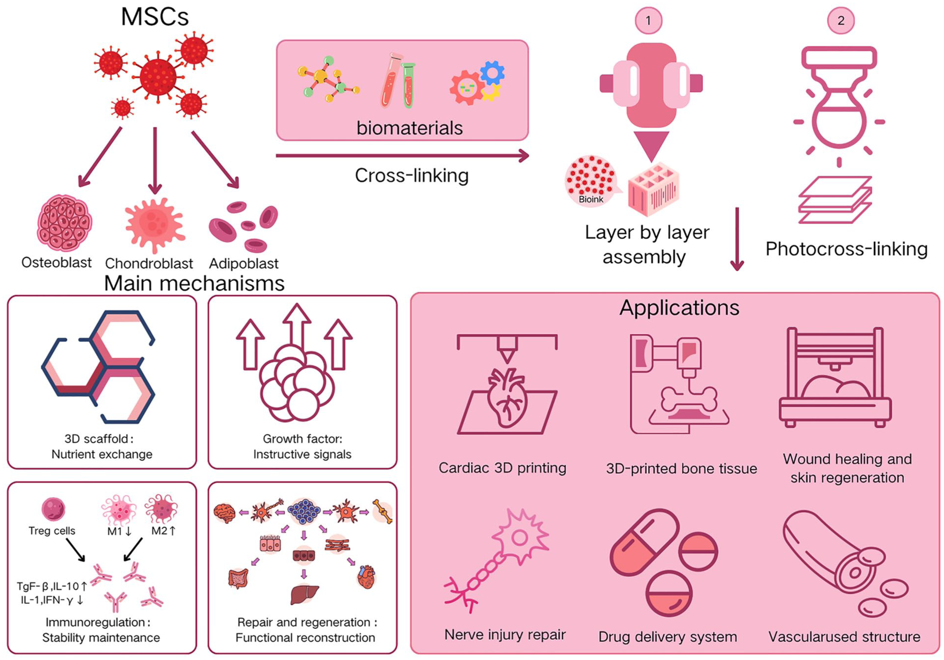
Recent years have witnessed rapid advancements in 3D bioprinting and the widespread application of mesenchymal stem cells (MSCs) across various medical disciplines. The synergistic integration of 3D bioprinting and MSCs has opened innovative avenues for tissue engineering and regenerative medicine, particularly in bone tissue repair and regeneration. However, the progress of 3D bioprinting in the field of MSCs research still requires further exploration, and there remains a scarcity of related bibliometric analyses in this domain. With the aim of addressing this existing gap, this research systematically searched the Web of Science Core Collection for publications spanning from January 2003 to October 2025. It employed CiteSpace for cluster and evolution analysis, VOSviewer for collaboration network and keyword co-occurrence analysis, and the R package “bibliometrix” for statistical evaluation of bibliometric indicators. This bibliometric analysis focused on tissue engineering research integrating 3D bioprinting with MSCs, encompassing 1,846 original articles. These articles were authored by 10,276 researchers from 2,024 institutions across 69 countries and published in 342 academic journals. From 2014 to 2023, the number of annual publications exhibited a fluctuating yet rapid upward trend. China and the United States emerged as the most influential countries, with China experiencing a particularly substantial increase in research output—though international collaborations among institutions and authors remained limited. Wu C.T. and Bose S. stood out as key contributors to this field, while journals such as
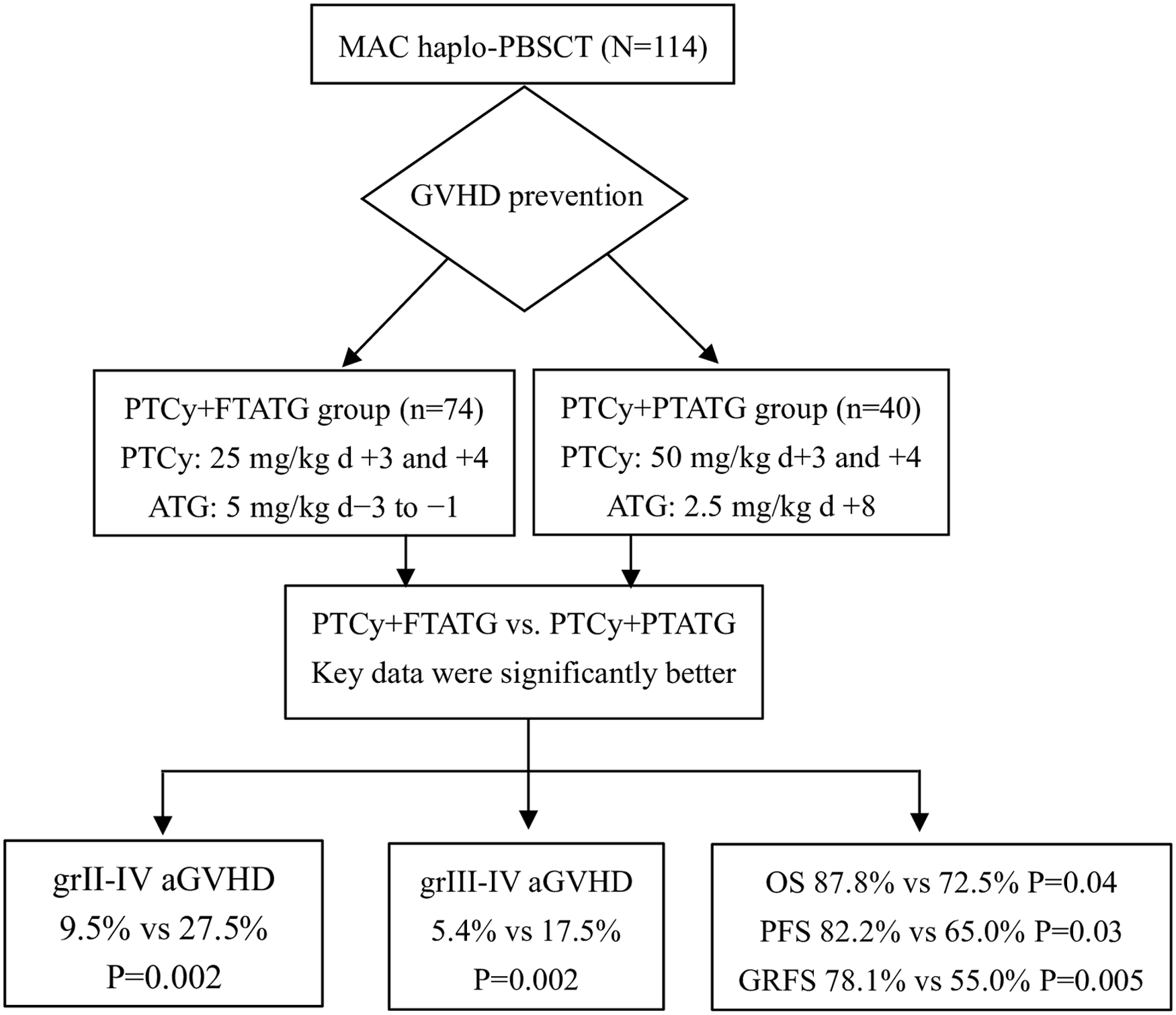
Open accessResearch articleFirst published 2026Although post-transplant cyclophosphamide (PTCy) is widely used to prevent graft-versus-host disease (GVHD), its protective effect remains inadequate in patients undergoing myeloablative haploidentical peripheral blood stem cell transplantation (haplo-PBSCT). We retrospectively evaluated the efficacy of PTCy combined with either pre-transplant or post-transplant antithymocyte globulin (ATG) for GVHD prevention in 114 haplo-PBSCT recipients. The PTCy+FTATG group (n = 74) received ATG at a total dose of 5 mg/kg on days −3 to −1, together with PTCy at 25 mg/kg on days +3 and +4. The PTCy+PTATG group (n = 40) received PTCy at 50 mg/kg on days +3 and +4, followed by ATG at 2.5 mg/kg on day +8. Both univariate and multivariate analyses showed that PTCy+FTATG prophylaxis significantly lowered the risk of grade II–IV acute GVHD (9.5% vs 27.5% [HR 0.24; 95% CI: 0.10–0.59;
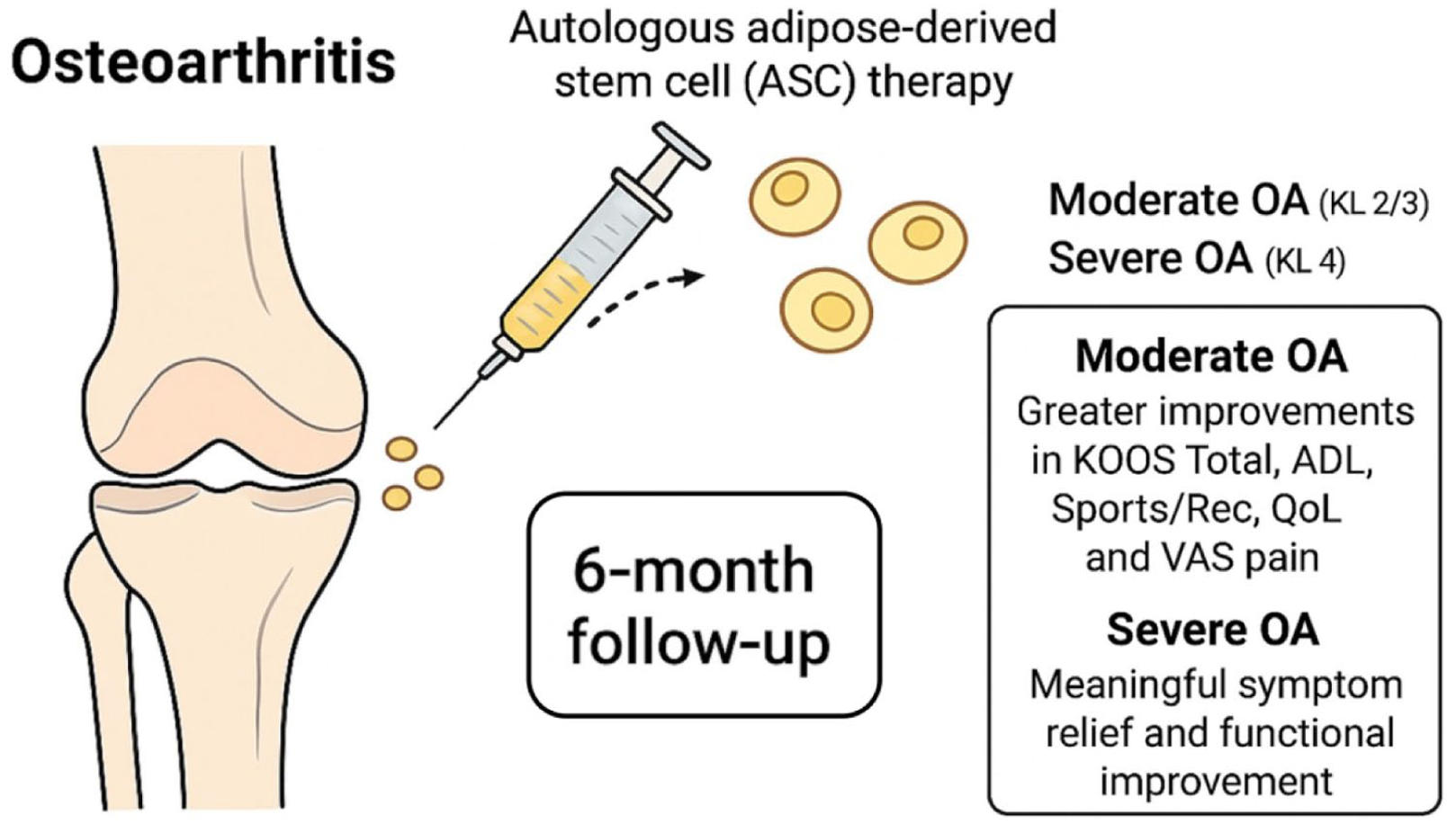
Open accessResearch articleFirst published 2026Knee osteoarthritis (OA) causes pain and disability, and autologous adipose-derived stem cell (ASC) therapy has emerged as a regenerative treatment option. This retrospective cohort study compared short-term outcomes of intra-articular ASC injections between patients with moderate (Kellgren–Lawrence [KL] 2/3) and severe (KL 4) OA. Among 242 treated patients, 98 in each group were analyzed after propensity score matching for age, sex, and body mass index. Pain (VAS) and Knee Injury and Osteoarthritis Outcome Score (KOOS) subscales were evaluated at baseline and 1, 3, and 6 months. Both groups showed significant improvements in pain and function, with KL 2/3 patients exhibiting greater gains in KOOS Total, Activities of Daily Living, Sports/Recreation, Quality of Life, and VAS pain compared with KL 4. KOOS pain and symptoms improved similarly in both groups. Analgesic effects increased over time, and no serious adverse events were observed. Mild transient swelling or discomfort occurred in about 5% to 6% of cases. ASC injections provided meaningful symptom relief and functional improvement, particularly in moderate OA, suggesting that preserved joint structure benefits therapeutic efficacy. In advanced OA, benefits were present but attenuated, indicating limited regenerative potential in end-stage disease.
Open accessResearch articleFirst published 2026To address the lack of effective treatments for germinal matrix hemorrhage-intraventricular hemorrhage (GMH-IVH), a devastating condition in preterm infants with poor prognosis, this study investigated the neuroprotective potential and underlying mechanism of human umbilical cord-derived mesenchymal stem cells (HUCMSCs). Using a collagenase VII-S-induced GMH neonatal rat model and lipopolysaccharide-stimulated BV2 microglial cells,
Open accessReview articleFirst published 2026Hematopoietic stem cell transplantation (HSCT) is frequently accompanied by severe inflammation-related complications, among which graft-versus-host disease (GVHD) stands out as one of the most common and life-threatening. As a systemic inflammatory disorder, GVHD arises when donor T cells recognize the recipient’s alloantigens and initiate an immune attack. Currently, effective second-line treatment options remain scarce for patients with antibiotic-resistant or steroid-refractory GVHD. Mesenchymal stem cells (MSCs) are non-hematopoietic cells widely distributed in fetal and adult tissues and organs, endowed with multipotent differentiation potential and prominent immunomodulatory properties. Emerging evidence suggests that impaired function or apoptosis of MSCs exacerbates HSCT-associated complications and significantly compromises hematopoietic stem cell engraftment. Over the past two decades, leveraging their potent anti-inflammatory and immunomodulatory capacities, MSCs have been rapidly integrated into HSCT research and clinical practice, where they play a pivotal role in promoting hematopoietic engraftment and preventing or treating GVHD. This review elaborates on the molecular basis of MSCs’ anti-inflammatory effects and the inflammatory pathological characteristics of HSCT-related complications. We conducted a systematic literature search in PubMed, Web of Science, and Embase databases up to December 2025. The search strategy combined the following terms: (“mesenchymal stem cells” OR “MSCs”) AND (“hematopoietic stem cell transplantation” OR “HSCT”) AND (“graft-versus-host disease” OR “GVHD”) AND (“inflammation” OR “immune regulation”). It systematically analyzes the molecular mechanisms underlying MSCs’ anti-inflammatory actions and their application progress in HSCT, aiming to provide a theoretical foundation and translational insights for the rational clinical application of MSCs in HSCT.
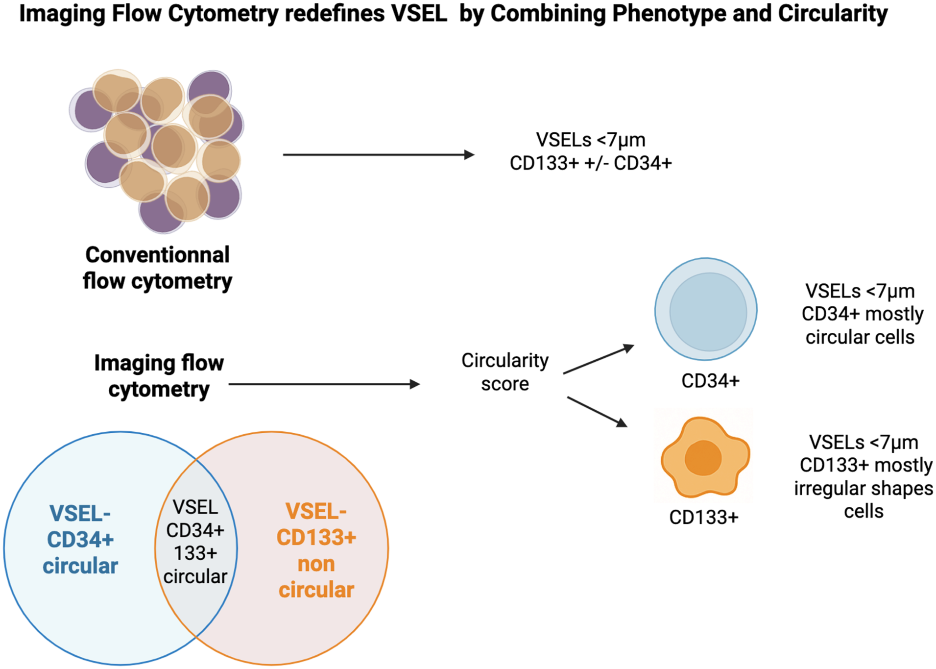
Open accessResearch articleFirst published 2026Very small embryonic-like stem cells (VSELs) have been identified as potential precursors of the endothelial lineage in humans. Traditionally characterized by CD34 and CD133 markers, these populations remain poorly delineated, particularly regarding their morphological traits. Leveraging recent advances in imaging flow cytometry (iFC), this study sought to refine the characterization of VSELs by analyzing CD34+ and CD133+ Lin⁻ CD45⁻ cells from various sources, including bone marrow (BM), mobilized blood (MB), peripheral blood (PB), and cord blood (CB). Using high-resolution iFC, we assessed cell circularity and size, identifying significant morphological differences within subpopulations smaller than 7 µm. CD133+ cells predominantly exhibited irregular shapes, whereas CD34+ cells, regardless of CD133 co-expression, were mostly circular. Quantification across multiple sources revealed an enrichment of small circular CD34+ cells, especially in BM, MB, and CB. A comparative analysis with conventional flow cytometry (cFC) showed similar counts for CD34+ cells between methods (
Open accessLetterFirst published 2026Donor-derived recipient-specific anti-HLA antibodies (RSAs) are rarely investigated in allogeneic hematopoietic stem cell transplantation (allo-HSCT). In this retrospective study, 185 haploidentical donor-recipient pairs were consecutively analyzed. Anti-HLA antibodies were detected in eight donors (4.3%) and 31 recipients (16.8%), with only one donor (0.5%) harboring RSAs. Donors were predominantly young, male, and transfusion-naïve, which likely contributed to the low antibody prevalence. No association was observed between donor antibody positivity and graft rejection or severe acute graft-versus-host disease (GVHD). However, recipients of antibody-positive grafts showed higher rates of Epstein–Barr virus (EBV) reactivation and transfusion refractoriness. Our findings suggest that donor-derived HLA antibodies, particularly RSAs, are infrequent and clinically limited under current haploidentical donor selection and immunosuppressive strategies, but may subtly influence post-transplant immune recovery.
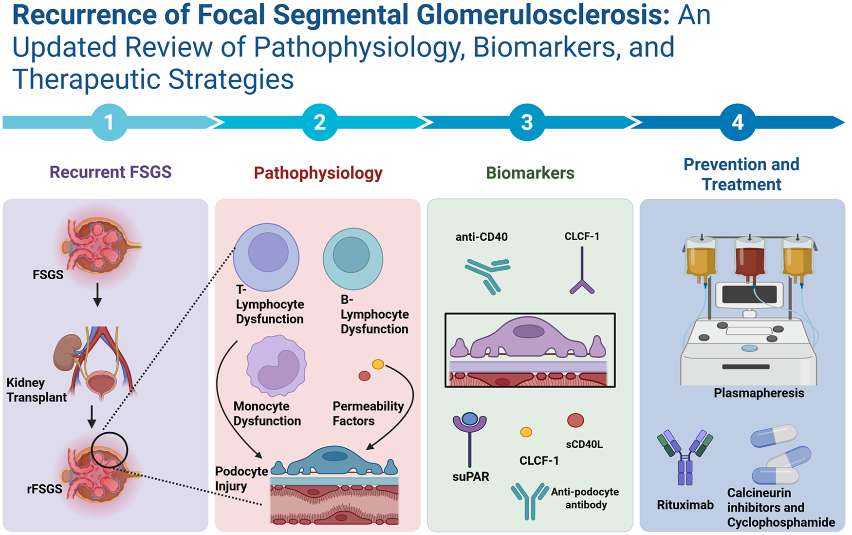
Open accessReview articleFirst published 2026Focal segmental glomerulosclerosis (FSGS) is one of the major causes of nephrotic syndrome, which can progress to end-stage renal disease, leading to kidney transplantation. Following renal transplantation, recurrence of FSGS (rFSGS) occurs in 30%–40% of patients with a high risk of graft loss. rFSGS typically presents with nephrotic-range proteinuria within days after post-transplantation. This review summarizes pathophysiology, biomarkers, and therapeutic strategies for rFSGS. Monogenic causes of FSGS, such as those caused by APOL1 mutation, show variable recurrence, while NPHS2 and ACTN4 show low recurrence of FSGS. Evidence suggests that idiopathic or primary FSGS is strongly associated with rFSGS, owing to podocyte structural damage caused by circulating permeability factors or immune dysfunction. Recent advances have identified biomarkers such as anti-nephrin antibodies, anti-CD40 antibodies, soluble tumor necrosis factor receptor 2 (sTNFR2), and soluble urokinase-type plasminogen activator receptor (suPAR) that help in early detection of recurrent FSGS. Post-transplant monitoring includes measuring urine protein-to-creatinine ratio (UPCR) and 24-h urine protein excretion, and a kidney biopsy. Preventive strategies, although including plasmapheresis and rituximab, show limited benefit and are not recommended for routine prophylaxis. Treatment options include plasmapheresis, immunoadsorption, and immunosuppressive drugs such as cyclophosphamide, rituximab, or calcineurin inhibitors. Recurrent FSGS is a clinical challenge with its multifactorial pathogenesis. Incorporating strategies such as genetic testing, risk stratification, and early detection with the help of biomarkers and early treatment can induce remission and preserve graft survival. Despite these advances, large prospective studies are still required for standardizing prevention and management strategies for rFSGS.
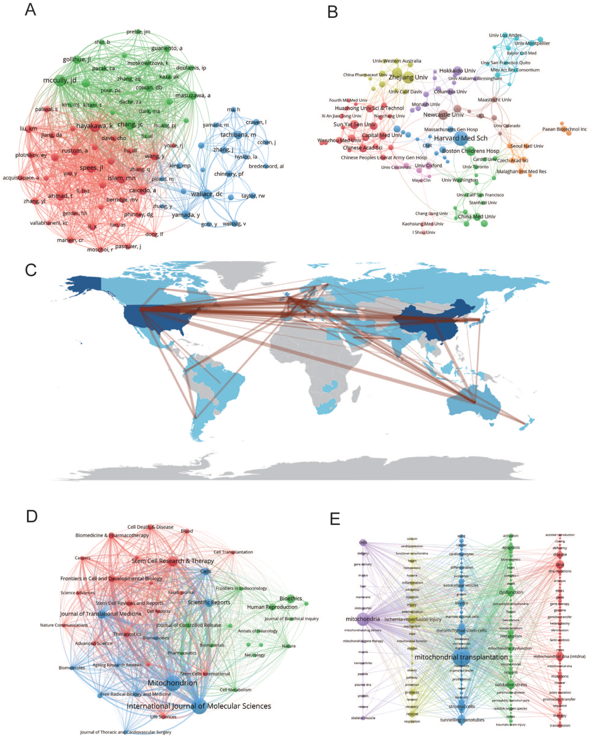
Open accessResearch articleFirst published 2026Recent years have witnessed rapid progress in mitochondrial transplantation (MT) as a novel strategy for restoring mitochondrial function in diverse pathological conditions, including somatic mitochondrial transfer and reproductive mitochondrial replacement therapy. With its expanding applications in regenerative medicine and disease modeling, systematic quantitative evaluation of the global MT research landscape remains limited. To address this gap, we performed a bibliometric analysis of publications indexed in the Web of Science Core Collection from 1996 to 2024, with cross-database validation using Scopus. CiteSpace, VOSviewer, and the R package
Open accessReview articleFirst published 2026, Xueyuan Li, Zijun Ye , [...]Bone defects arise from trauma, infection, tumor resection, or congenital deformities, with large defects often progressing to nonunion. Autologous bone grafting is considered the gold standard therapy due to its osteoconductive, osteoinductive, and osteogenic properties, but it is limited by insufficient graft volume and donor-site complications. Allogeneic grafts and early synthetic bone substitutes partially addressed these limitations but typically provided only osteoconductive function and performed poorly in complex microenvironments. Bone tissue engineering utilizes bioactive scaffolds, cells, and factors to promote bone repair through mechanical support, osteogenic regulation, and cellular adhesion/proliferation/differentiation mechanisms. This review synthesizes current evidence on bioactive scaffold materials and bone marrow enrichment strategies for bone defect repair within a scaffold–bone marrow coupling framework. It also highlights key roles of pore architecture, angiogenesis, osteoimmunomodulation, and defect-specific material selection, while critically discussing current limitations, evidence gaps, and future translational directions.
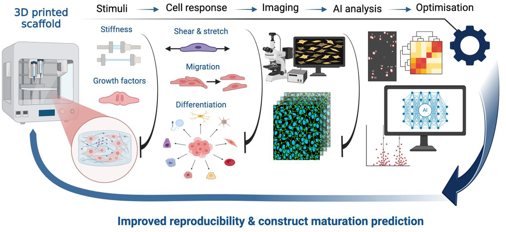
Open accessReview articleFirst published 2026This review examines how mechanical and chemical stimuli shape stem-cell behavior and how live-cell imaging combined with artificial intelligence can support the evaluation and optimization of 3D bioprinting workflows. Current evidence indicates that substrate stiffness, stretch, shear stress, compression, and soluble factors such as transforming growth factor beta (TGF-β), bone morphogenetic protein-2 (BMP-2), vascular endothelial growth factor (VEGF), and basic fibroblast growth factor (FGF-2) influence viability, migration, morphology, and lineage commitment in biopolymeric constructs. Imaging modalities including phase-contrast, fluorescence, confocal, two-photon, and light-sheet microscopy enable dynamic observation of these responses, while computational pipelines for segmentation, tracking, and feature extraction improve scalability and reproducibility of analysis. Across the reviewed studies, image-derived readouts such as cell distribution, motility, adhesion patterns, and early morphological changes emerge as promising indicators of construct quality and maturation potential. At the same time, broader application remains limited by phototoxicity, imaging depth constraints, data-processing demands, incomplete standardization of metrics, and restricted model generalizability. Overall, imaging-based cell tracking integrated with AI-assisted analysis offers a practical framework for post-print evaluation and iterative optimization in 3D bioprinting, with potential to strengthen quality control, reproducibility, and biological interpretation in regenerative engineering.
Open accessCase reportFirst published 2026Chimeric antigen receptor T-cell (CAR-T) therapy is a breakthrough in cancer treatment that can induce durable remission in patients with hematological malignancies who have failed prior therapies. As the use of CAR-T cell therapy increases, treatment-related renal complications deserves careful attention, particularly in patients with a solitary kidney, in whom renal complications may further increase clinical risk. Here, we report the case of a living kidney donor who was diagnosed with diffuse large B-cell lymphoma (DLBCL). After CAR-T cell therapy, the patient developed only mild cytokine release syndrome (CRS), without signs of acute kidney injury or renal impairment, and subsequently achieved a favorable metabolic response approaching complete remission.
Open accessResearch articleFirst published 2026Human pancreatic islets are essential for studies in β-cell biology, cell transplantation, and tissue engineering, yet access to viable human islets remains limited because conventional isolation protocols primarily rely on whole pancreases from deceased donors. Surgical pancreatectomy specimens may represent an accessible alternative source, but factors influencing successful isolation and functional preservation remain poorly defined. In this study, we evaluated a standardized method for isolating human islets from pancreatic tissue obtained from surgical pancreatectomy specimens and investigated patient-, specimen-, and surgery-related factors affecting islet yield and functionality. Between March and October 2024, 50 consecutive islet isolations were performed from pancreatic specimens obtained during surgical resections. Following enzymatic digestion, tissue fractions were cultured for 24 h before handpicking of morphologically intact islets. Islet yield was quantified as islet equivalents (IEQ), and functional integrity was assessed by glucose-stimulated insulin secretion assays on culture days 1, 3, and 5. A mean yield of 6690 IEQ/g pancreatic tissue was obtained (range 0–56,500 IEQ/g). Exploratory analyses suggested potential associations between islet yield and factors such as younger patient age, shorter surgical duration, and preserved pancreatic parenchyma. However, these findings should be interpreted cautiously given the variability of the specimens and the exploratory design of the study. These findings support the feasibility of using surgical pancreatectomy specimens as an accessible source of functional human islets for experimental and translational research.
Open accessReview articleFirst published 2026, Paritosh TayadeType 1 diabetes (T1D) is a chronic autoimmune disease characterized by the destruction of pancreatic beta cells (β-cell), leading to lifelong dependence on exogenous insulin. Despite advances in insulin delivery systems and glucose monitoring, achieving tight glycemic control remains a challenge for many patients. In recent years, stem cell–derived β-cell therapy has emerged as a promising strategy for restoring endogenous insulin production. This review explores the progress in differentiating human pluripotent stem cells, such as embryonic stem cells and induced pluripotent stem cells (iPSCs), into functional insulin-secreting β-like cells. We highlight recent breakthroughs in improving cell maturity, graft survival, and immune protection. Delivery approaches, encapsulation technologies, and transplantation strategies are discussed alongside an overview of ongoing clinical trials. The review also addresses current limitations, including immune rejection, cell dedifferentiation, and safety concerns. Finally, we examine future directions, such as genome editing, personalized cell therapy, and the integration of artificial intelligence in monitoring and optimizing treatment. Stem cell–derived β-cell therapy holds transformative potential as a curative approach for T1D, but further research is essential to overcome remaining barriers to its widespread clinical application.
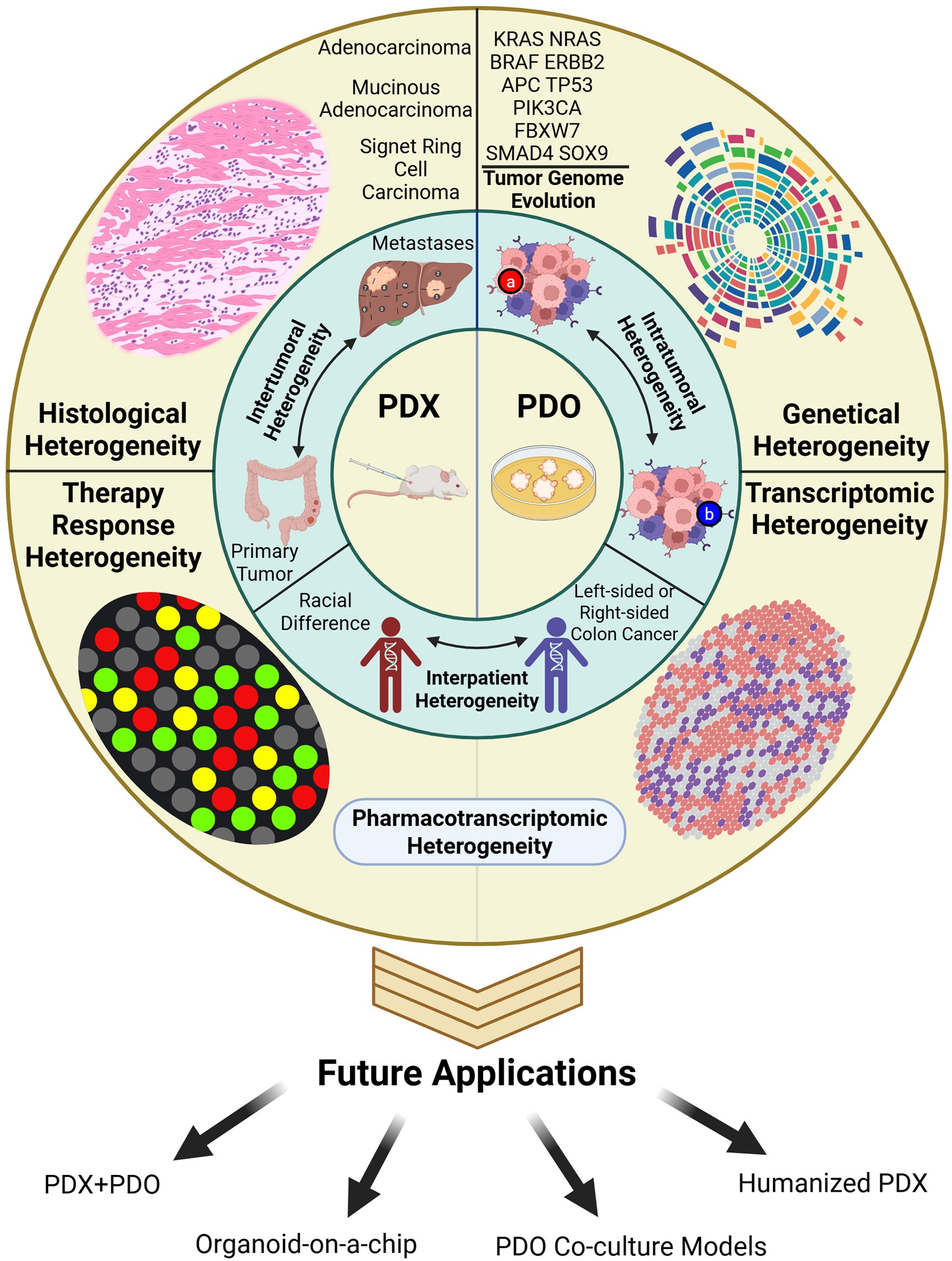
Stem Cell–Derived β-Cell Replacement Therapy for Type 1 Diabetes: From Differentiation to Clinical Translation. This graphical abstract summarizes how stem cell–derived β-cells are generated, protected from immune rejection, and translated into clinical therapies for type 1 diabetes. It highlights the convergence of stem cell biology, bioengineering, immunomodulation, and clinical trials, outlining a roadmap toward durable and potentially curative β-cell replacement.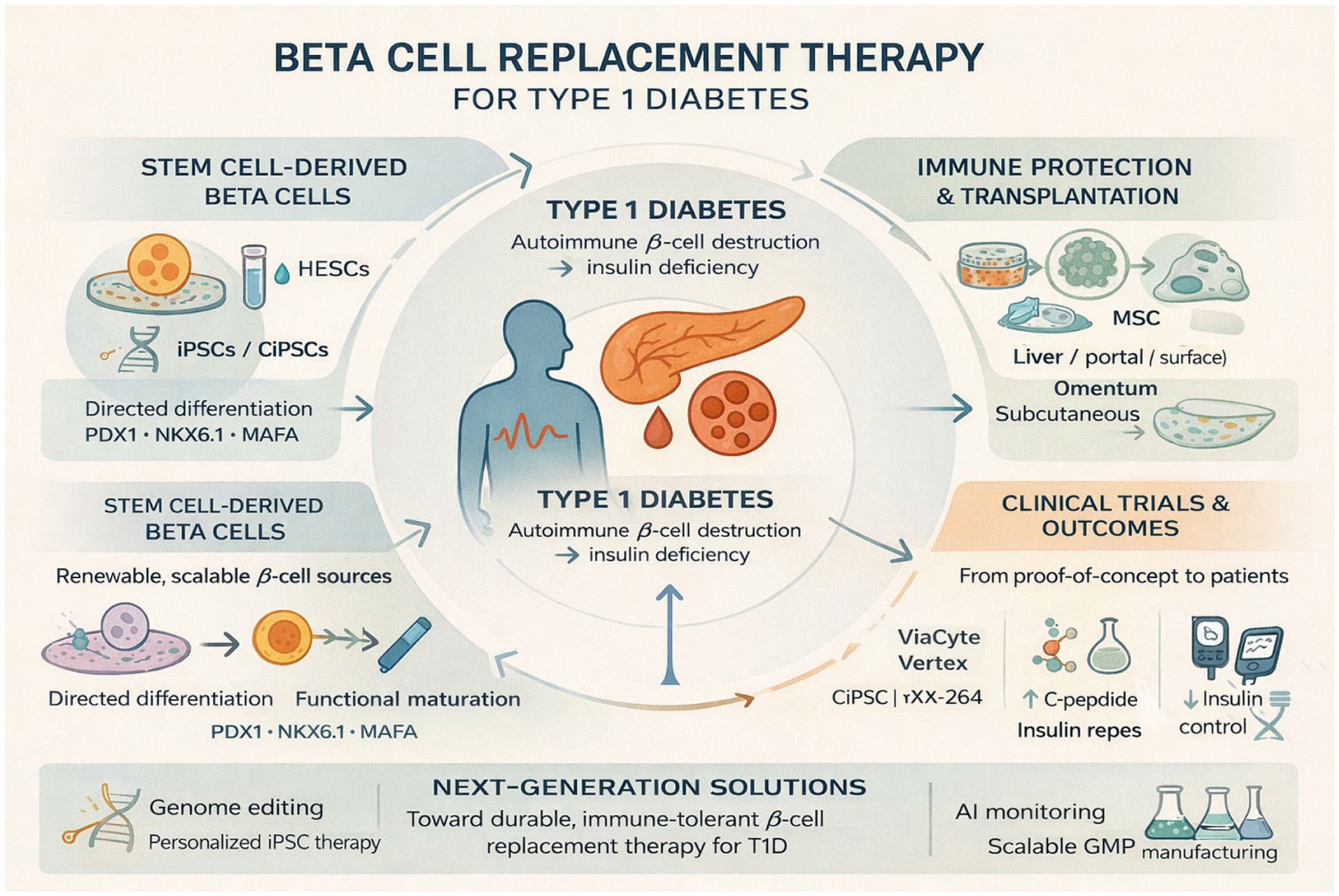
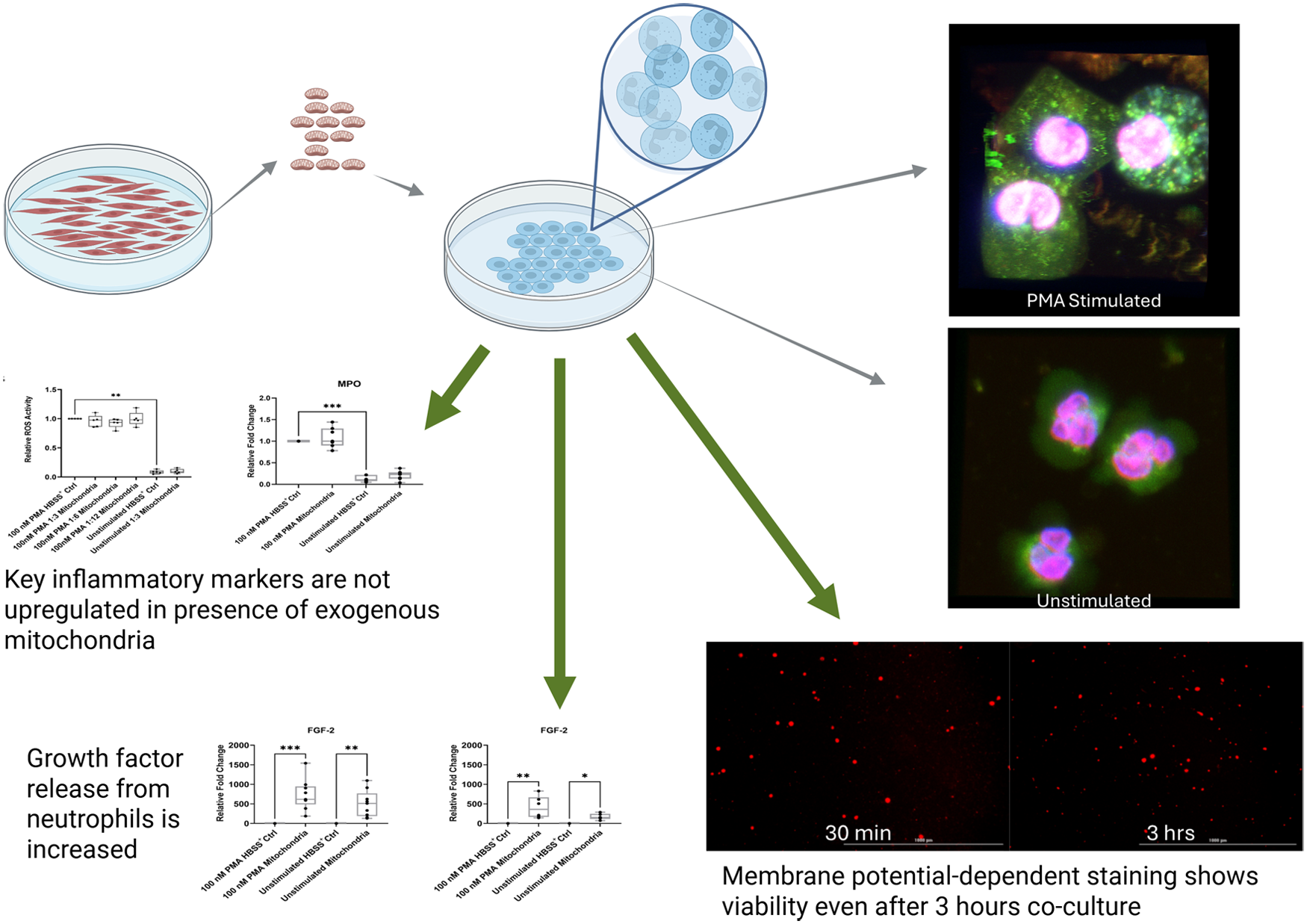
Open accessResearch articleFirst published 2026Following biomaterial implantation, modulation of the acute immune response is essential for tissue regeneration. Polymorphonuclear leukocytes (PMNs) are critical effector cells in wound healing, and PMN dysfunction is mediated by mitochondrial dysfunction and can lead to prolonged inflammation and tissue damage. It was hypothesized that mitochondrial transplantation could be applied to PMNs in pro-inflammatory states as a means of upregulating regenerative proteins. Primary human PMNs were isolated from donor blood. Isolated PMNs and exogenous mitochondria were co-incubated to induce mitochondrial transplantation. Resulting interactions were assessed through microscopy to confirm initial uptake and mitochondria membrane potential retention, intracellular reactive oxygen species (ROS) analyses (n = 5), and PMN secretome quantification (n = 10) using multiplex protein analysis. Human PMNs were able to successfully uptake delivered mitochondria, and regenerative factors essential for tissue repair and immune cell recruitment including fibroblast growth factor-2 (FGF-2), interleukin (IL)-22, monocyte chemoattractant protein-1 (MCP-1), and granulocyte colony-stimulating factor (G-CSF) were significantly upregulated, indicating that exogenous mitochondria represent promising modulators of PMN function with broad clinical potential.
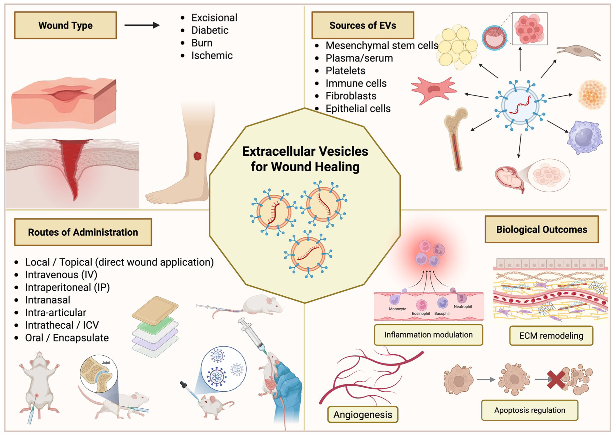
Open accessReview articleFirst published 2026, Paul Bigliardi, Julianna Goenaga , [...]Normal wound healing is a complex, yet critical process. Unfortunately, delayed or impaired wound healing presents a significant burden for numerous patients worldwide. Recent research has demonstrated that exosomes, a subtype of extracellular vesicles, may play a pivotal role in mediating cell-cell communication during wound healing. This narrative review aims to explore the use of exosomes in wound healing, with a focus on
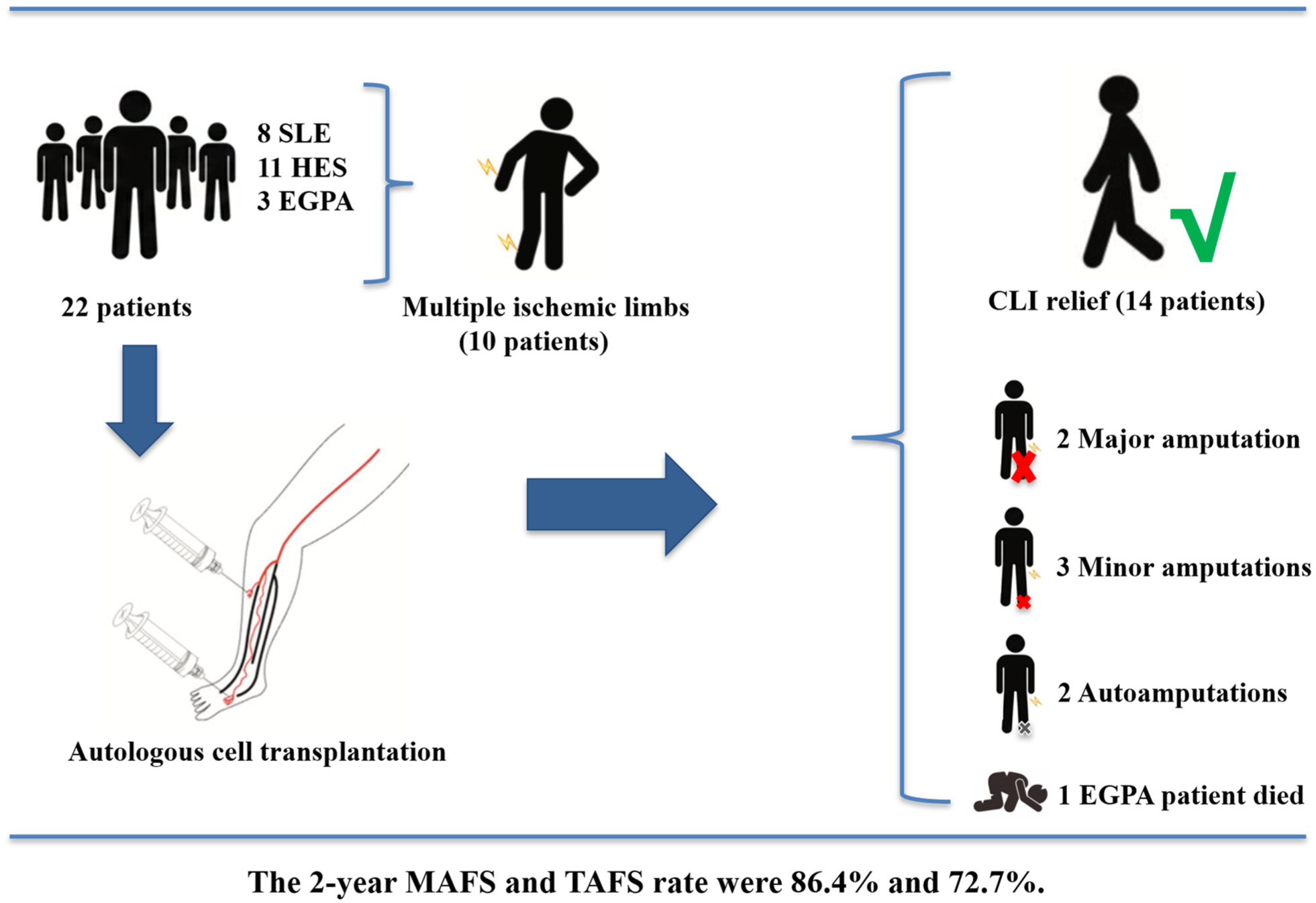
Open accessResearch articleFirst published 2026Autoimmune disease–related critical limb ischemia (AD-CLI) represents a significant portion of no-option critical limb ischemia (NO-CLI). Despite the demonstrated safety and efficacy of cell transplantation for NO-CLI, most studies focused on thromboangiitis obliterans (TAO). There remains a scarcity of reports on cell transplantation for AD-CLI induced by systemic lupus erythematosus (SLE), hypereosinophilic syndrome (HES), and so on. From May 2011 to May 2024, 22 patients with 36 ischemic limbs were enrolled. The primary outcome was amputation, whereas secondary outcomes included toe-brachial index (TBI), Wong-Baker Faces Pain Rating Scale (WBFPS), transcutaneous oxygen pressure (TcPO2), and so on. Among them, 8 had SLE, 11 had HES, and 3 had eosinophilic granulomatous polyangiitis (EGPA). During the follow-up, one EGPA patient died, two patients underwent major amputation, and five underwent minor amputation. The 2-year major and total amputation-free survival rates were 86.4% and 72.7%, respectively. Critical limb ischemia (CLI) relief was observed in 14 patients, with a 1-year cumulative CLI-free survival rate of 61.0%. Significant improvements were noted in postoperative TBI and WBFPS. Autologous cell transplantation showed satisfactory safety and efficacy outcomes for non-TAO AD-CLI patients. Validation of the conclusions awaits more evidence based on the long-term outcomes of a larger number of patients.
Open accessResearch articleFirst published 2026, Guanzheng Cui, Ziqiang Dai , [...]Cardiac xenotransplantation is advancing rapidly from basic research to early clinical use, but its research landscape has not been comprehensively mapped. We conducted a bibliometric and science-mapping analysis of English-language articles and reviews in the Web of Science Core Collection (1964–2025). Using VOSviewer, CiteSpace, and Bibliometrix, we assessed publication growth, collaboration networks, citation impact, and topic trends. The field has grown steadily and has accelerated in recent years in parallel with key clinical developments. Collaboration is strongest among institutions in the United States, Europe, and East Asia, and a small number of authors and centers contribute a large share of publications. Most studies are published in specialty transplantation journals, whereas major breakthroughs often appear in general medical journals. Research topics continue to focus on rejection and complement biology, while newer work highlights gene-edited donors, biosafety, clinical trial readiness, and pig-to-human models. Overall, the field appears to be maturing toward clinical implementation, and these findings can help guide research priorities, funding decisions, and policy planning.
Open accessLetterFirst published 2026, Chika Miyagi-Shiohira, Takuya Sadahira , [...] Open accessLetterFirst published 2026
 Open accessResearch articleFirst published 2026, Jiaping Wang, Xue Hu , [...]
To identify the optimal transplantation route for enhancing homing of mesenchymal stem cells (MSCs) to the kidney, thereby ameliorating rat Adriamycin nephropathy (AN).
We used different approaches to transplant MSCs. Under ultrasound-mediated conditions, the MSCs transplanted via the renal artery significantly improved the renal damage in rats with doxorubicin-induced nephropathy through the JAK2/STAT3 signaling pathway.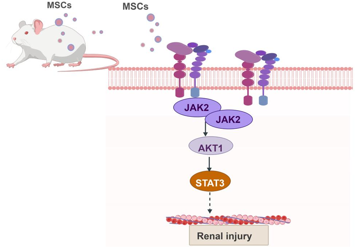
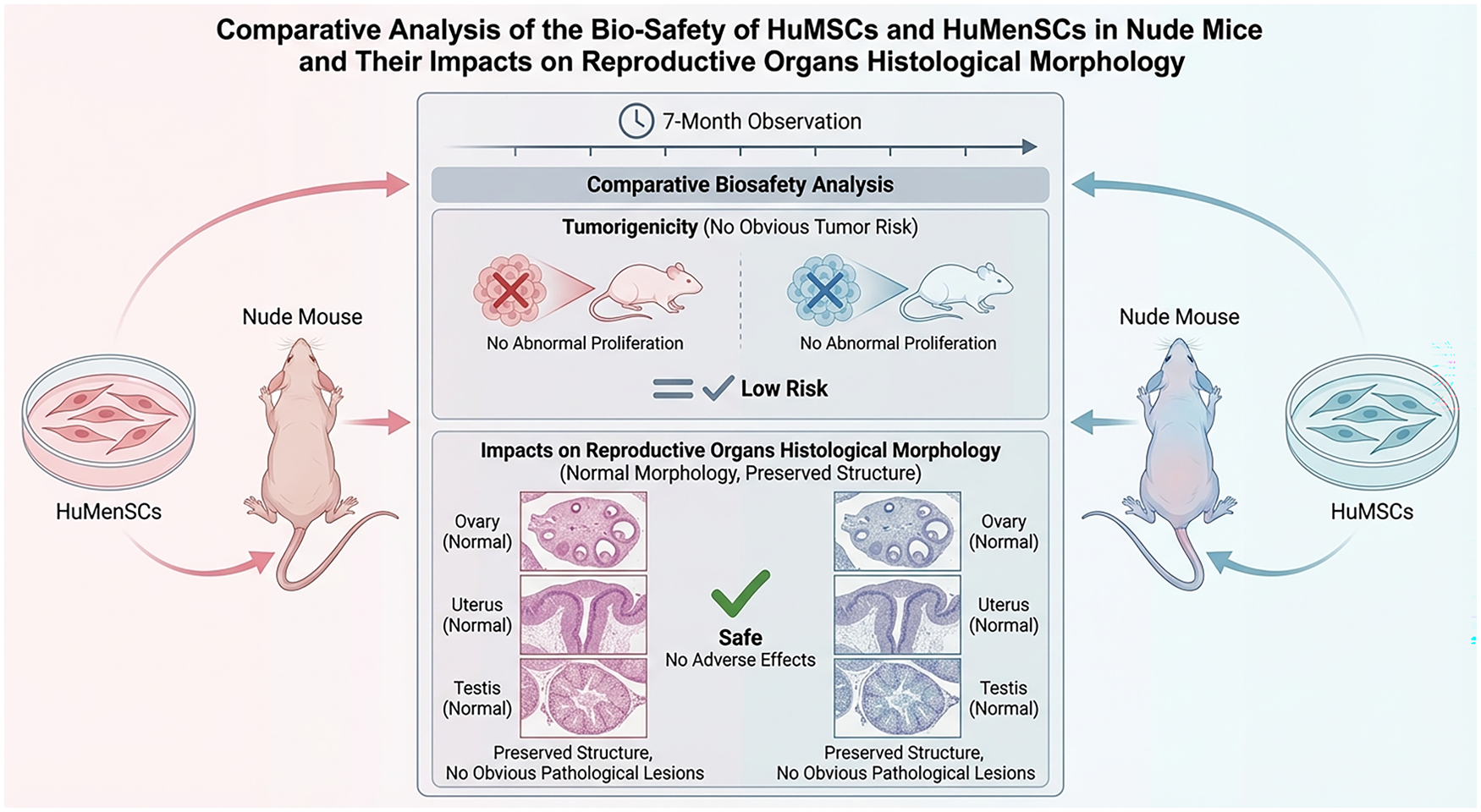
Open accessResearch articleFirst published 2026The tumorigenesis and long-term bio-function of the implanted mesenchymal stromal cells (MSCs) were needed before clinical applications. We herein observed 7 months and investigated the viable tumors formation, the survival rate, the distribution of the injected cells in different reproductive organs after intravenous (IV) injection of the human menstrual blood–derived MSCs (LXT- and CDH-huMenSCs), and the human umbilical MSCs (huMSCs). The survival rate was not different among the huMenSCs, the huMSCs, and the negative control (IV injection of PBS) group. The longevities of the mice died within the observation period of the LXT and the huMSCs groups were significantly longer than that in the other groups; the positive control (IV injection of LLC (Lewis lung carcinoma cells)) group had the shortest longevity. The number of visible tumors in the huMenSCs, huMSCs, and the negative control group was lower than that in the positive control group. Human-sourced DNA was only detected in the ovaries of the huMSCs-injected mice. The histological morphology of the CDH- group had more normal follicles than that in other groups; the CDH- and the huMSCs group had more atretic follicles than that in other groups. In the huMenSCs and the huMSCs groups, the testis seminiferous tubules were fuller and more orderly arranged than the negative control group. The endometrial thickness of the LXT- group was thicker than that of the huMSCs- and the negative control groups, whereas the number of uterine glands was similar among all groups. The results suggested that IV administrations of xenogeneic huMenSCs and huMSCs in nude mice did not cause tumor formation. The injected huMenSCs from different donors functioned differentially on the mice’s life-span, endometrial thickness, and the number of follicles.
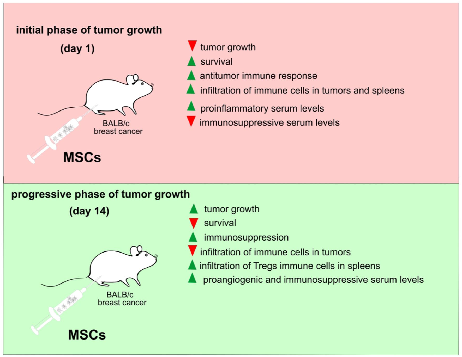
Open accessResearch articleFirst published 2026, Danijela Niciforovic , [...]Mesenchymal stem cells (MSCs) are recognized for their capacity to modulate immune responses, including those directed against tumors. In this study, we investigated the temporal effects of MSCs administration on anti-tumor immunity in a murine 4T1 breast cancer model. BALB/c mice were intraperitoneally injected with MSCs either 24 h (MSC1d) or 14 days (MSC14d) after orthotopic implantation of 4T1 mammary carcinoma cells. Early MSC administration (MSC1d) exhibited changes in immune cell phenotypes consistent with enhanced antitumor potential, including increased activity of natural killer (NK) cells, dendritic cells (DCs), macrophages, and T lymphocytes. These immunological changes correlated with reduced tumor growth and prolonged survival. Mice in the MSC1d group exhibited elevated serum levels of pro-inflammatory and anti-tumor cytokines (TNF-α, IFN-γ, IL-6, and IL-17), alongside decreased concentrations of immunosuppressive cytokines (TGF-β and IL-10). Tumor tissue analysis revealed increased infiltration of NK cells expressing markers associated with antitumor activity (IFN-γ-producing CD178⁺), CD80⁺/CD86⁺/I-A⁺ TNF-α-producing DCs, Th1-type CD4⁺ T cells, and Granzyme B-expressing CD8⁺ cytotoxic T lymphocytes (CTLs). Additionally, spleens of MSC1d-treated mice displayed significantly elevated populations of CD11c⁺ DCs, TNF-α/IFN-γ-secreting NK cells, CD4⁺ Th1 and Th17 cells, and CD8⁺ CTLs expressing markers associated with cytotoxic function (TNF-α, IFN-γ, and IL-17). Conversely, late MSCs administration (MSC14d) was associated with immunosuppression. Tumors from MSC14d-treated mice showed a decreased presence of IFN-γ⁺ and IL-17⁺ NK1.1⁺ cells, F4/80⁺ macrophages, IL-12⁺ DCs, and cytotoxic T cells. Spleens from these mice revealed a significant expansion of regulatory T cell (Treg)-like populations, including CD25⁻, FoxP3⁻, CD25⁺FoxP3⁻ cells, and TGF-β/IL-10-producing CD3⁺ and CD4⁺ T cells. Furthermore, serum levels of immunosuppressive mediators TGF-β and vascular endothelial growth factor (VEGF) were significantly elevated in the MSC14d group. Collectively, these findings demonstrate that the immunomodulatory effects of MSCs on breast cancer are highly dependent on the timing of their administration. Mesenchymal stem cells delivered during early tumor development enhance phenotypes consistent with antitumor potential and suppress tumor progression, whereas administration during later stages promotes immune evasion and tumor growth.
Open accessResearch articleFirst published 2026, Yinsheng Xi, Zhihong Yang , [...]This study investigated donor and islet isolation procedural factors influencing the islet yield during human islet isolation for transplantation. We retrospectively analyzed 133 islet isolations from deceased donors performed over 15 years at a single center. Isolations were stratified by post-purification islet yield (≥400,000 islet equivalents [IEQ], successful; <400,000 IEQ, unsuccessful) and by intent (clinical vs research). Higher donor body mass index and height over 170 cm were independently associated with successful isolation, whereas an enzyme perfusion temperature exceeding 14°C for ≥50% of the perfusion duration emerged as an independent risk factor for isolation failure. Clinically intended isolations exhibited tighter thermal control during Phase 1 digestion, greater digestion efficiency, and lower undigested tissue weight. A logistic regression model incorporating body surface area, packed tissue volume, and phase 1 digestion time (the interval from the start of warm recirculation to the collection phase) showed a moderate predictive value for isolation success (area under the curve = 0.755). Subgroup analysis revealed that longer relative phase 2 digestion time (the interval from the start of collection to its end) and higher North American Islet Donor Scores were associated with higher islet yield. These findings highlight the importance of donor anthropometrics, procedural consistency, and thermal regulation during islet isolation. Optimizing donor selection and controlling intra-procedural variables can improve islet yield and increase the likelihood of achieving the required clinical transplantation islet dose.
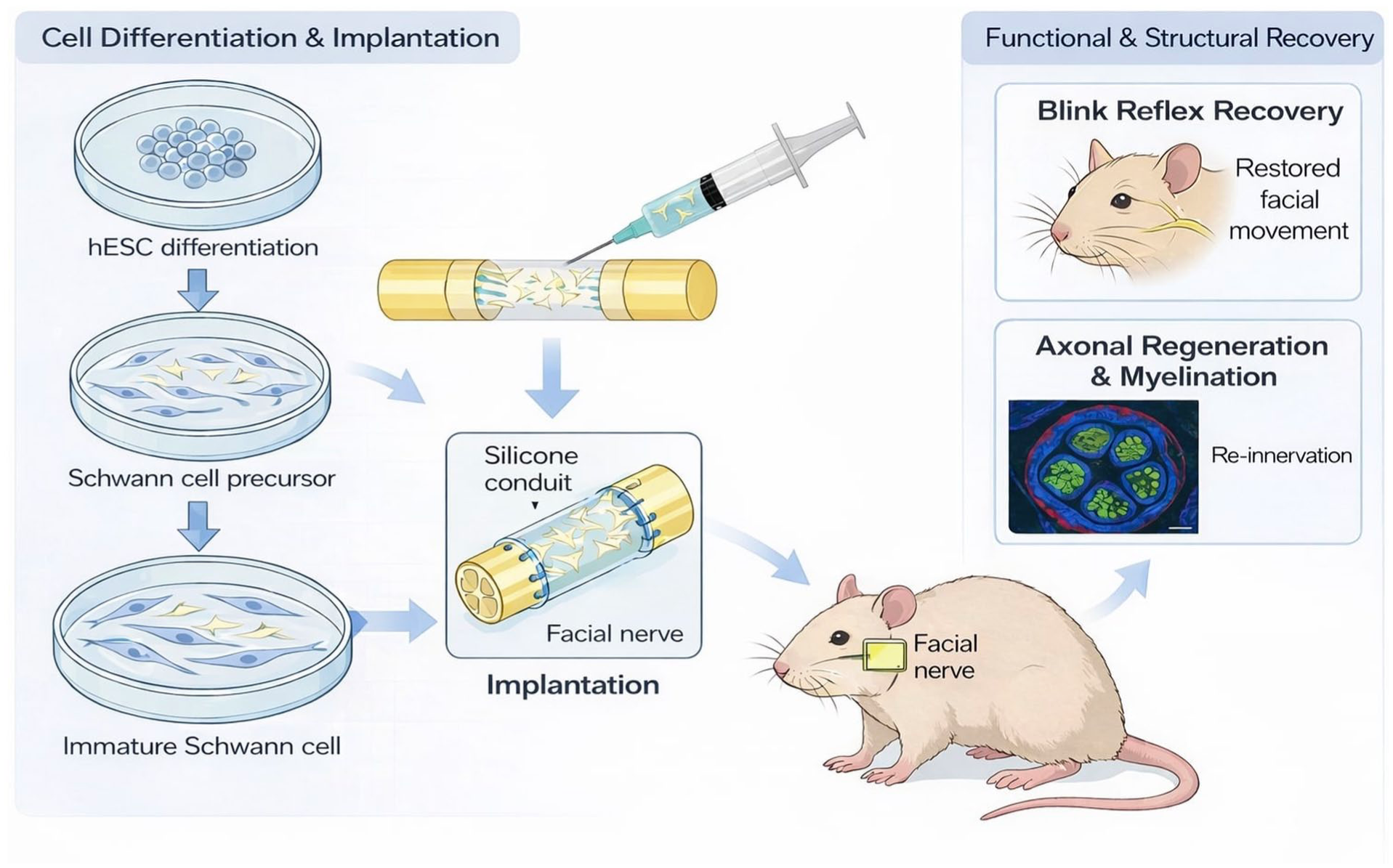
Open accessResearch articleFirst published 2026, Jong Ha Lee, Jae Min Lee , [...]Facial nerve transection frequently results in incomplete functional recovery despite microsurgical repair due to axonal misalignment and limited intrinsic regenerative capacity. Schwann cells (SCs) are essential for peripheral nerve repair, supporting axon elongation, remyelination, and neurotrophic signaling. Human embryonic stem cell–derived Schwann cells (hESC-SCs), including Schwann cell precursors (SCPs) and immature SCs, constitute a scalable source of cells for tissue-engineered nerve grafts. This study investigated whether silicone conduits loaded with hESC-SCs could enhance facial nerve regeneration following complete transection in rats. hESCs were differentiated into SCPs or immature SCs using a stepwise
Open accessResearch articleFirst published 2026Currently, there is very limited large-scale real-world data on the use of total marrow irradiation (TMI) as conditioning for allogeneic hematopoietic stem cell transplantation (allo-HSCT). The primary objective of this study was to evaluate the real-world feasibility, efficacy, and safety of TMI-based conditioning in a large multicenter cohort. We retrospectively included consecutive patients undergoing allo-HSCT with TMI-based conditioning across four Chinese centers (2017–2024). The primary endpoint was overall survival (OS); secondary endpoints included disease-free survival (DFS), graft-versus-host disease–free/relapse-free survival (GRFS), nonrelapse mortality (NRM), graft-versus-host disease (GVHD), and safety. Among 205 patients, acute lymphoblastic leukemia (ALL) was the predominant diagnosis (81.5%). With a median follow-up of 19.3 months among survivors, 2-year OS, DFS, and GRFS were 71.7%, 60.0%, and 37.8%, respectively. One-year cumulative incidences of relapse and NRM were 20.2% and 13.8%. Day-100 grade II–IV acute GVHD occurred in 32.9%, and 1-year moderate-to-severe chronic GVHD in 19.1%. In multivariable analyses, acute myeloid leukemia (AML) diagnosis, haploidentical donor, and Eastern Cooperative Oncology Group (ECOG) performance status >1 were associated with inferior outcomes. Exploratory dose-stratified analyses were performed. Severe toxicities within 100 days were infrequent, predominantly infections (21.46%). In this large multicenter cohort, TMI-based conditioning demonstrated real-world feasibility with encouraging survival and manageable safety outcomes.
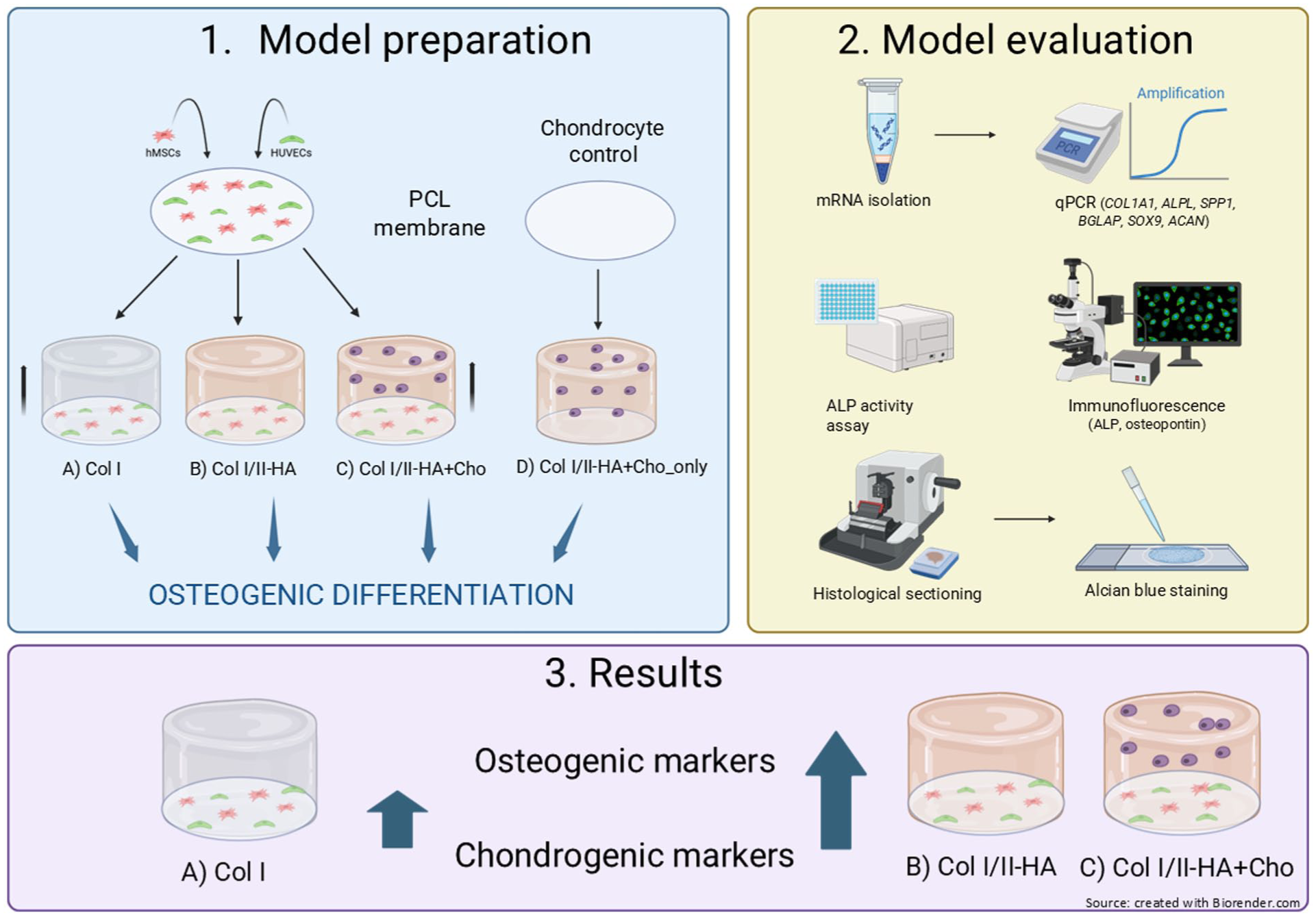
Open accessResearch articleFirst published 2026Tissue engineering strategies aim to create bone models to enhance our understanding of bone development and improve bone repair. Endochondral ossification (EO), where cartilage is replaced by bone, is difficult to replicate. We developed a collagen–hyaluronic acid (HA) hydrogel system for a three-dimensional (3D) co-cultivation of human chondrocytes, human mesenchymal stem cell (hMSCs), and human umbilical vein endothelial cells (HUVECs) to model the early stages of EO. To our knowledge, this specific tri-culture configuration within such a hydrogel composition has not been previously reported, providing a novel platform for studying EO-related cellular and matrix interactions
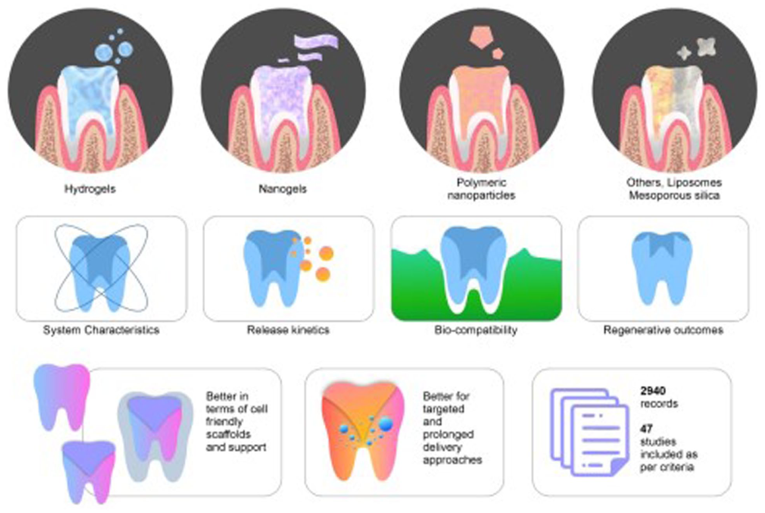
Open accessResearch articleFirst published 2026This systematic review examines emerging delivery systems for bioactive molecules within regenerative endodontic therapy (RET) where hydrogels, nanogels, and polymeric nanoparticles along with advanced nanocarriers such as liposomes aquasomes, vesosomes, and mesoporous silica nanoparticles form the primary focus. The extensive literature search in PubMed, Scopus, and Web of Science databases (until August 2025) yielded a total of 47 eligible articles, including
Open accessResearch articleFirst published 2026, Yibo Wu, Tingting Yang , [...]Open accessResearch articleFirst published 2026, Junjie Liu, Wenlong GaoLigament injuries can lead to lasting instability and early joint degeneration despite rehabilitation or surgery. Stem cell therapies may aid regeneration, but studies are highly variable. Bibliometric analysis can summarize trends, hotspots, and research gaps. We searched the Web of Science Core Collection for English-language original articles on stem cell therapy for ligament injuries from 2001 to 2025 on April 2, 2025, excluding non-original publications. Publication and citation trends were summarized, and co-authorship, co-citation, and keyword networks were mapped using Excel, VOSviewer, CiteSpace, and Bibliometric.com. A total of 599 articles were included, with steadily increasing annual publications. The United States and China were leading contributors, with strong collaboration networks involving major institutions such as the University of Pittsburgh and Zhejiang University. Keyword analyses identified seven main themes spanning mechanisms, clinical translation, tissue engineering, surgery/grafting, injury models, biomechanics, and biomaterials. Recent hotspots included “inflammation” and “artificial ligament”. Stem cell research for ligament injuries is increasingly focused on mechanisms and biomaterials, but evidence is still mainly preclinical and inconsistent. Better comparative clinical studies and standardized, well-defined biomaterial–cell approaches are needed.

Open accessResearch articleFirst published 2026This study aimed to define the incidence of
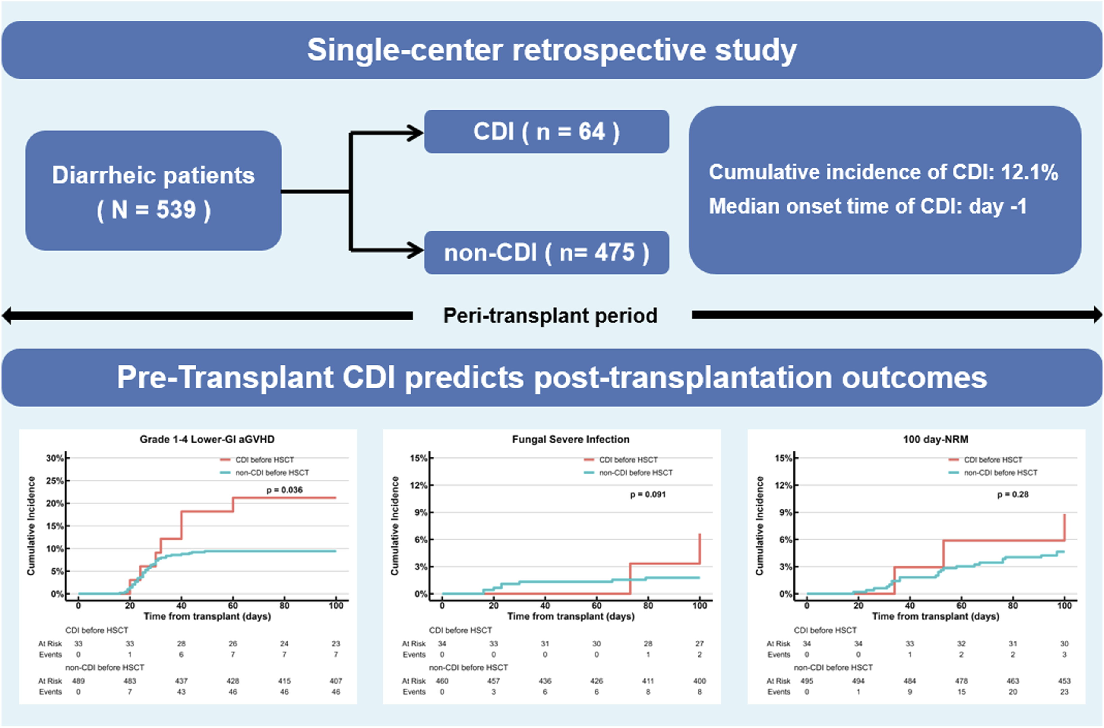
Open accessResearch articleFirst published 2026Amniotic epithelial cells (AECs) have immunomodulatory and anti-inflammatory properties that may improve outcomes in cell transplantation. However, their effect on islet engraftment after intraportal co-transplantation remains unclear. We evaluated the impact of co-transplanting syngeneic 600 islet equivalents (IEQs) with human AECs (hAECs) via the portal vein in a rat streptozotocin-induced diabetes model. The co-transplantation (Co-Tx) group showed normalization of blood glucose levels within 7 days after transplantation, sustained normoglycemia thereafter, and achieved a higher diabetes reversal rate than controls (100% vs. 71.4%, p < 0.01). Serum CXCL1 levels were significantly lower in the Co-Tx group indicating suppression of early inflammatory responses. Thrombin-antithrombin complex (TAT) levels also tended to be lower, raising the possibility of attenuation of the instant blood-mediated inflammatory reaction (IBMIR). In contrast, no significant differences were observed in VEGF levels or intrahepatic microvascular density. Co-transplantation with hAECs enhances islet engraftment likely through suppression of early inflammation, highlighting their potential as an adjunctive cellular therapy in islet transplantation.
Open accessResearch articleFirst published 2026Peripheral nerve injury (PNI) often results in persistent functional deficits, and current treatments remain suboptimal. This study developed a tissue-engineered graft by integrating Cdc42-modified bone marrow-derived mesenchymal stem cell (BMSC)-derived exosomes (Exos-Cdc42) with an acellular nerve allograft (ANA) and evaluated its therapeutic potential for nerve regeneration and functional recovery. Exosomes were isolated from BMSCs, and Exos-Cdc42 were generated by transfecting these cells with Cdc42 overexpression vectors. In vitro, Exos-Cdc42 significantly enhanced Schwann cell proliferation, migration, and secretion of neurotrophic factor (BDNF, NGF, CNTF), while upregulating repair-associated markers and downregulating myelination-related markers. In vivo, the combination of Exos-Cdc42 and ANA improved functional recovery of the sciatic nerve, as evidenced by higher sciatic functional index scores and increased muscle weight. Histological analyses demonstrated enhanced axonal regeneration and myelination, characterized by thicker myelin sheaths and larger axon diameters. These findings suggest that Exos-Cdc42 enhance the therapeutic efficacy of ANA by promoting Schwann cell-mediated repair responses, representing a promising strategy for peripheral nerve regeneration.
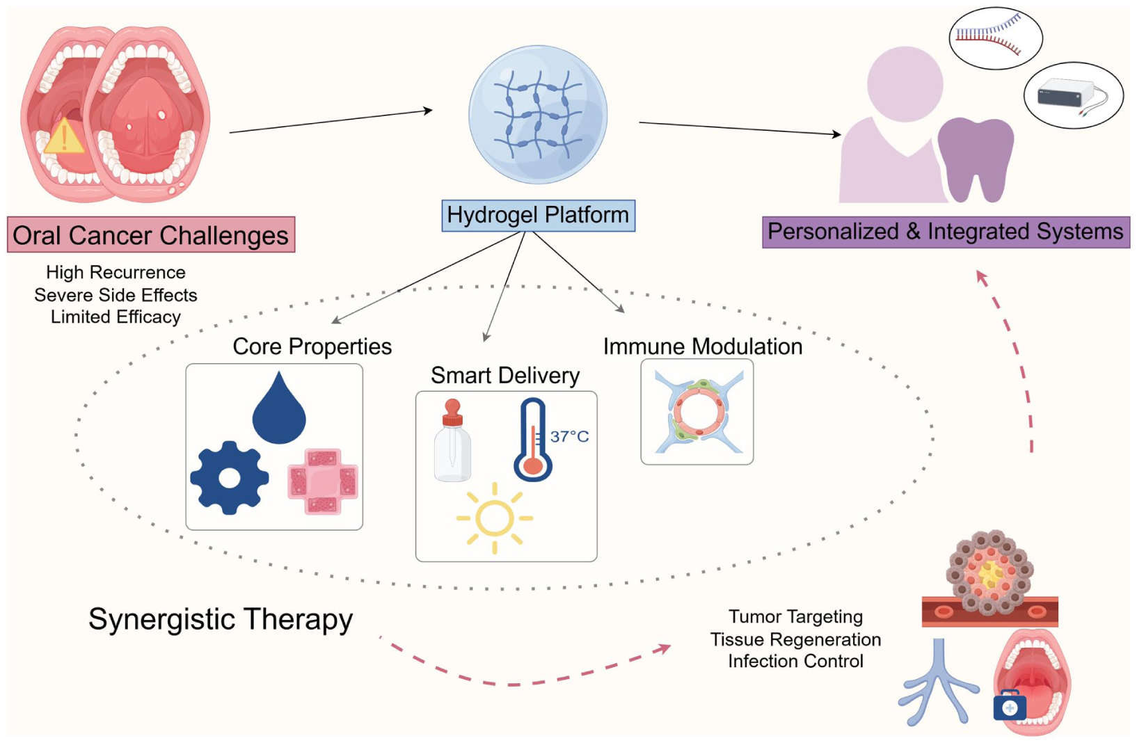
Open accessReview articleFirst published 2026, Huan-huan He, Tianqi Wu , [...]Oral cancer ranks among the most prevalent malignant tumors of the head and neck, comprising 1%–2% of all global malignancies. Despite advancements in surgery, radiotherapy, chemotherapy, and immunotherapy, challenges remain in oral cancer treatment, including high recurrence rates, severe side effects, and limited efficacy. Hydrogels, biocompatible materials with a three-dimensional network structure, have emerged as promising candidates in oral cancer therapy due to their dual roles in drug delivery and immune modulation. This review examines the contribution of hydrogels to oral cancer treatment through their synergistic control of drug release and localized immune activation. By enabling targeted, stimuli-responsive drug release, and enhancing local immune responses, hydrogels reduce systemic toxicity and improve treatment precision. When integrated with advanced approaches such as gene therapy and immune modulation, hydrogels hold potential to further amplify therapeutic efficacy. Although preclinical results are promising, clinical translation requires addressing scalability, quality control, and long-term safety concerns. With ongoing development, hydrogels are positioned to become vital tools in the treatment of oral cancer, periodontitis, and other oral diseases.
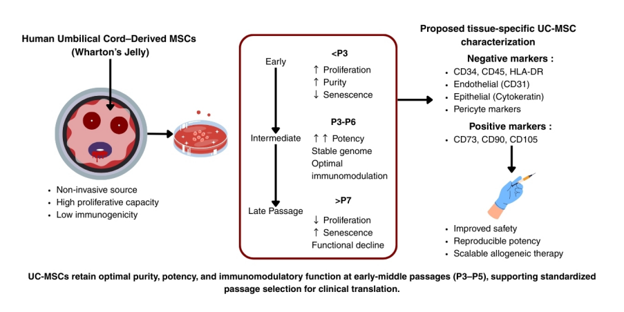
Open accessReview articleFirst published 2026, Maimonah Eissa Al-Masawa , [...]Umbilical cord mesenchymal stromal cells (UC-MSCs) are emerging as leading stem cells in regenerative medicine due to their high proliferative capacity, potent immunomodulatory effects, and non-invasive collection. However, the absence of standardized guidance on optimal passage number and tissue-specific characterization criteria limits clinical translation, raising concerns about variability in potency, genomic stability, and safety. This review synthesizes evidence on how
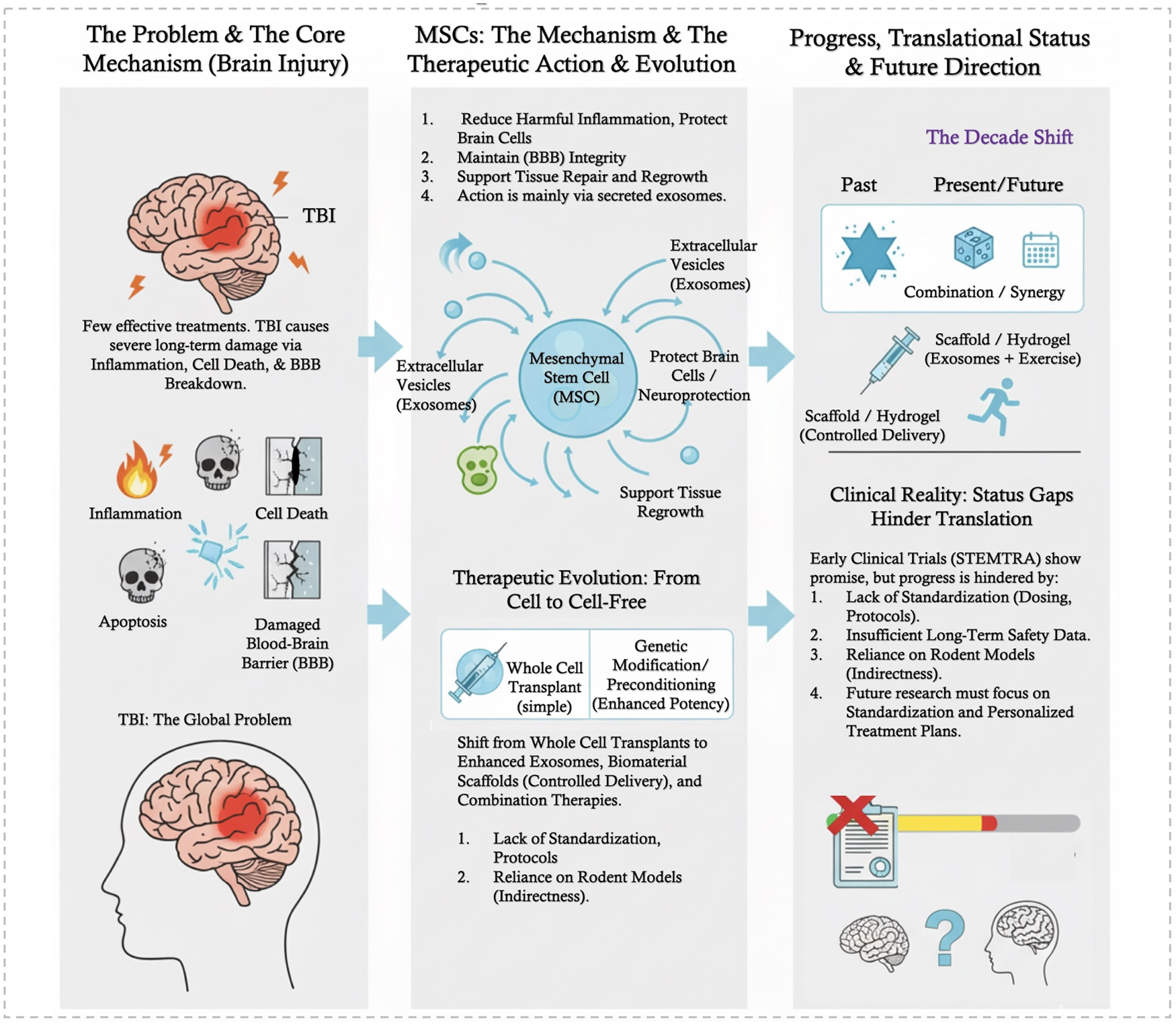
Open accessReview articleFirst published 2026, Yufei GaoTraumatic brain injury (TBI) remains a major global health challenge with limited effective treatments. This systematic review documents the significant evolution of mesenchymal stem cell (MSC) therapies from traditional cell transplants to advanced cell-free products and engineered delivery systems. A systematic search was conducted across Web of Science, Embase, and PubMed/Medline for studies published from January 2015 to June 2025, resulting in the inclusion of 80 studies for qualitative synthesis. The review identifies four primary therapeutic mechanisms: reducing inflammation, protecting brain cells, maintaining the blood–brain barrier, and supporting tissue repair/regrowth. This PROSPERO-registered systematic review followed Preferred Reporting Items for Systematic Reviews and Meta-Analyses (PRISMA) guidelines. Methodological quality and risk-of-bias were rigorously assessed using a multi-tool approach tailored to study design (Cochrane RoB, SYRCLE, NOS/JBI). Recent clinical evidence (e.g. STEMTRA trial) suggests these treatments are safe and can improve function in chronic TBI patients. Despite this, critical research gaps persist in establishing standardized protocols, optimal dosing, and long-term safety data. Emerging trends include (1) shifting toward enhanced exosomes/secretomes, (2) integrating MSCs/derivatives with advanced biomaterials for controlled delivery, (3) exploring alternative MSC sources, and (4) developing combination therapies. With a deepening mechanistic understanding and positive early clinical results, future research must prioritize standardization and personalized treatment plans to accelerate clinical translation.
Open accessReview articleFirst published 2026Neurodegenerative dementias, including Alzheimer’s disease and vascular dementia, have long been viewed through a neuron-centric lens. However, growing evidence highlights the indispensable and multifaceted roles of glial cells, astrocytes, microglia, and oligodendrocytes in both the onset and progression of these disorders. While prior reviews have cataloged glial dysfunction in isolation, this review offers a novel, integrative framework that maps the interconnected roles of glial subtypes across molecular, cellular, and circuit-level pathology in dementia. We critically synthesize recent advances in single-cell RNA sequencing, spatial transcriptomics, and glial imaging to redefine glial heterogeneity and function in disease states. Special emphasis is placed on the dynamic cross talk between glial populations and the feedback loops that govern their dual roles in neuroprotection and neurodegeneration. Furthermore, we examine emerging therapeutic strategies targeting glial-specific pathways, including NF-κB, JAK/STAT, CSF1R, and TREM2 signaling, as well as remyelinating agents and stem cell–based interventions. By integrating glial biology with therapeutic innovation, this review positions glial cells not as supporting actors but as central regulators and potential gatekeepers of dementia pathogenesis and treatment.
Open accessResearch articleFirst published 2026Post-transplant relapse remains the chief therapeutic challenge in Ph-negative B cell acute lymphoblastic leukemia (Ph- B-ALL). This retrospective study evaluated whether short-course blinatumomab for measurable resident disease (MRD) eradication could improve transplant outcomes. We compared 23 patients receiving pre-transplant short-course blinatumomab (2-week) for MRD eradication with 46 chemotherapy-only controls. All achieved MRD-negative before allogeneic peripheral blood stem cell transplantation (allo-PBSCT). Only two patients developed grade 2 cytokine release syndrome with blinatumomab. The neutrophil and platelet engraftment times were similar between the two groups. The blinatumomab cohort had a significantly lower 18-month cumulative incidence (CI) of relapse (
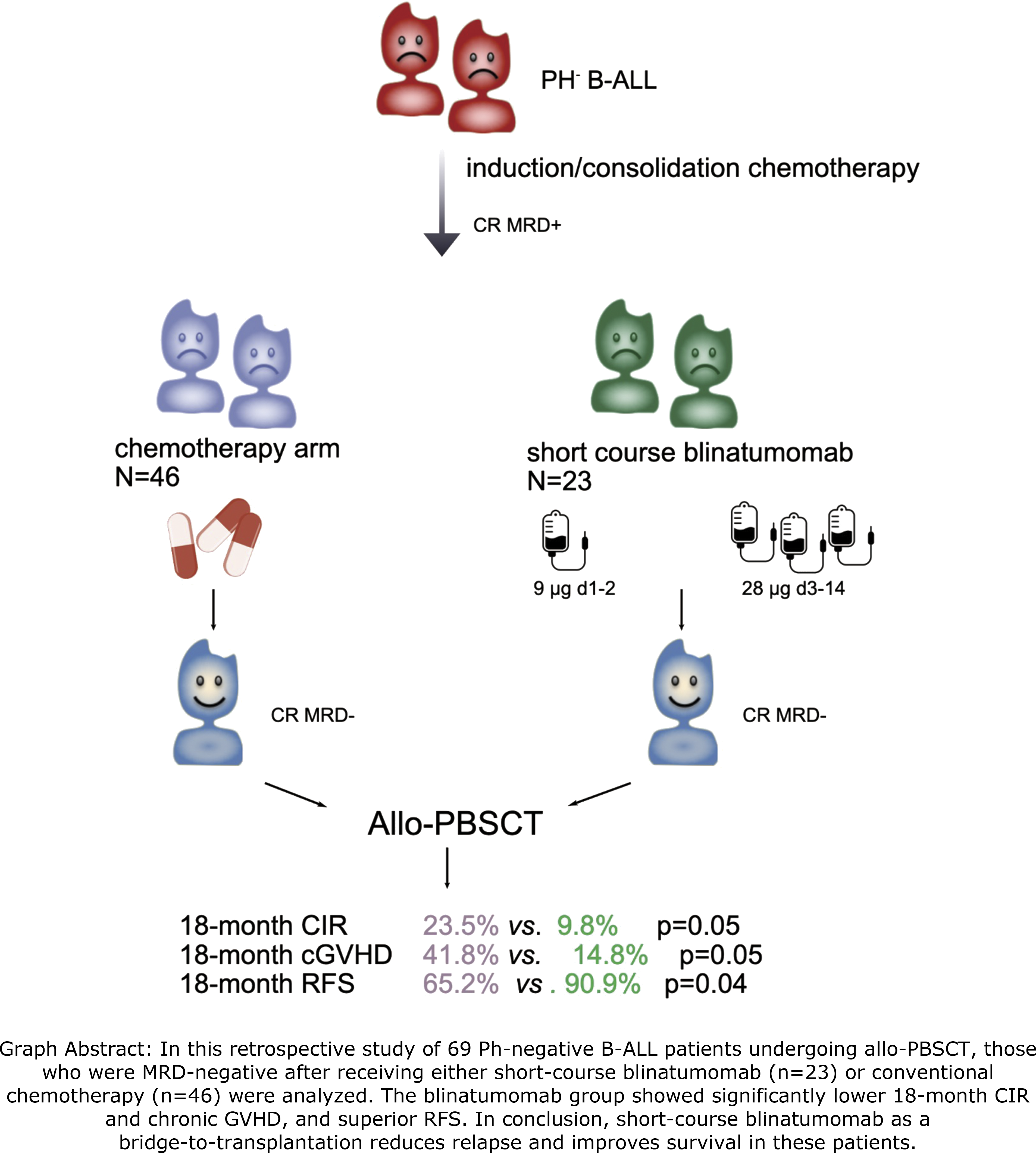
Open accessReview articleFirst published 2026Stromal vascular fraction (SVF)-based therapies and autologous fat grafting have emerged as promising regenerative strategies due to their pro-angiogenic, immunomodulatory, and trophic properties. However, despite encouraging preclinical and clinical findings, therapeutic outcomes remain highly heterogeneous, with marked variability in graft retention and functional efficacy between patients. Increasing evidence suggests that this variability cannot be explained solely by procedural factors or cellular composition, but may also depend on host-related immune and microenvironmental determinants. This review explores the biological mechanisms governing SVF engraftment and introduces the emerging concept of “SVF therapy resistance,” defined as the failure of autologous regenerative therapies resulting from maladaptive interactions between transplanted stromal cells and the host tissue environment. Particular attention is given to sterile inflammation, innate immune activation, and early graft–host interactions. Following transplantation, tissue injury and ischemia induce the release of danger-associated molecular patterns (DAMPs), triggering neutrophil recruitment, macrophage activation, complement signaling, and inflammatory remodeling. While controlled inflammatory responses may support tissue repair and angiogenesis, excessive neutrophil activation, neutrophil extracellular trap (NET) formation, persistent pro-inflammatory macrophage polarization, and impaired vascular adaptation may compromise graft survival and regenerative efficacy. The review further discusses how SVF processing, inflammatory priming, stromal cell heterogeneity, and donor-related factors—including obesity, aging, metabolic dysfunction, and chronic inflammation—may influence therapeutic responsiveness. Emerging evidence from mesenchymal stromal cell biology suggests that stromal cells are highly sensitive to inflammatory licensing and microenvironmental cues. Candidate biomarkers and immune profiling strategies capable of identifying responders and non-responders to SVF-based therapies are also reviewed. Finally, these mechanisms are discussed in spinal cord injury, a condition characterized by chronic inflammation and vascular dysfunction. Overall, this review proposes a translational framework linking innate immunity, sterile inflammation, angiogenesis, and stromal cell heterogeneity to the variability of SVF therapy outcomes, highlighting the need for personalized regenerative medicine approaches.
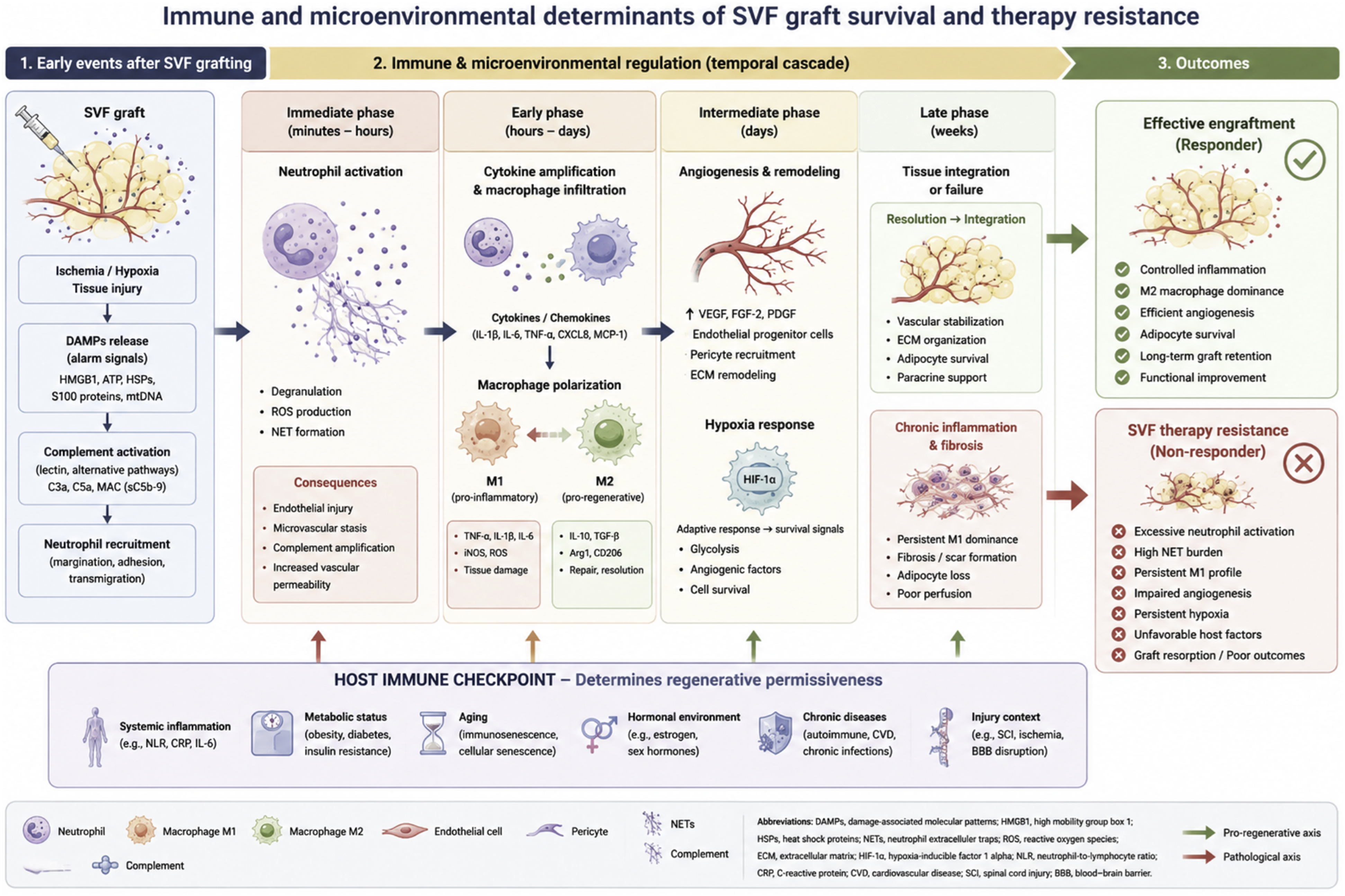
Open accessResearch articleFirst published 2026 , [...]Androgenic alopecia (AGA) is a common hair follicle miniaturization disease driven by androgens. Fibroblasts, especially dermal papilla (DP) cells, are considered the core of the pathogenesis of AGA, but there is a lack of systematic synthesis of evidence.
To systematically review the role of fibroblasts (DP cells and dermal sheath cells) in the pathogenesis of AGA, with a focus on androgen signaling transduction, paracrine signaling, disruption of stem cell niche, inflammation, and fibrosis.
PubMed and Web of Science, covering the period from August 23, 2025 to May 28, 2026, including human basic research, animal model studies, and clinical pathology studies related to AGA fibroblast function. Two reviewers independently screened literature, extracted data, and evaluated the risk of bias.
Through retrieval, a total of 1592 articles were identified, and 43 articles were ultimately included. DP cells consistently express high levels of androgen receptor (AR) and 5α-reductase type II. Activation of androgens (dihydrotestosterone) in DP cells leads to: (1) downregulation of Wnt/β-catenin signaling and upregulation of TGF-β/BMP pathway; (2) Changes in secretion profile (decrease in VEGF/IGF-1 and increase in DKK-1/TGF-β1); (3) DP cell aggregation behavior is impaired; (4) Destruction of hair follicle stem cell activation; (5) Inducing inflammation and fibrosis around hair follicles. The evidence for dermal sheath cells as a DP cell bank is still limited.
DP cells are the main transducers of androgen signaling in AGA, driving follicle miniaturization through multiple interrelated mechanisms. The current evidence supports DP cell dysfunction as a therapeutic strategy.
Open accessReview articleFirst published 2026, Rima Haifa, Angliana Chouw , [...]Mesenchymal stromal cell (MSC)–based therapies have demonstrated broad therapeutic potential across inflammatory, degenerative, and neurological disorders; however, clinical outcomes remain highly variable, largely due to heterogeneity in cell source, dosing strategies, routes of administration, treatment timing, and patient-specific factors. This review evaluates how MSC dose and delivery route influence clinical efficacy and safety, and whether specific administration routes require tailored dosing strategies. A systematic literature search of PubMed, Scopus, Web of Science, and the Cochrane Library (2015–2025) identified 29 eligible clinical trials employing intravenous (n = 13), intrathecal (n = 10), intramuscular (n = 4), intranasal (n = 1), and multi-route (n = 1) administration, with doses ranging from 2 × 10⁵ cells/kg body weight to fixed doses exceeding 1 × 10⁸ cells. Intravenous delivery was predominantly used for systemic indications but was limited by pulmonary first-pass sequestration, whereas intrathecal and intranasal routes enhanced central nervous system targeting and intramuscular administration supported prolonged local persistence and paracrine activity. Although MSC therapy was generally well tolerated across studies, substantial outcome variability underscores the need for harmonized frameworks for dose selection, route optimization, and manufacturing consistency. This review highlights critical gaps in dose–route optimization and supports the development of standardized clinical guidelines and advanced strategies, including MSC priming and engineering, to improve therapeutic predictability and translational success.
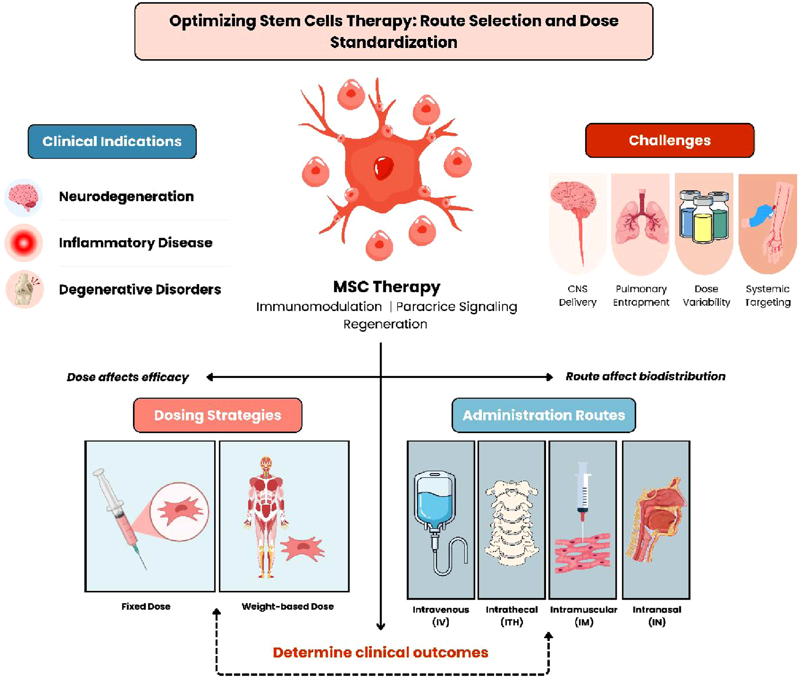
Open accessResearch articleFirst published 2026 , [...]Organ-on-a-chip (OoC) platforms are microengineered systems that combine microfluidic control with living cells to emulate the physiological functions of human tissues and organs in vitro. OoC has become a transformative tool in pharmaceutical research, offering unprecedented capabilities for predicting drug efficacy, pharmacokinetics, and toxicity with human-relevant precision. This study presents a comprehensive scientometric and patent landscape analysis of OoC studies in pharmaceutical sciences spanning 2008–2025. Using CiteSpace, we mapped 1,786 publications to identify influential authors, landmark works, and temporal shifts in thematic focus. Keyword burst and clustering analyses reveal emerging frontiers in multi-organ integration, disease modeling, and drug screening. Patent data indicate a rapid expansion since 2016, led by China and the United States, underscoring a translational trajectory from fundamental research to applied biotechnology. These findings delineate the evolving intellectual and technological framework of organ-on-a-chip research in drug development and highlight future priorities in multi-organ systems, biomaterials optimization, and clinical translation.
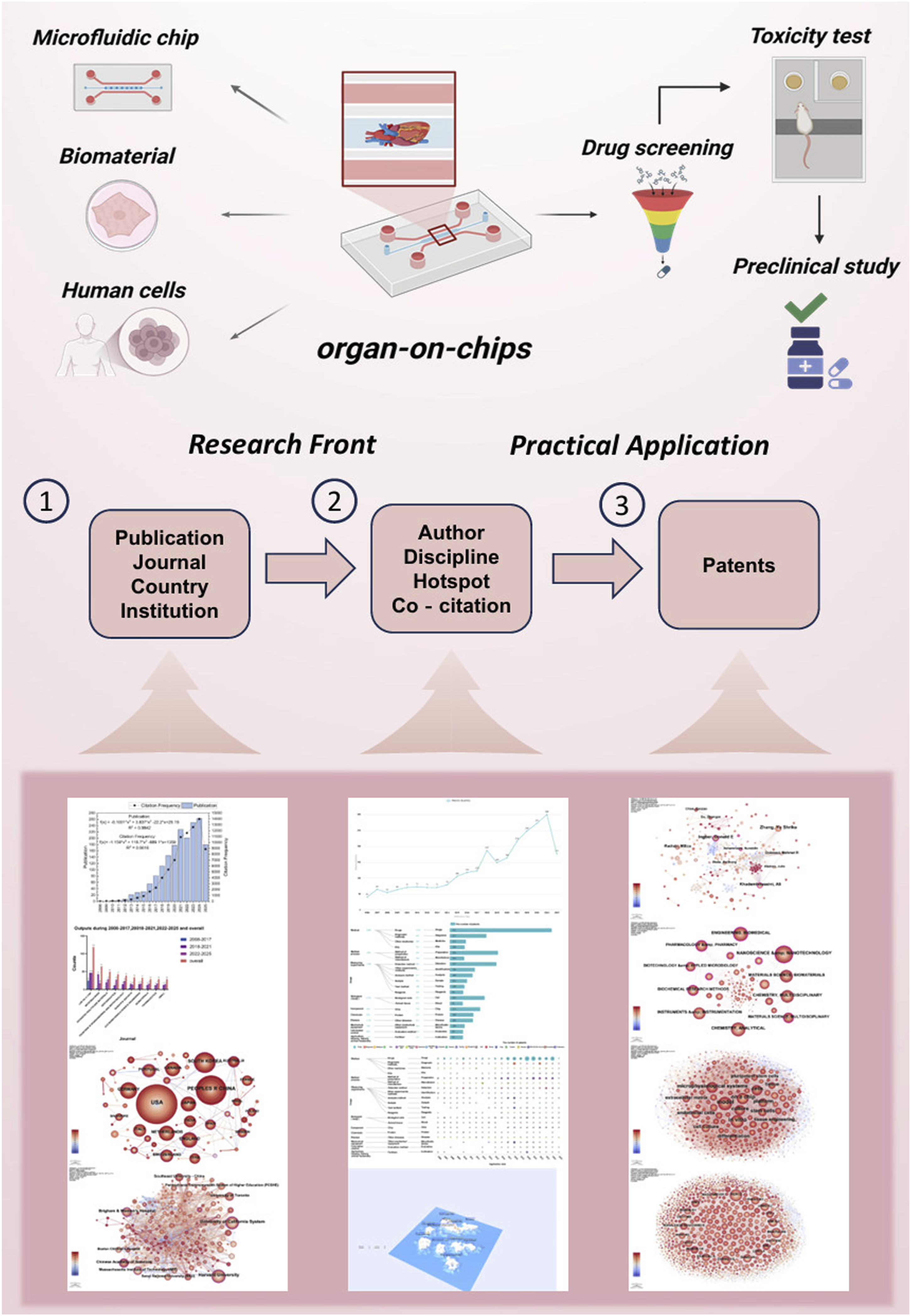
Open accessReview articleFirst published 2026Metastatic colorectal cancers (mCRCs) exhibit substantial heterogeneity at the genetic, transcriptomic, histological, and microenvironmental levels, which contributes to therapeutic resistance and variable clinical outcomes. Patient-derived organoids (PDOs) and patient-derived xenografts (PDXs) have emerged as powerful platforms for modeling this complex disease. PDOs faithfully recapitulate tumor architecture, molecular features, and heterogeneity, enabling high-throughput drug screening and personalized treatment response prediction. In addition, PDX models maintain tumor–stroma interactions